

 PDF
PDF ePub
ePub Citation
 Print
Print


Abstract
Purpose
The spontaneous extensor pollicis longus (EPL) tendon rupture is a well-documented complication of non-displaced or minimally displaced distal radius fracture. Authors analyzed the radiographs of patients treated for closed EPL rupture after distal radius fracture.
Materials and Methods
Twenty-eight patients (21 females, 7 males; average age of 58 years) with tendon transfer for spontaneous rupture of EPL after distal radius fracture were included. Wrist radiographs were taken at the first visit with EPL rupture. On the lateral view, posterior cortical displacement, distance from highest point in Lister's tubercle to fracture line, and height of the Lister's tubercle were measured. The distance from the lunate facet to the fracture line was measured on anteroposterior view. Radiologic change at the time of EPL rupture around the Lister's tubercle was evaluated by comparing it with the contra lateral wrist radiograph. Radial beak fracture pattern was also identified.
Results
The interval between the injury and the spontaneous EPL rupture varied from 2 to 20 weeks, with an average of 6.7 weeks. There were 25 cases of non-displacement, 3 cases of mean 2.0 mm cortical displacement. The average distance from the lunate facet to the fracture line was 9.1 mm (3-12.1 mm), from the highest point in Lister's tubercle to the fracture line was 3.0 mm toward proximal radius (1.7-4.9 mm). The average height of the Lister's tubercle was 3.4 mm in the injured wrist and 3.1 mm in the opposite wrist. Radial beak fracture pattern was shown at 11 cases.
Figures and Tables
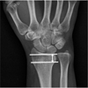
 | Fig. 3Height of the Lister's tubercle is measured from the dorsal aspect of the radial metaphysis to the highest point in the Lister's tubercle.
|
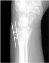
 | Fig. 4Distance from Lister's tubercle to the fracture line is measured from the highest point in Lister's tubercle to the fracture line.
|
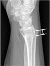
 | Fig. 5Radial beak fracture pattern shows that the fracture line deviates from transverse to proximal at the radial side.
|
Table 1
Descriptive Values of Patients (n=28)

![]()
Table 2
Fractures Included in Analysis

![]()
References
1. Duplay S. Rupture sous-cutanée du tendon du long extenseur du pouce de le main droite, au niveau de la tabatière anatomique. Flexion permanente du pouce. Rétablissement de la faculté d'extension par une opération (suture de l'extrémité de tendon rompu avec le primer radial externe). Bulletins et Mémoires de la Societé Chirurgie de Paris. 1876; 2:788.
2. Leslie BM, Carlson G, Ruby LK. Results of extensor carpi ulnaris tenodesis in the rheumatoid wrist undergoing a distal ulnar excision. J Hand Surg Am. 1990; 15:547–551.

3. Huang HW, Strauch RJ. Extensor pollicis longus tenosynovitis: a case report and review of the literature. J Hand Surg Am. 2000; 25:577–579.
4. Hunt JR. Paralysis of the ungual phalanx of the thumb from spontaneous rupture of the extensor pollicis longus: the socalled drummer's palsy. JAMA. 1915; 64:1138–1140.
5. McMaster PE. Late ruptures of extensor and flexor pollicis longus tendons following Colles' fracture. J Bone Joint Surg. 1932; 14:93.
6. White BD, Nydick JA, Karsky D, Williams BD, Hess AV, Stone JD. Incidence and clinical outcomes of tendon rupture following distal radius fracture. J Hand Surg Am. 2012; 37:2035–2040.
7. Smith FM. Late rupture of extensor policis longus tendon following Colles's fracture. J Bone Joint Surg Am. 1946; 28:49–59.
8. Skoff HD. Postfracture extensor pollicis longus tenosynovitis and tendon rupture: a scientific study and personal series. Am J Orthop (Belle Mead NJ). 2003; 32:245–247.
9. McKay SD, MacDermid JC, Roth JH, Richards RS. Assessment of complications of distal radius fractures and development of a complication checklist. J Hand Surg Am. 2001; 26:916–922.
10. Wolfe SW. Distal radius fractures. In : Wolfe SW, Hotchkiss RN, Pederson WC, Kozin SH, Cohen MS, editors. Green's operative hand surgery. . Philadelphia: PA Elsevier;2017. p. 576.
11. Denman EE. Rupture of the extensor pollicis longus: a crush injury. Hand. 1979; 11:295–298.
12. Engkvist O, Lundborg G. Rupture of the extensor pollicis longus tendon after fracture of the lower end of the radius: a clinical and microangiographic study. Hand. 1979; 11:76–86.
13. Helal B, Chen SC, Iwegbu G. Rupture of the extensor pollicis longus tendon in undisplaced Colles' type of fracture. Hand. 1982; 14:41–47.
14. Tubiana R. The hand. Philadelphia: WB Saunders;1981.
15. Bonatz E, Kramer TD, Masear VR. Rupture of the extensor pollicis longus tendon. Am J Orthop (Belle Mead NJ). 1996; 25:118–122.
16. Hirasawa Y, Katsumi Y, Akiyoshi T, Tamai K, Tokioka T. Clinical and microangiographic studies on rupture of the E.P.L. tendon after distal radial fractures. J Hand Surg Br. 1990; 15:51–57.
17. Cho NY, Seo CY, Kim MS, Kim HS, Lee KB. Extensor pollicis longus rupture after distal radius fracture. J Korean Fract Soc. 2012; 25:52–57.
18. Hove LM. Delayed rupture of the thumb extensor tendon. A 5-year study of 18 consecutive cases. Acta Orthop Scand. 1994; 65:199–203.
19. Roth KM, Blazar PE, Earp BE, Han R, Leung A. Incidence of extensor pollicis longus tendon rupture after nondisplaced distal radius fractures. J Hand Surg Am. 2012; 37:942–947.
20. Belsole RJ, Hess AV. Concomitant skeletal and soft tissue injuries. Orthop Clin North Am. 1993; 24:327–331.
21. Trevor D. Rupture of the extensor pollicis longus tendon after Colles fracture. J Bone Joint Surg Br. 1950; 32:370–375.
22. Koh S, Andersen CR, Buford WL Jr, Patterson RM, Viegas SF. Anatomy of the distal brachioradialis and its potential relationship to distal radius fracture. J Hand Surg Am. 2006; 31:2–8.
23. Iwamoto A, Morris RP, Andersen C, Patterson RM, Viegas SF. An anatomic and biomechanic study of the wrist extensor retinaculum septa and tendon compartments. J Hand Surg Am. 2006; 31:896–903.
24. Diep GK, Adams JE. The prodrome of extensor pollicis longus tendonitis and rupture: rupture may be preventable. Orthopedics. 2016; 39:318–322.
25. Noordanus RP, Pot JH, Jacobs PB, Stevens K. Delayed rupture of the extensor pollicis longus tendon: a retrospective study. Arch Orthop Trauma Surg. 1994; 113:164–166.
26. Navaratnam AV, Ball S, Eckersley R. Prophylactic decompression of extensor pollicis longus to prevent rupture. BMJ Case Rep. 2013; 2013:pii: bcr2013010196.
 XML Download