

 PDF
PDF ePub
ePub Citation
 Print
Print


Abstract
The wrist joint is a complicated structure composed of many bones and ligaments. Therefore, understanding the anatomy and the biomechanics of the wrist is important in order to administer proper treatment for patients. To easily understand the complicated structure, there were many trials to unite the complicated structure with a simple group such as the carpal row concept and the carpal column concept. Movement and load transfer along the wrist joint occurs with balanced action between carpal bones. To evaluate this static equilibrium, measuring tools such as carpal height ratio are used. When wrist flexion/extension occurs, each carpal row moves synchronously with action of the scaphoid. In contrast with flexion/extension, when wrist radial deviation/ulnar deviation occurs, the proximal carpal row moves in the sagittal plane, instead of the coronal plane. Recently, the dart throwing motion which occurred from the position of dorsiflexion with radial deviation to volar flexion with ulnar deviation is considered the main movement of the wrist joint.
Figures and Tables
Fig. 1
Row concept of carpal bones. The joints with adjacent bones which do not have gross motion, considered as a single motor unit. The proximal row is composed of scaphoid, lunate, and triquetrum. The distal row is composed of trapezium, trapezoid, capitate, and hamate.

Fig. 2
Column concept of carpal bones. The lateral column is composed of scaphoid, trapezium, and trapezoid. The middle column is composed of lunate and capitate, and the medial column is composed of triquetrum and hamate.
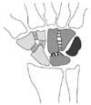
Fig. 3
Scaphoid nonunion advanced collapse. Left wrist simple radiograph of a 53-year-old man. The advanced degenerative changes are evident around the radiocarpal joint with scaphoid nonunion.
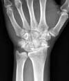
Fig. 4
Carpal height ratio. (A) Carpal height ratio is calculated by dividing carpal height (b) with length of the 3rd metacarpal bone (a). (B) A simple radiograph of measurement of carpal height ratio.
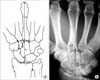
Fig. 5
Movement of carpal rows during radial deviation and ulnar deviation. (A) During radial deviation of the wrist joint, the proximal carpal row is flexed volarly and deviated radially, and the distal carpal row is flexed dorsally. (B) During ulnar deviation of the wrist joint, the proximal carpal row is flexed dorsally and deviated ulnarly, and the distal carpal row is flexed volarly.
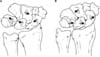
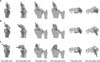
Fig. 6
Computed tomography based three dimensional kinematic comparison of the scaphoid during dart throwing motion between distal radius fracture malunion and contralateral normal side. (A) Radial deviation and dorsal tilt position. (B) Mid-range of dart throwing motion. (C) Ulnar deviation and volar tilt position. The orientations of the helical axes (bold line of each figure) of the scaphoid are different between two sides.
References
1. Erhart S, Schmoelz W, Arora R, Lutz M. The biomechanical effects of a deepened articular cavity during dynamic motion of the wrist joint. Clin Biomech (Bristol, Avon). 2012; 27:557–561.

2. Berdia S, Wolfe SW. Effects of scaphoid fractures on the biomechanics of the wrist. Hand Clin. 2001; 17:533–540. vii–viii.
3. Moritomo H, Apergis EP, Garcia-Elias M, Werner FW, Wolfe SW. International Federation of Societies for Surgery of the Hand 2013 Committee's report on wrist dart-throwing motion. J Hand Surg Am. 2014; 39:1433–1439.
4. Evarts CM. Surgery of the musculoskeletal system. New York: Churchill Livingstone;1983.
5. Tang JB. General concepts of wrist biomechanics and a view from other species. J Hand Surg Eur Vol. 2008; 33:519–525.
6. Taleisnik J. Classification of carpal instability. Bull Hosp Jt Dis Orthop Inst. 1984; 44:511–531.
7. Palmer AK, Werner FW. Biomechanics of the distal radioulnar joint. Clin Orthop Relat Res. 1984; 187:26–35.
8. Strauch RJ. Scapholunate advanced collapse and scaphoid nonunion advanced collapse arthritis: update on evaluation and treatment. J Hand Surg Am. 2011; 36:729–735.
9. Youm Y, Flatt AE. Kinematics of the wrist. Clin Orthop Relat Res. 1980; 149:21–32.
10. Wang YC, Tseng YC, Chang HY, Wang YJ, Chen CJ, Wu DY. Gender differences in carpal height ratio in a taiwanese population. J Hand Surg Am. 2010; 35:252–255.
11. Schuind F, Eslami S, Ledoux P. Kienbock's disease. J Bone Joint Surg Br. 2008; 90:133–139.
12. Bouman HW, Messer E, Sennwald G. Measurement of ulnar translation and carpal height. J Hand Surg Br. 1994; 19:325–329.
13. Malone PS, Cooley J, Morris J, Terenghi G, Lees VC. The biomechanical and functional relationships of the proximal radioulnar joint, distal radioulnar joint, and interosseous ligament. J Hand Surg Eur Vol. 2015; 40:485–493.
14. Ishii S, Palmer AK, Werner FW, Short WH, Fortino MD. Pressure distribution in the distal radioulnar joint. J Hand Surg Am. 1998; 23:909–913.
15. Hotchkiss RN, An KN, Sowa DT, Basta S, Weiland AJ. An anatomic and mechanical study of the interosseous membrane of the forearm: pathomechanics of proximal migration of the radius. J Hand Surg Am. 1989; 14:256–261.
16. Moojen TM, Snel JG, Ritt MJ, Venema HW, Kauer JM, Bos KE. In vivo analysis of carpal kinematics and comparative review of the literature. J Hand Surg Am. 2003; 28:81–87.
17. Wolfe SW, Neu C, Crisco JJ. In vivo scaphoid, lunate, and capitate kinematics in flexion and in extension. J Hand Surg Am. 2000; 25:860–869.
18. Landsmeer JM. Studies in the anatomy of articulation. II. Patterns of movement of bi-muscular, bi-articular systems. Acta Morphol Neerl Scand. 1961; 3:304–321.
19. Chung MS, Seong SC, Choi IH. Textbook of fractures. 3rd ed. Seoul: Koonja publishing Co;2008. p. 285–307.
20. Nanno M, Buford WL Jr, Patterson RM, Andersen CR, Viegas SF. Three-dimensional analysis of the ligamentous attachments of the first carpometacarpal joint. J Hand Surg Am. 2006; 31:1160–1170.
21. Viegas SF, Yamaguchi S, Boyd NL, Patterson RM. The dorsal ligaments of the wrist: anatomy, mechanical properties, and function. J Hand Surg Am. 1999; 24:456–468.
22. Kijima Y, Viegas SF. Wrist anatomy and biomechanics. J Hand Surg Am. 2009; 34:1555–1563.
23. Rajan PV, Day CS. Scapholunate Interosseous Ligament Anatomy and Biomechanics. J Hand Surg Am. 2015; 40:1692–1702.
24. Berger RA, Imeada T, Berglund L, An KN. Constraint and material properties of the subregions of the scapholunate interosseous ligament. J Hand Surg Am. 1999; 24:953–962.
25. Patterson RM, Yazaki N, Andersen CR, Viegas SF. Prediction of ligament length and carpal diastasis during wrist flexion-extension and after simulated scapholunate instability. J Hand Surg Am. 2013; 38:509–518.
26. Salva-Coll G, Garcia-Elias M, Hagert E. Scapholunate instability: proprioception and neuromuscular control. J Wrist Surg. 2013; 2:136–140.
27. Tang JB, Ryu J, Han JS, Omokawa S, Kish V, Wearden S. Biomechanical changes of the wrist flexor and extensor tendons following loss of scaphoid integrity. J Orthop Res. 1997; 15:69–75.
28. Moritomo H, Murase T, Goto A, Oka K, Sugamoto K, Yoshikawa H. Capitate-based kinematics of the midcarpal joint during wrist radioulnar deviation: an in vivo three-dimensional motion analysis. J Hand Surg Am. 2004; 29:668–675.
29. Wolfe SW, Crisco JJ, Orr CM, Marzke MW. The dartthrowing motion of the wrist: is it unique to humans? J Hand Surg Am. 2006; 31:1429–1437.
30. Lee S, Kim YS, Park CS, et al. CT-based three-dimensional kinematic comparison of dart-throwing motion between wrists with malunited distal radius and contralateral normal wrists. Clin Radiol. 2014; 69:462–467.
31. Leventhal EL, Moore DC, Akelman E, Wolfe SW, Crisco JJ. Carpal and forearm kinematics during a simulated hammering task. J Hand Surg Am. 2010; 35:1097–1104.
32. Werner FW, Short WH, Palmer AK, Sutton LG. Wrist tendon forces during various dynamic wrist motions. J Hand Surg Am. 2010; 35:628–632.
33. Brigstocke GH, Hearnden A, Holt C, Whatling G. In-vivo confirmation of the use of the dart throwers motion during activities of daily living. J Hand Surg Eur Vol. 2014; 39:373–378.
 XML Download