

 PDF
PDF ePub
ePub Citation
 Print
Print


Abstract
Shoulder dislocation is the most common dislocation presenting to the emergency department. In old age, the attempt of closed reduction is made with caution in order to prevent iatrogenic fracture around the shoulder. We report two cases of iatrogenic fractures of humeral shaft and anatomical neck in female patients older than 70 years old, which occurred during the manual closed reduction. One patient was proved as first-time and the other was recurrent. In addition, the second case had a massive irreparable rotator cuff tear. Those patients were treated successfully with humeral nailing and reverse total shoulder arthroplasty, respectively.
Figures and Tables
Fig. 1
(A, B) Shoulder radiographs show a subglenoid type of anterior glenohumeral dislocation. (C) The humeral shaft fracture occurred when attempting closed reduction detected in a post reduction radiograph. (D) A radiograph taken five months later shows fixation with humeral nailing. Postoperatively, dislocation was reduced and there were no specific complications. Bone union was observed at 5 months of final follow-up.
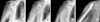
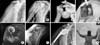
Fig. 2
(A, B) Shoulder radiographs show a subacromial type of anterior glenohumeral dislocation. (C) Anterior dislocation with humeral anatomical neck fracture was observed on 3-dimensional computed tomography images. (D) Intraoperative photograph shows a humeral anatomical neck fracture. (E) Coronal oblique, T2-weighted magnetic resonance imaging (MRI) scan of a full thickness tear and medial retraction of the supraspinatus tendon (white arrow). (F) And supraspinatus muscle (SSP) with Goutallier classification grade III was seen on a sagittal, T1-weighted MRI scan. (G, H) Postoperative with a reverse total shoulder arthroplasty (Zimmer, Cowpens, SC, USA) 4 months later, radiograph and clinical photograph, there were no specific complications and favorable range of motion of the right shoulder was gained at 4 months of follow-up.
References
1. Court-Brown CM, Heckman JD, McQueen MM, Tornetta P. Rockwood and Green's fractures in adults. 8th ed. Philadelphia: Lippincott Williams & Wilkins Co;2015. p. 1531.
2. Atoun E, Narvani A, Even T, et al. Management of first-time dislocations of the shoulder in patients older than 40 years: the prevalence of iatrogenic fracture. J Orthop Trauma. 2013; 27:190–193.

3. Court-Brown CM, Caesar B. Epidemiology of adult fractures: a review. Injury. 2006; 37:691–697.
4. Sayegh FE, Kenanidis EI, Papavasiliou KA, Potoupnis ME, Kirkos JM, Kapetanos GA. Reduction of acute anterior dislocations: a prospective randomized study comparing a new technique with the Hippocratic and Kocher methods. J Bone Joint Surg Am. 2009; 91:2775–2782.
5. Hersche O, Gerber C. Iatrogenic displacement of fracture-dislocations of the shoulder. A report of seven cases. J Bone Joint Surg Br. 1994; 76:30–33.
6. McLaughlin HL, MacLellan DI. Recurrent anterior dislocation of the shoulder. II. A comparative study. J Trauma. 1967; 7:191–201.
7. Gumina S, Postacchini F. Anterior dislocation of the shoulder in elderly patients. J Bone Joint Surg Br. 1997; 79:540–543.
8. Shin DI, Shin DJ, Choi S, Park JH. Reduction of anterior shoulder dislocations by acromion upholding method (Shin's method): technical note. J Korean Orthop Assoc. 2013; 48:471–474.
9. Beattie TF, Steedman DJ, McGowan A, Robertson CE. A comparison of the Milch and Kocher techniques for acute anterior dislocation of the shoulder. Injury. 1986; 17:349–352.
10. Saitoh S, Nakatsuchi Y, Latta L, Milne E. Distribution of bone mineral density and bone strength of the proximal humerus. J Shoulder Elbow Surg. 1994; 3:234–242.
 XML Download