

 PDF
PDF ePub
ePub Citation
 Print
Print


Introduction
Midshaft clavicle fractures account for 80% of all types of clavicle fractures.1234567) The mainstay of treatment for displaced midshaft clavicle fractures is an open reduction and plate fixation.28) It is more beneficial than conservative treatment in terms of the incidence of malunion and nonunion and early motion.78910) However, there might be some complications with regard to open reduction and plate fixation such as infections, hypoesthesia, irritation of the skin, and hardware failure.11) Clavicular hypoesthesia is a common complication after surgery for clavicle midshaft fractures and its effect on clinical outcome is known to be insignificant with respect to function.261112) The permanent hypoesthesia might occur in some patients and they might severely suffer from this complication and could be unsatisfied with those complications.61113) However, we are still less informed about the clinical implications of clavicular hypoesthesia.
Supraclavicular nerve injury during the operation is known to be a pathology of clavicular hypoesthesia.61415) The supraclavicular nerve arises from the roots of C3 and C4 and the intermediate and lateral branches of the supraclavicular nerve are the main branches of innervation around the shoulder.614) The anatomical safe zone without any disruption of the supraclavicular nerve during clavicle surgery is known to be located at the narrow area near the sternoclavicular (SC) joint and the acromioclavicular joint irrespective of anatomical variations.14) The vertical incision and the minimally invasive approach rather than the transverse incision along the long axis of the clavicle are known to reduce the incidence of clavicular hypoesthesia.6) However, the minimally invasive exposure and the vertical incision might be insufficient to expose the long spiral type of fractures.
We hypothesized that clavicular hypoesthesia might be clinically correlated with the satisfaction of patients rather than the other functional problems and there might be a anatomical correlation between the location of the clavicular plate and the incidence of hypoesthesia. We postulated that the extent of the approach was proportionally related to the length of the plate that we used during surgery. Therefore, the analysis regarding the extent of the approach could be performed by the radiological analysis of plate location. The aims of this study were to verify the correlation between the location of clavicular plates and the incidence of clavicular hypoesthesia and to identify clinical features of clavicular hypoesthesia including the patient's satisfaction and pain.
Materials and Methods
The institutional review board of the Seonam University College of Medicine Myongji Hospital approved this study (MJH 15-013).
1. Cohort
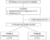
Ninety-eight patients who received the open reduction and internal fixation for the displaced midshaft clavicle fractures between March 2013 and October 2014 at two hospitals were found to be eligible for this retrospective study. Exclusion criteria were ipsilateral upper extremity fractures such as scapular fracture (n=4) and humeral shaft fracture (n=1), patients with infection (n=1) and fractures in disabled patients, and 14 patients were excluded due to loss of follow-up. A total of 78 patients were finally enrolled in this study (Fig. 1). Mean age of the 78 enrolled patients (58 men, 20 women) was 41.0±17.6 years (range, 15-71 years). The mean follow-up period was 12.6±3.0 months (range, 6-18 months). Demographic characteristics and radiological variables of patients in groups A and B are summarized in Table 1.
2. Operation technique
We performed the operation with the patients in the beach chair position. Before the start of the operation, the fracture site was marked after confirmation by the c-arm. Then, a transverse skin incision along the long axis of the clavicle was performed such that the center of the incision was at the center of the fracture. During the operation, the procedure for identification of every branches of the supraclavicular nerve was not performed. We used the lag screws for comminuted fractures, and locking compression plates (Acumed, Portland; Synthes, Oberdorf, Netherlands) were applied to stabilize the fractures.
3. Postoperative rehabilitation
Arm sling was applied for 2 weeks and the pendulum and active assisted exercises were started immediately after the operation. However, weight bearing onto the affected shoulder and contact sports were prohibited until postoperative 12 weeks.
4. Radiological evaluation
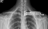
Radiologic evaluation was performed by using the clavicle anteroposterior (AP), cephalic and caudal tilt views. The location of the clavicular plate was assessed by two 10-year experienced orthopedic surgeons (J.Y.K., S.H.K.) in shoulder surgery on a clavicle AP X-ray. The total clavicular length (A), the distance to the medial end of the plate from the SC joint (B), and the distance to the lateral end of the plate from the SC joint (C) were measured and the distance to the medial end of the plate to the total length of the clavicle (B/A) and the distance to the lateral end of the plate to the total length o f the clavicle (C/A) w ere calculated (Fig. 2). T he bone union was evaluated at 4, 6, 12, 18, and 24 weeks postoperatively. Union was defined as callus formation or filling of hairline like gap during the follow-up.
5. Clinical outcome evaluation
Hypoesthesia was evaluated before the surgery and at postoperative 4 weeks of follow-up.13) We asked the patients whether they could identify the light touch sensation caused by the finger contact just distal to the clavicle or incision site and anterior chest and compared it to that on the contralateral side. We tried to differentiate the hypoesthesia with tingling sensation or hyperesthesia. The severity of hypoesthesia was also assessed in three grades or as mild, moderate, and severe, and the location of hypoesthesia was assessed as medial or lateral. The recovery of hypoesthesia was assessed at a month interval. Pain visual analogue scale (VAS; 0 to 10, with 10 defined as the worst pain), range of motion (ROM) of the shoulder, and satisfaction VAS (0 to 10, with 10 defined as highest satisfaction) were evaluated at the final follow-up (minimum 6 months). Patients without hypoesthesia were designated as group A and patients with hypoesthesia were designated as group B.
6. Sample size calculation
At the start of the study, the sample size was calculated to detect 10% difference of satisfaction VAS of patients with clavicular hypoesthesia. The 10% difference was determined to be clinically significant based on the pilot study in the author's hospital that included 20 patients. A sample size of 96 patients was required for a power of 80% at a type I error level of 0.05 and for an expected dropout rate of 20%. We also performed the post-hoc analysis with the results of satisfaction VAS in each group and the power was calculated 73%.
7. Statistical analyses
All statistical analyses were performed using the PASW software package ver. 18.0 (IBM Co., Armonk, NY, USA). Student t-test was used to evaluate differences between the groups for continuous variables, and the chi-square or Fisher's exact test was used for comparison of categorical variables.
A p<0.05 was considered to indicate statistical significance.
Results
There were no significant demographic differences between the two groups (all p>0.05). We found that every case of enrolled clavicle fractures revealed no preoperative hypoesthesia, and the incidence of postoperative hypoesthesia was 32.1% (25 out of 78 patients). Twenty-two patients reported that their symptoms were mild rather than moderate (p=0.917). The locations of hypoesthesia were evenly distributed (medial in 12 cases, lateral in 13 cases). The recovery from hypoesthesia was observed in 23 out of 25 patients (p=0.008) and mean recovery time was 8.2±4.1 months (range, 3-15 months).
The medial end location of the plate was at a mean 17.5%±16.0% of the total length of the clavicle and the lateral end location of the plate was at a mean 51.0%±44.3% of the total length of the clavicle. No correlation was observed with respect to the location of the clavicular plate and the incidence of clavicular hypoesthesia (p=0.666 at the medial end and p=0.369 at the lateral end).
Hypoesthesia was related to the satisfaction VAS (p=0.002). Interestingly, we observed that the pain VAS was also related to the incidence of hypoesthesia (p=0.022). There was no significant difference with respect to the shoulder ROM between the two groups (Table 2).
Discussion
According to the results of our study, there was no correlation between the location of the plate and the incidence of clavicular hypoesthesia. The incidence of clavicular hypoesthesia in this study was 32.1% and most of the cases of hypoesthesia recovered finally, which was similar to the previous results.13)
The universal transverse skin incision along the long axis of the clavicle might cause injury to branches of the supraclavicular nerve more often than the other approaches such as vertical incision and minimally invasive incision and it might cause permanent neurologic damage or worsen the clinical outcomes.6) Some of the anatomical studies found that there might be two (medial and lateral) or three branches (medial, intermediate, and lateral) of the supraclaviclular nerve that vertically pass over the clavicle and the anatomical locations of these branches to the whole length of the clavicle were documented to be predictable.61415) However, the clinical correlation between incidence of supraclavicular nerve injury and anatomical location of the plate or extent of the approach during clavicle operation has not enoughly studied.
We hypothesized that there might be some correlation between plate location and the incidence of clavicular hypoesthesia as well as the clavicular hypoesthesia was related to the satisfaction of patients or other clinical variables such as pain of the shoulder.
In the current study, clavicular plates were located between mean 5.0±1.4 cm and 8.8±5.9 cm or between mean 17.5%±16% and 51%±44.3% from the SC joint. We observed that both the absolute distance of the lateral end of the plate from the SC joint (B) and the relative location of the lateral end of the plate to the whole length of the plate (B/A) were in wide range; however, the incidence of hypoesthesia was not affected by these variations in the location of the plate or extent of the approach. We considered that this might be somewhat due to the presence or absence of the intermediate branch of the supraclavicular nerve.14) According to Nathe et al.,14) the intermediate branch was observed in about half of the normal population and the distance from t he S C joint to t he i ntermediate branch w as b etween 26.9% and 64.3% the length of the clavicle. These characteristics related to the intermediate branch of the supraclavicular nerve might have resulted in the unpredictable incidence of hypoesthesia.
In this study, the satisfaction VAS score was statistically decreased in patients with clavicular hypoesthesia. However, according to the results of a previous study comparing the overall satisfaction between the vertical and horizontal incisions during the clavicle fracture operation, the patients were significantly more satisfied with the horizontal incision, and it might be due to the difference in satisfaction with the scar.6) Previous studies reported that there were several patients who suffered from severe hypoesthesia after the clavicle operation.613) However, we observed that most of the patients enrolled in this study reported mild hypoesthesia. Only two out of twenty-five patients reported moderate hypoesthesia. The high prevalence of mild hypoesthesia rather than moderate and severe hypoesthesia might be due to the late evaluation period at postoperative 4 weeks as well as the possible bias among the physicians' evaluations. The patients might represent diminished symptoms at postoperative 4 weeks compared to the postoperative two week period and physicians might evaluated the degree of hypoesthesia as mild to acquire better surgical results.
Interestingly, we observed that the pain VAS in group B (with hypoesthesia) was significantly worse than that in group A (without hypoesthesia) at the final follow-up. We thought this might be due to the neuralgic pain during recovery that occurred in some cases of injury to branches of the supraclavicular nerve. Previous studies reported that hypoesthesia was not related to the shoulder function which was composed of pain, shoulder motion, and muscle power. In this study, we did not perform functional evaluation by using scores; however, the pain VAS and ROM of the shoulder were evaluated. No significant differences with ROMs were observed between groups.
There are some limitations to this study. First, we did not identify the every branch of the supraclavicular nerve during surgery; therefore, we could not verify the actual relationship between injury to branches of the supraclavicular nerve and clavicular hypoesthesia. Second, the degree of hypoesthesia was evaluated in only three categories such as mild, moderate, and severe which might be inappropriate for evaluating the exact degree of hypoesthesia. Finally, the post-hoc analysis revealed the 73% of power which meant that we might not made the strong conclusion with this study regarding satisfaction of patients.
To the best of the authors' knowledge, this is the first study t o investigate t he c orrelation b etw een t he l ocation of the plate or the extent of approach and the incidence of hypoesthesia although radiological evaluation was performed by using the clavicle AP X-ray.
Conclusion
We could not predict the incidence of hypoesthesia by the location of clavicular plates. Clinically, the satisfaction and pain of patients might be related to the incidence of clavicular hypoesthesia and larger size cohort study might be needed in the future to get the strong conclusion regarding clavicular hypoesthesia.


 XML Download