

 PDF
PDF ePub
ePub Citation
 Print
Print


Abstract
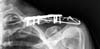
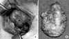
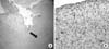
Pins and wires are still used frequently in surgeries of the shoulder; however, these can cause breakage or migration to surrounding tissues, leading to complications. We report on case of a patient with a neck mass who had a past history of pulmonary tuberculosis and distal clavicle fracture with internally fixated state. She was misdiagnosed as tuberculous cervical lymphadenopathy and treated for approximately one year, but was finally revealed as granulation tissue around the internally fixated distal clavicle fracture site, thus, mass excision and metal removal was performed. This case shows the importance of a proper selection device, internal fixation technique, duration, and close follow-up after the operation.
Figures and Tables
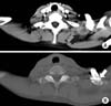 | Fig. 2Axial cervical computed tomography scans. About 2.5 cm sized rim enhancing low density mass was seen at supraclavicular area. The tip of K-wire was in the mass. Black arrow: rim enchanced mass. White arrow: K-wire. (A) Lung setting, (B) bone setting.
|
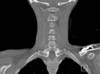 | Fig. 3Coronal cervical computed tomography scans confirmed migration of the K-wire into the mass, showing irritation of the mass by tip of the K-wire.
|
References
1. Lyons FA, Rockwood CA Jr. Migration of pins used in operations on the shoulder. J Bone Joint Surg Am. 1990; 72:1262–1267.

2. Sharma H, Taylor GR, Clarke NM. A review of K-wire related complications in the emergency management of paediatric upper extremity trauma. Ann R Coll Surg Engl. 2007; 89:252–258.
3. Cheon SJ, Lee JM. Early intrathoracic migration of K-wire used for fixation of proximal humerus fracture. J Korean Orthop Assoc. 2011; 46:167–171.
4. Lee BI, Kim YB, Choi HS, Kim CH, Ji JW. Spinal canal migration of a K-wire used for fixation of a distal clavicular fracture. J Korean Orthop Assoc. 2013; 48:231–235.
5. Jean JL, Lee CH, Chu CM, Hsieh SP, Tang HL. Pseudocyst formation after intertrochanteric fracture fixation: a case report. J Formos Med Assoc. 2001; 100:285–288.
6. Bisbinas I, Mikalef P, Gigis I, Beslikas T, Panou N, Christoforidis I. Management of distal clavicle fractures. Acta Orthop Belg. 2010; 76:145–149.
7. Meda PV, Machani B, Sinopidis C, Braithwaite I, Brownson P, Frostick SP. Clavicular hook plate for lateral end fractures: a prospective study. Injury. 2006; 37:277–283.
8. Wu K, Chang CH, Yang RS. Comparing hook plates and Kirschner tension band wiring for unstable lateral clavicle fractures. Orthopedics. 2011; 34:e718–e723.
9. Lee YS, Lau MJ, Tseng YC, Chen WC, Kao HY, Wei JD. Comparison of the efficacy of hook plate versus tension band wire in the treatment of unstable fractures of the distal clavicle. Int Orthop. 2009; 33:1401–1405.
10. Wang SG. Differential diagnosis and treatment of neck masses. J Korean Med Assoc. 2007; 50:613–625.
 XML Download