

 PDF
PDF ePub
ePub Citation
 Print
Print


Figures and Tables
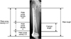
Fig. 1
Dynamic compression principle. Since the screw is inserted into an inclined and transverse cylindrical holes in DCP, the horizontal movement of the screw head can occur along the screw hole. This movement results in movement of the bone fragment relative to the plate and leads to compression of the fracture site. DCP: Dynamic compression plate.
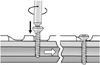
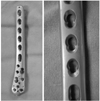
Fig. 4
(A-D) A 46-year-old male with comminuted fractures of the distal femur, patella and proximal tibia.
(E, F) Distal femur fracture was fixed with locking compression plate (LCP), using bridging plating technique and bone union was achieved one year after surgery.
(G, H) Patella was fixed with vertical and cerclage wiring. Intraarticular proximal tibia fracture was fixed with LCP and a lag screw using a compression plating technique. Bony union was uneventfully achieved.
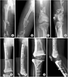
Fig. 5
(A) A 32-year-old female with simple fracture of the distal tibia and fibula.
(B) Distal tibia was reduced and fixed with LCP using MIPO technique. Five months after operation, delayed union was seen.
(C) Complete union was achieved at one year postsurgery.
LCP: Locking compression plate, MIPO: Minimal invasive percutaneous plate osteosynthesis.
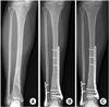
Fig. 6
(A, B) A 71-year-old female with periprosthetic fracture of the distal femur above the femoral component after total knee arthroplasty.
(C) The fracture was percutaneously reduced and fixed with periarticular LCP.
LCP: Locking compression plate.
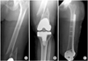
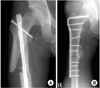
Fig. 7
(A, B) A 74-year-old female with long spiral fracture of the distal femur with the fracture extension into a lateral femoral condyle (black arrow).
(C) The fracture was fixed with LCP using MIPO technique.
(D) Two months after surgery, she slipped down and peri-plate fracture occurred at the proximal end screw site.
LCP: Locking compression plate, MIPO: Minimal invasive percutaneous plate osteosynthesis.
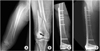
Fig. 8
(A) A 64-year-old male with multifragmentary fracture of the femur shaft with canal diameter of 7 mm.
(B) The fracture was reduced using percutaneous wiring and fixed with LCP using MIPO technique.
(C) One year after surgery, union was obtained around fracture segments.
LCP: Locking compression plate, MIPO: Minimal invasive percutaneous plate osteosynthesis.
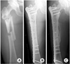
Fig. 9
The schematic drawing showing the plate length and plate screw density in a bridging plating technique using LCP in a comminuted fracture of the distal femur. The overall plate screw density of the construct in this example is 0.47. LCP: Locking compression plate.
References
1. Ahmad M, Nanda R, Bajwa AS, Candal-Couto J, Green S, Hui AC. Biomechanical testing of the locking compression plate: when does the distance between bone and implant significantly reduce construct stability? Injury. 2007. 38:358–364.

2. Allgöwer M, Ehrsam R, Ganz R, Matter P, Perren SM. Clinical experience with a new compression plate "DCP". Acta Orthop Scand Suppl. 1969. 125:45–61.
3. Bottlang M, Doornink J, Byrd GD, Fitzpatrick DC, Madey SM. A nonlocking end screw can decrease fracture risk caused by locked plating in the osteoporotic diaphysis. J Bone Joint Surg Am. 2009. 91:620–627.
4. Doornink J, Fitzpatrick DC, Boldhaus S, Madey SM, Bottlang M. Effects of hybrid plating with locked and nonlocked screws on the strength of locked plating constructs in the osteoporotic diaphysis. J Trauma. 2010. 69:411–417.
5. Egol KA, Kubiak EN, Fulkerson E, Kummer FJ, Koval KJ. Biomechanics of locked plates and screws. J Orthop Trauma. 2004. 18:488–493.
6. Farouk O, Krettek C, Miclau T, Schandelmaier P, Guy P, Tscherne H. Minimally invasive plate osteosynthesis: does percutaneous plating disrupt femoral blood supply less than the traditional technique? J Orthop Trauma. 1999. 13:401–406.
7. Frigg R. Development of the locking compression plate. Injury. 2003. 34:Suppl 2. B6–B10.
8. Frigg R, Appenzeller A, Christensen R, Frenk A, Gilbert S, Schavan R. The development of the distal femur less invasive stabilization system (LISS). Injury. 2001. 32:Suppl 3. SC24–SC31.
9. Fulkerson E, Egol KA, Kubiak EN, Liporace F, Kummer FJ, Koval KJ. Fixation of diaphyseal fractures with a segmental defect: a biomechanical comparison of locked and conventional plating techniques. J Trauma. 2006. 60:830–835.
10. Gardner MJ, Griffith MH, Demetrakopoulos D, et al. Hybrid locked plating of osteoporotic fractures of the humerus. J Bone Joint Surg Am. 2006. 88:1962–1967.
11. Gautier E, Sommer C. Guidelines for the clinical application of the LCP. Injury. 2003. 34:Suppl 2. B63–B76.
12. Goyal T, Nag HL, Tripathy SK. Dynamization of locked plating on distal femur fracture. Arch Orthop Trauma Surg. 2011. 131:1331–1332.
13. Greiwe RM, Archdeacon MT. Locking plate technology: current concepts. J Knee Surg. 2007. 20:50–55.
14. Helfet DL, Shonnard PY, Levine D, Borrelli J Jr. Minimally invasive plate osteosynthesis of distal fractures of the tibia. Injury. 1997. 28:Suppl 1. A42–A47.
15. Kääb MJ, Frenk A, Schmeling A, Schaser K, Schütz M, Haas NP. Locked internal fixator: sensitivity of screw/plate stability to the correct insertion angle of the screw. J Orthop Trauma. 2004. 18:483–487.
16. Kessler SB, Deiler S, Schiffl-Deiler M, Uhthoff HK, Schweiberer L. Refractures: a consequence of impaired local bone viability. Arch Orthop Trauma Surg. 1992. 111:96–101.
17. Kinast C, Bolhofner BR, Mast JW, Ganz R. Subtrochanteric fractures of the femur. Results of treatment with the 95 degrees condylar blade-plate. Clin Orthop Relat Res. 1989. (238):122–130.
18. Krettek C, Schandelmaier P, Miclau T, Tscherne H. Minimally invasive percutaneous plate osteosynthesis (MIPPO) using the DCS in proximal and distal femoral fractures. Injury. 1997. 28:Suppl 1. A20–A30.
19. Lill H, Hepp P, Korner J, et al. Proximal humeral fractures: how stiff should an implant be? A comparative mechanical study with new implants in human specimens. Arch Orthop Trauma Surg. 2003. 123:74–81.
20. Mast J, Jakob R, Ganz R. Planning and reduction technique in fracture surgery. 1989. Berlin; New York: Springer-Verlag;–254.
21. Miller DL, Goswami T. A review of locking compression plate biomechanics and their advantages as internal fixators in fracture healing. Clin Biomech (Bristol, Avon). 2007. 22:1049–1062.
22. Oh JK, Hwang JH, Lee SJ, Kim JI. Dynamization of locked plating on distal femur fracture. Arch Orthop Trauma Surg. 2011. 131:535–539.
23. Owsley KC, Gorczyca JT. Fracture displacement and screw cutout after open reduction and locked plate fixation of proximal humeral fractures [corrected]. J Bone Joint Surg Am. 2008. 90:233–240.
24. Perren SM, Buchanan JS. Basic concepts relevant to the design and development of the point contact fixator (PC-Fix). Injury. 1995. 26:Suppl 2. B1–B4.
25. Perren SM, Cordey J, Rahn BA, Gautier E, Schneider E. Early temporary porosis of bone induced by internal fixation implants. A reaction to necrosis, not to stress protection? Clin Orthop Relat Res. 1988. 232:139–151.
26. Perren SM, Klaue K, Pohler O, Predieri M, Steinemann S, Gautier E. The limited contact dynamic compression plate (LC-DCP). Arch Orthop Trauma Surg. 1990. 109:304–310.
27. Rockwood CA, Green DP, Bucholz RW. Rockwood and Green's fractures in adults. 2010. 7th ed. Philadelphia: Wolters Kluwer Health/Lippincott Williams & Wilkins.
28. Rozbruch SR, Müller U, Gautier E, Ganz R. The evolution of femoral shaft plating technique. Clin Orthop Relat Res. 1998. (354):195–208.
29. Rüedi T, Kolbow H, Allgöwer M. Experiences with the dynamic compression plate (DCP) in 418 fresh fractures of the tibial shaft (author's transl). Arch Orthop Unfallchir. 1975. 82:247–256.
30. Rüedi TP, Murphy WM. AO principles of fracture management. 2000. Stuttgart, New York: Thieme; AO Pub..
31. Schütz M, Haas NP. LISS-internal plate fixator. Kongressbd Dtsch Ges Chir Kongr. 2001. 118:375–379.
32. Sommer C, Babst R, Müller M, Hanson B. Locking compression plate loosening and plate breakage: a report of four cases. J Orthop Trauma. 2004. 18:571–577.
33. Stanbury SJ, Salo A, Elfar JC. Biomechanical analysis of a volar variable-angle locking plate: the effect of capturing a distal radial styloid fragment. J Hand Surg Am. 2012. 37:2488–2494.
34. Stoffel K, Dieter U, Stachowiak G, Gächter A, Kuster MS. Biomechanical testing of the LCP-how can stability in locked internal fixators be controlled? Injury. 2003. 34:Suppl 2. B11–B19.
35. Stoffel K, Klaue K, Perren SM. Functional load of plates in fracture fixation in vivo and its correlate in bone healing. Injury. 2000. 31:Suppl 2. S-B37–S-B50.
36. Tan SL, Balogh ZJ. Indications and limitations of locked plating. Injury. 2009. 40:683–691.
37. Uhthoff HK, Poitras P, Backman DS. Internal plate fixation of fractures: short history and recent developments. J Orthop Sci. 2006. 11:118–126.
38. Wagner M. General principles for the clinical use of the LCP. Injury. 2003. 34:Suppl 2. B31–B42.
 XML Download