

 PDF
PDF ePub
ePub Citation
 Print
Print


Abstract
Purpose
To discriminate the characteristics between reverse obliquity fractures in the elderly and that of young adults using three-dimensional computed tomography (3D CT).
Materials and Methods
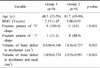
Eighteen patients who had reverse obliquity intertrochanteric fractures were enrolled from January 2007 to March 2012. The fracture pattern was analyzed using the 3D CT. The area showing low density (bone defect) of trochanter and femoral neck region was measured. Patients were divided into two groups: Group I, less than 65 years old and Group 2, 65 years and over.
Results
In all 9 cases of group 1, the proximal fragment had a 'V' shape with an average of 5.6 cm below the vastus ridge; however, the fracture of 8 cases (88.97%) in group 2 had a 'Λ' shape of the distal fragment at the level of vastus ridge and an additional fracture line extending to the greater trochanter tip. The bone defect volume of the trochanter and femoral neck region was larger significantly in group 2 than in group 1.
Conclusion
Reverse obliquity intertrochanteric fracture in the elderly demonstrated a pattern of bursting fracture with 4 parts, which had different patterns from that of young patients. We believe that the larger volume of bone defects resulted in the difference of fracture patterns between the two groups.
Figures and Tables
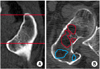 | Fig. 1
(A) Region of interest in trochanter from the greater trochanter tip to the upper part of lesser trochanter.
(B) Measurement of bone defect of the trochanter and femoral neck region.
|
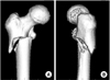 | Fig. 2Three-dimensional image of reverse obliquity fracture in the young group (group 1).
(A) Anterior-posterior view showing a typical reverse obliquity pattern.
(B) Lateral view showing a 'V' shape apex of proximal fragment.
|
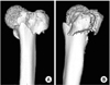 | Fig. 3Three-dimensional image of reverse obliquity fracture in the elderly group (group 2).
(A) Anterior-posterior view showing a typical reverse obliquity pattern with additional fracture line along the trochanteric line.
(B) Lateral view showing a 'Λ' shape apex of distal fragment and additional vertical splitting of the greater trochanter.
|
References
1. Bergman GD, Winquist RA, Mayo KA, Hansen ST Jr. Subtrochanteric fracture of the femur. Fixation using the Zickel nail. J Bone Joint Surg Am. 1987; 69:1032–1040.

2. Gnudi S, Ripamonti C, Lisi L, Fini M, Giardino R, Giavaresi G. Proximal femur geometry to detect and distinguish femoral neck fractures from trochanteric fractures in postmenopausal women. Osteoporos Int. 2002; 13:69–73.
3. Haidukewych GJ, Israel TA, Berry DJ. Reverse obliquity fractures of the intertrochanteric region of the femur. J Bone Joint Surg Am. 2001; 83:643–650.
4. Hansen S, Jensen JE, Ahrberg F, Hauge EM, Brixen K. The combination of structural parameters and areal bone mineral density improves relation to proximal femur strength: an in vitro study with high-resolution peripheral quantitative computed tomography. Calcif Tissue Int. 2011; 89:335–346.
5. Holzer G, von Skrbensky G, Holzer LA, Pichl W. Hip fractures and the contribution of cortical versus trabecular bone to femoral neck strength. J Bone Miner Res. 2009; 24:468–474.
6. Im GI, Lim MJ. Proximal hip geometry and hip fracture risk assessment in a Korean population. Osteoporos Int. 2011; 22:803–807.
7. Ito M, Wakao N, Hida T, et al. Analysis of hip geometry by clinical CT for the assessment of hip fracture risk in elderly Japanese women. Bone. 2010; 46:453–457.
8. Kim JW, Chang JS, Lee H, Bae JY, Kim JJ. Clinical results of femoral subtrochanteric fractures. J Korean Hip Soc. 2010; 22:222–226.
9. Madsen JE, Naess L, Aune AK, Alho A, Ekeland A, Strømsøe K. Dynamic hip screw with trochanteric stabilizing plate in the treatment of unstable proximal femoral fractures: a comparative study with the Gamma nail and compression hip screw. J Orthop Trauma. 1998; 12:241–248.
10. Orthopaedic Trauma Association. Fracture and dislocation compendium. Orthopaedic Trauma Association Committee for Coding and Classification. J Orthop Trauma. 1996; 10:suppl 1. v–ix. 1–154.
11. Szulc P, Duboeuf F, Schott AM, et al. Structural determinants of hip fracture in elderly women: re-analysis of the data from the EPIDOS study. Osteoporos Int. 2006; 17:231–236.
12. Willoughby R. Dynamic hip screw in the management of reverse obliquity intertrochanteric neck of femur fractures. Injury. 2005; 36:105–109.
 XML Download