

 PDF
PDF ePub
ePub Citation
 Print
Print


Arterial injury is a rare complication of hip fracture surgery. The few reported cases have been caused by penetration of the artery by a drill, screw, retractor, or, less commonly, displaced fracture fragments1~3,5,8,11,12).
We present a case in which an arterial injury was discovered during closed reduction and intramedullary nail fixation of a subtrochanteric hip fracture. Preoperative thigh circumcumference was increased due to severe swelling and the vascular injury was located substantially proximal to the fracture and the area of instrumentation, leading us to presume that it occurred at the time of fracture or during 2 weeks of neglection. The patient and the guardians were informed that data concerning the case would be submitted for publication.
CASE REPORT
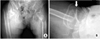
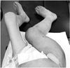
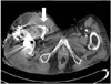
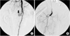
An 82-year-old woman, previously in bedridden state due to general weakness, was brought to the emergency department by guardians with right hip pain and severe thigh swelling. She was hospitalized at local hospital for 2 years and during stay, only sitting position was possible. Two weeks prior to our hospital visit, she had a history of fall-down from bed but was neglected. The radiographs demonstrated a subtrochanteric hip fracture (Seinsheimer classification grade 2b) with abrupt angulation of the proximal fragment and artherosclerotic vessel (Fig. 1). Additional findings included 30~40 degrees of flexion contracture of knee with severe swelling of the injured limb, more than twice the circumference compared to the contralateral limb (Fig. 2). There were ecchymoses in the anteromedial aspect of the right proximal thigh. The femoral and pedal pulses were symmetric and palpable in both lower limbs. On admission, the blood pressure was 100/80 mmHg with the heart rate of 90 bpm. The initial hemoglobin was 9.5 g/dl with the hematocrit of 0.22. The patient was managed with Buck's traction overnight. She was taken to the operating room the next day and gently placed on the fracture table under spinal anesthesia. The proximal fragment was severely abducted, externally rotated and flexed, making the nail entry into the greater trochanter difficult. Small (3 cm) incision at greater trochanter level was made in order to insert the reduction clamp. Large amount of hematoma was evacuated right after deep fascia incision. Reduction clamp was placed through an incision to reduce the proximal fragment prior to nailing. But, soon after the reduction against the deforming force, active bleeding was observed. The bleeding point was unidentifiable due to massive bleeding and inadequate operative field. Vital sign became unstable with systolic blood pressure dropping to 50 mmHg. Massive gauze packing on the suspected bleeding site was done with rapid intramedullary nail insertion (Proximal Femur Nail, Synthes, Davos, Switzerland). Skin closure and compressive dressing was performed to stabilize the vital sign. Popliteal and pedal pulse was weak. In order to identify the injured vessel, computed tomographic angiography was performed, and active bleeding in one of the branch vessel of right deep femoral artery was suspected (Fig. 3). To further evaluate the injured vessel, emergent digital subtraction angiography was performed. At first, the left common femoral artery was punctured by using a Seldinger technique. Extravasations of contrast medium was identified on the extremity angiogram, originating from one of the minor proximal branch of right deep femoral artery, while filling a 2 cm sized pseudoaneurysm (Fig. 4A). At this stage, coil embolization was attempted to stop the ongoing bleeding. The suspected branch of right deep femoral artery was superselected by using a 2.4 Fr microcatheter (Progreat, Terumo, Tokyo, Japan). Via the microcatheter, attempts were made to completely embolize the injured artery with 13 platinum-made microcoils (Tornado®, Cook Inc., Bloominton, Minnesota, USA), including four 6/2 mm, one 5/2 mm, six 4/2 mm and two 3/2 mm microcoils. Immediately after the embolization procedure, digital subtraction angiography was performed. The angiograms demonstrated complete occlusion of the feeding vessel and the cessation of the active bleeding (Fig. 4B). No other branch except the injured terminal branch was occluded on the angiogram. The day after embolization, the packed gauze was removed surgically.
DISCUSSION
This case shows the importance of prompt diagnosis and intervention of vascular injury for preventing severe complications, including massive hemorrhage and death. A painful swollen hip and thigh with severe bruises accompanied by hip fracture should alert the surgical team to a likely vascular injury. Although not performed in this case, the preoperative angiogram should be taken whenever the vascular injury is suspected. This could prepare the surgical team for vascular intervention either prior or during operation.
The incidence of vascular injury in pertrochanteric hip fracture has increased over the years6). The deep femoral artery and its branches are most commonly affected5). The complication may be caused by drilling injuries, locking screws extending too far outside the bone thereby puncturing the vessels, or by the sharp ends of the Kirschner wires or a retractor, or attributed to hardware eroding into the vessel over time1,6,14). Late complications occurring between 6 weeks to 10 years after arterial injury are false aneurysm, arteriovenous fistula, and ischemia of the leg distal to the injured vessel. Two cases of vascular laceration due to a spike of the fractured fragment; lesser trochanter in intertrochanteric fractures, have been reported13,15). However, laceration of the deep femoral vessel due to a bony spike of the proximal fragment in subtrochanteric hip fracture has not been reported.
When the artery is injured, extensive blood loss usually occurs, and immediate surgery is required10). If angiography is performed to localize the bleeding vessel, treatment by percutaneous transcatheter embolization may be an option, depending on the type of injury, the surgical risk, and the experience of the interventional radiologist10). Embolization may be performed by placing coils in the damaged vessel. The bleeding can also be treated by placing intravascular stents or grafts, thereby occluding the opening in the vessel that causes the hemorrhage9). In a large series involving 107 arterial injuries, percutaneous transcatheter embolization was performed successfully in more than 80% of cases, with surgery performed in the remainder10). Proximal lesions concerning the deep femoral artery, in which repair is possible, are generally recommended to reconstruct surgically. In the lower extremity, superior or inferior gluteal, distal profunda femoral, circumflex femoral, or muscular branches of the superficial femoral arteries are considered candidates for embolization10).
Computed tomographic angiography and duplex ultrasonography are methods used to evaluate arterial trauma as noninvasive imaging studies4,7). Although computed tomographic angiography has been shown to be 90% to 95% sensitive and 98% to 100% specific for the detection of arterial injury in young patients who have sustained trauma, it has a major disadvantage that a concomitant therapeutic intervention cannot be undertaken7). Duplex ultrasonography is also limited by errors in the identification of injuries to secondary branches and the documented increased difficulty of this operator-dependent technique in the presence of severe atherosclerosis4). There were two reasons that computed tomographic angiography was taken prior to interventional angiogram in our case. First, we were not sure which vessels were injured. If major vessel such as superficial femoral artery is injured, emergent surgery should have been planned. Second, intervention angiography is dependent on radiologic specialist who is not always available as emergent basis in every hospital.
Arterial trauma associated with hip fracture treatment is still a rare complication, but the surgeon must be aware that vessel injury may occur not only during or after operation but also at the time of injury. Routine suspection of vascular injury is always recommended before or after any type of fracture around hip region. Interventional arteriography is preferred in every case of suspected injury, and embolization may be performed as a less invasive therapy.
 XML Download