

 PDF
PDF ePub
ePub Citation
 Print
Print


Abstract
Purpose
To report the clinical results of surgical treatment of clavicle shaft fracture by percutaneous reduction with towel clips and percutaneous intramedullary pin fixation.
Materials and Methods
This study reviewed the results of 80 cases of clavicle shaft fracture treated by percutaneous reduction with towel clips and percutaneous intramedullary pin fixation with Steinmann pins from January 2002 to August 2010, after follow-up for 12 months or more. We evaluated the clinical results, such as union time and complications.
Figures and Tables
Fig. 1
The picture and the radiograph shows the percutaneous insertion of Steinmann pin into the intramedullary canal under the fluoroscopic guidance from the fracture side to the medial end.
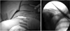

Fig. 3
The picture and the radiograph shows retrograde reinsertion of Steinmann pin after reduction of the fracture site under the fluoroscopic guide.

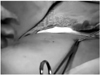
Fig. 5
(A) The radiograph shows the displaced clavicle shaft fracture as Robinson classification type 2B1
(B) Immediate postoperative radiograph shows well reducted clavicle with percutaneous Steinmann pin fixation.
(C) Postoperative 12 weeks radiograph shows bone union.
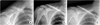
Fig. 6
(A) Preoperative 3D computerized tomography shows displaced clavicle shaft fracture with double butterfly fragment as Robinson classification type 2B2.
(B) Immediate postoperative radiograph shows well reducted clavicle fracture with double butterfly fragments with Steinmann pin.
(C) Postoperative 12 weeks radiograph shows complete bone union with butterfly fragments and complete remodeling of
clavicle fracture.

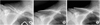
Fig. 7
(A) The radiograph shows the displaced clavicle shaft fracture as Robinson classification type 2B1.
(B) The radiograph shows the bended Steinmann pin due to slip down on 2 weeks after operation.
(C) Postoperative 12 weeks radiograph shows bone union and remodeling without any other procedure.


References
1. Adams F. The Genuine works of hippocrates. 1886. New York: William Wood & Co.
2. Byun YS. Minimally invasive plate osteosynthesis, MIPO. J Korean Fract Soc. 2007. 20:99–114.

3. Copland SM. Total resection of the clavicle. Am J Surg. 1946. 72:280.
4. Craig EV. Rockwood CA, Matsen FA, editors. Fractures of the clavicle. The shoulder. 1990. Philadelphia: WB Saunders;367–412.
5. Harnroongroj T, Jeerathanyasakun Y. Intramedullary pin fixation in clavicular fractures: a study comparing the use of small and large pins. J Orthop Surg (Hong Kong). 2000. 8:7–11.
6. Hill JM, McGuire MH, Crosby LA. Closed treatment of displaced middle-third fractures of the clavicle gives poor results. J Bone Joint Surg Br. 1997. 79:537–539.
7. Johnson EW Jr, Collins HR. Nonunion of the clavicle. Arch Surg. 1963. 87:963–966.
8. Jupiter JB, Leffert RD. Non-union of the clavicle. Associated complications and surgical management. J Bone Joint Surg Am. 1987. 69:753–760.
9. Kang KS, Ahn JI, Oh HY, Kang YS, Lee SJ. Clinical study of clavicle fracture. J Korean Orthop Assoc. 1984. 19:367–372.
10. Kim BH, Im JI, Yim UK, Kim JJ. Operative treatment of clavicle fracture. J Korean Soc Fract. 1998. 11:658–664.
11. Lee SH, Shin DM, Rim GS. Treatment of the fractures of clavicle with butterfly fragment. J Korean Soc Fract. 1992. 5:43–49.
12. Neer CS 2nd. Nonunion of the clavicle. J Am Med Assoc. 1960. 172:1006–1011.
13. Neviaser RJ, Neviaser JS, Neviaser TJ, Neviaser JS. A simple technique for internal fixation of the clavicle. A long term evaluation. Clin Orthop Relat Res. 1975. 109:103–107.
14. Paffen PJ, Jansen EW. Surgical treatment of clavicular fractures with Kirschner wires: a comparative study. Arch Chir Neerl. 1978. 30:43–53.
15. Robinson CM. Fractures of the clavicle in the adult. Epidemiology and classification. J Bone Joint Surg Br. 1998. 80:476–484.
16. Robinson CM, Court-Brown CM, McQueen MM, Wakefield AE. Estimating the risk of nonunion following nonoperative treatment of a clavicular fracture. J Bone Joint Surg Am. 2004. 86:1359–1365.
17. Rowe CR. An atlas of anatomy and treatment of midclavicular fractures. Clin Orthop Relat Res. 1968. 58:29–42.
18. Schwarz N, Höcker K. Osteosynthesis of irreducible fractures of the clavicle with 2.7-MM ASIF plates. J Trauma. 1992. 33:179–183.
19. White RR, Anson PS, Kristiansen T, Healy W. Adult clavicle fractures: relationship between mechanism of injury and healing. Orthop Trans. 1989. 13:514–515.
20. Wood VE. The results of total claviculectomy. Clin Orthop Relat Res. 1986. 207:186–190.
 XML Download