

 PDF
PDF ePub
ePub Citation
 Print
Print


Abstract
Fractures of the femoral shaft with marked bowing face some obstacles in fixation of the fracture such as difficulty in insertion of the intramedullary nail (IM nail) or exact contouring plate. Locking compression plates (LCP) are an option to manage this problem. However, we experienced consecutive breakage of LCP twice and IM nail once in an 80-year-old female. Finally, union of the fracture was achieved after fixation of the IM nail and additional plate together. Fractures of the femur shaft with marked bowing are thought to have different biomechanical properties; therefore, we present this case with a review of the literature.
Figures and Tables
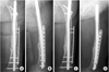
 | Fig. 1
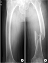
(A) Right femur has marked anterior and lateral bowing.
(B) Initial radiographs show the distal shaft fracture of the left femur (Winquist-Hansen type III).
|
 | Fig. 2
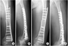
(A, B) Postoperative radiographs show diminished lateral bowing and gap of medial cortex.
(C, D) X-ray at postoperative 3 months shows breakage of the locking compression plate.
|
 | Fig. 3
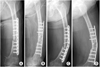
(A, B) A second operation was performed using locking compression plate (LCP).
(C, D) But 6 weeks later, the LCP was broken again.
|
 | Fig. 4
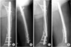
(A, B) Third postoperative radiographs show the fracture treated with an antegrade femur nail that preserved the anterior and lateral bowing of the femur.
(C, D) 9 months after the third operation, a fatigue fracture occurred at the most proximal hole among the distal locking holes.
|
References
1. Beaupré GS, Schneider E, Perren SM. Stress analysis of a partially slotted intramedullary nail. J Orthop Res. 1984. 2:369–376.

2. Bucholz RW, Ross SE, Lawrence KL. Fatigue fracture of the interlocking nail in the treatment of fractures of the distal part of the femoral shaft. J Bone Joint Surg Am. 1987. 69:1391–1399.
3. Dencker H. Errors in technique and complications specific to intramedullary nailing. A study based on 459 nailed femoral shaft fractures. Acta Orthop Scand. 1964. 35:164–169.
4. Frigg R. Development of the locking compression plate. Injury. 2003. 34:Suppl 2. B6–B10.
5. Oh JK, Oh CW, Park SH, Roh KJ, Jeong CW. Conformity of the LCP-DF (locking compression plate-distal femur) in Korean adult femur: a cadaver study. J Korean Fract Soc. 2005. 18:399–404.
6. Ostrum RF, Levy MS. Penetration of the distal femoral anterior cortex during intramedullary nailing for subtrochanteric fractures: a report of three cases. J Orthop Trauma. 2005. 19:656–660.
7. Sommer C, Gautier E, Müller M, Helfet DL, Wagner M. First clinical results of the Locking Compression Plate (LCP). Injury. 2003. 34:Suppl 2. B43–B54.
8. Yau WP, Chiu KY, Tang WM, Ng TP. Coronal bowing of the femur and tibia in Chinese: its incidence and effects on total knee arthroplasty planning. J Orthop Surg (Hong Kong). 2007. 15:32–36.
 XML Download