

 PDF
PDF ePub
ePub Citation
 Print
Print


The clavicle is one of the most commonly fractured bones, accounting for about 5% of all fractures and comprising 44% of all shoulder girdle injuries2). Among these, a clavicle shaft fracture accounts for the majority of fractures, followed by a distal clavicle fracture, and a proximal end fracture is rare. However, simultaneous ipsilateral fractures of the proximal, distal or middle-third clavicle are extremely rare; only seven cases have been reported in the English and Japanese literatures3~8,10). We report a case of an ipsilateral distal and proximal clavicle fracture caused by a fall and describe its presumed mechanism, diagnosis, and treatment, accompanied by a review of the literature.
CASE REPORT
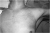
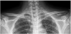
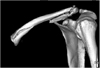
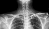
A 33-year-old male who fell from a bridge in a drunken state was admitted to the emergency room. He was unconscious when he arrived at our hospital. A physical examination revealed ecchymosis and swelling around the left shoulder and chest (Fig. 1). He was diagnosed with an epidural hemorrhage and underwent emergency surgery. A plain radiograph revealed an extra-articular fracture of the distal clavicle which was classified to Neer type II with inferior bone fragment (Fig. 2). A computed tomography (CT) scan demonstrated an intra-articular fracture of the proximal clavicle, Craig classification type III, with two displaced fragments that were separated into superior and inferior fragments (Fig. 3)9).
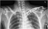
An open reduction and internal fixation was performed 13 days after the trauma. A 15-cm transverse incision was made from the acromioclavicular joint to the sternoclavicular joint parallel with the clavicle, and soft tissue was dissected until both fracture sites were encountered. The distal clavicle fracture was reduced and fixed using an AO hook plate (LCP clavicle hook plate, Synthes®, Switzerland). The proximal clavicle fracture was of the intra-articular type, and superior and inferior fragments were separated by a transverse fracture line. Fracture fragments were fixed using the lag-screw technique, and, later, a 3.5-mm LCP reconstruction plate (Synthes®, Switzerland) was applied to the anterior aspect of the clavicle. The two plates were placed to overlap each other to prevent the stress concentration between them (Fig. 4).
The patient was given an arm sling and allowed to perform range-of-motion exercises of the elbow and wrist and pendulum exercises of the shoulder joint. At 4 weeks after the operation, the patient had nearly full range of motion of the shoulder: 180° forward flexion and abduction, and 70° external and internal rotation. At 5 months after the operation, radiographs revealed that a callus had formed on both fracture sites, and the patient had no symptoms or limitations on daily activities (Fig. 5).
DISCUSSION
Among all clavicular fractures, 69% occur in the middle-third, 28% at the distal end, and the remaining 3% at the proximal end9). Although ipsilateral proximal and distal clavicle fractures can be classified as segmental fractures according to the number of fracture fragments, several studies have described a "double clavicle fracture"3,5,7,8,10). The double clavicle fracture is an even rarer injury; indeed, only four cases have been reported in the English literature3,4,6,8). One case was fractures of the middle and distal clavicle3) and the other case was the proximal and distal clavicular fractures with a extra-articular fracture4). Two cases were middle-third clavicle fractures in children, with a proximal clavicular physis injury6,8). On the other hand, three cases of fractures of the middle and distal clavicle have been reported in Japan5,7,10). However, in our unusual case, the fractures occurred simultaneously in the distal portion and proximal end of the clavicle, involving the articular surface of the sternoclavicular joint; such a case has not previously been reported.
Two separate mechanisms produce clavicular fractures. One is direct force on the point of the shoulder girdle due to a fall9). The other is an indirect force, which may give rise to clavicle fractures by the shearing force delivered from the humerus to the sternum via the shoulder joint and the clavicle when patients fall on their out-stretched arms1). Although a definite mechanism for simultaneous fractures of the proximal and distal clavicle has not been clearly defined, it seems that either separate or sequential forces result in such fractures. In our case, we presumed that direct force on the shoulder joint led to the distal clavicle fracture, especially considering the head injury. The proximal clavicle fracture was less likely to be caused by direct force, given its relationship with surrounding structures. The mechanism of injury to the proximal clavicle is similar to that of injury to the sternoclavicular joint. A proximal clavicle fracture can occur when either direct or indirect force is applied, either on the anterolateral or posterolateral aspect of the shoulder joint3). Therefore, in this case, we supposed that direct force applied to the shoulder joint resulted in the distal clavicle fracture, and then the sequential blow was transferred to the medial aspect of the clavicle, which caused the proximal clavicle fracture.
Fractures of the proximal clavicle are difficult to visualize because overlying structures in the chest obscure the view of the proximal end. For rare proximal fractures, particularly those that extend into the sternoclavicluar joint, a Hobb's view or a serendipity view may be helpful to access the fractures and identify their relationship to the sternoclavicular joint. However, proximal fractures require a computed tomography scan for adequate visualization. A proximal clavicular fracture combined with a distal one can be overlooked, and two studies have reported that proximal fractures in a double clavicle fracture were initially missed4,6). Therefore, a careful analysis is required when several signs of a suspected fracture of the proximal clavicle are presented, including tenderness, swelling, and subcutaneous hemorrhage on the anterior chest. A distal and proximal clavicle fracture can be treated either nonoperatively or operatively. Although proximal clavicular fractures heal well with nonoperative treatment, we believe that more active treatment through a surgical intervention should be considered in cases of a segmental fracture, considering that the clavicle forms one of the struts of the suspension ring of the shoulder girdle. In this case, plating with lag screws was applied to the proximal clavicular fracture in order to protect bending and torsional force to the clavicle. However, lag screws without plating for proximal clavicular fracture could be an alternative to the open plating so as to prevent worrisome problem, such as extensive soft tissue disruption and nonunion.
A concurrent distal and proximal clavicle fracture is a rare injury and may be difficult to diagnose. Therefore, when a distal clavicle fracture is diagnosed, a close physical examination and radiographic evaluation of the proximal clavicle fracture is necessary to prevent late complications such as a nonunion.
 XML Download