

 PDF
PDF ePub
ePub Citation
 Print
Print


Abstract
Purpose
To evaluate the radiographic results of patients with subtrochanteric femoral fracture using minimal incision and cephalomedullary nail technique.
Materials and Methods
This study was performed on 54 patients, 54 cases of hip, recruited among patients who underwent minimal incision and Cephalomedullary nail from September 2005 to August 2008 and were available for 1-year or longer follow up. The gender ratio was 37 males and 17 females, and the mean age at the time of surgery was 57.4 years (range; 16~81 years). According to injury mechanism, traffic accident was 29 cases, fall down form high height was 18 cases, slip down was 7 cases. In classification by Seinsheimer, type II was 23 cases (m/c), type III was 18 cases, type IV was 13 cases. Average follow up period was 14 months (12~18). Radiographic evaluation was performed for time taking union, mal-union and complication.
Results
53 of the 54 cases united. 39 of 54 reductions were anatomic. 19 fractures had a monir varus deformity of proximal fragment (between 2° and 5°). There was no varus deformity of more than 5°. 1 case that had been treated with PFN had nail breakage without trauma. There were no other complications.
Conclusion
Surgical treatment of subtrochanteric fractures with minimal incision and Cephalomedullary nail technique can reslut in excellent reduction without complications including inflammation & malunion. Careful attention to detail for using Lowman clamp is demanding to decrease soft tissue injury.
Figures and Tables
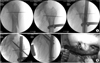 | Fig. 1Surgical technique.
(A) Temporary fixation by using Lowman clamp.
(B) Nail insertion & screw fixation through previous proximal incision site.
|
References
1. Afsari A, Liporace F, Lindvall E, Infante A Jr, Sagi HC, Haidukewych GJ. Clamp-assisted reduction of high subtrochanteric fractures of the femur. J Bone Joint Surg Am. 2009. 91:1913–1918.

2. Bedi A, Toan Le T. Subtrochanteric femur fractures. Orthop Clin North Am. 2004. 35:473–483.
3. Blatter G, Janssen M. Treatment of subtrochanteric fractures of the femur: reduction on the traction table and fixation with dynamic condylar screw. Arch Orthop Trauma Surg. 1994. 113:138–141.
4. Celebi L, Can M, Muratli HH, Yagmurlu MF, Yuksel HY, Bicimoğlu A. Indirect reduction and biological internal fixation of comminuted subtrochanteric fractures of the femur. Injury. 2006. 37:740–750.
5. Gugenheim JJ, Probe RA, Brinker MR. The effects of femoral shaft malrotation on lower extremity anatomy. J Orthop Trauma. 2004. 18:658–664.
6. Haidukewych GJ, Berry DJ. Nonunion of fractures of the subtrochanteric region of the femur. Clin Orthop Relat Res. 2004. 419:185–188.
7. Hasenboehler EA, Agudelo JF, Morgan SJ, Smith WR, Hak DJ, Stahel PF. Treatment of complex proximal femoral fractures with the proximal femur locking compression plate. Orthopedics. 2007. 30:618–623.
8. Kim JJ, Kim JW. Subtrochanteric fracture: intramedullary nailing. J Korean Fract Soc. 2009. 22:114–122.
9. Kim SK, Rhee KB, Oh SJ, Lee SC. The clinical study of subtrochanteric fractures of the femur. J Korean Orthop Assoc. 1992. 27:1006–1013.
10. Kinast C, Bolhofner BR, Mast JW, Ganz R. Subtrochanteric fractures of the femur. Results of treatment with the 95 degrees condylar blade-plate. Clin Orthop Relat Res. 1989. 238:122–130.
11. Krettek C, Schandelmaier P, Miclau T, Tscherne H. Minimally invasive percutaneous plate osteosynthesis MIPPO using the DCS in proximal and distal femoral fractures. Injury. 1997. 28:Suppl 1. A20–A30.
12. Mahomed MN, Harrington IJ, Hearn TC. Biomechanical analysis of the Medoff sliding plate. J Trauma. 2000. 48:93–100.
13. Neher C, Ostrum RF. Treatment of subtrochanteric femur fractures using a submuscular fixed low-angle plate. Am J Orthop (Belle Mead NJ). 2003. 32:9 Suppl. 29–33.
14. Oh CW. Minimally invasive plate osteosynthesis of subtrochanteric femoral fractures. J Korean Fract Soc. 2009. 22:123–129.
15. Pai CH. Dynamic condylar screw for subtrochanteric femur fractures with greater trochanteric extension. J Orthop Trauma. 1996. 10:317–322.
16. Ruff ME, Lubbers LM. Treatment of subtrochanteric fractures with a sliding screw-plate device. J Trauma. 1986. 26:75–80.
17. Sanders R, Regazzoni P. Treatment of subtrochanteric femur fractures using the dynamic condylar screw. J Orthop Trauma. 1989. 3:206–213.
18. Siebenrock KA, Müller U, Ganz R. Indirect reduction with a condylar blade plate for osteosynthesis of subtrochanteric femoral fractures. Injury. 1998. 29:Suppl 3. C7–C15.
19. Yoon HG. Surgical treatment of subtorchanteric fractures. J Korean Hip Soc. 2007. 19:283–291.
20. Yoon TR, Rowe SM, Song EK, Seol JY, Gyoo SS. Treatment of subtrochanteric fracture - Comparison of treatment efficacy according to internal fixation device -. J Korean Soc Fract. 2001. 14:189–199.
 XML Download