

 PDF
PDF ePub
ePub Citation
 Print
Print


Abstract
Purpose
The purpose of this article is to show the efficacy of a biodegradable plate for treating lateral malleolar fractures in the ankle joint.
Materials and Methods
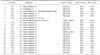
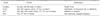
The 20 patients who underwent an open reduction and internal fixation for lateral malleolar fractures in the ankle joint from February, 2006 to February, 2007 in our hospital were enrolled into the study. The average age of the patients was 49.7 years and the average follow-up period was 5.6 months. The cases were analyzed by radiological bone union time and clinical results according to the criteria of Meyer et al.
Figures and Tables
Fig. 1
Photographs of biodegradable plate and screw and surgical steps.
(A) 6 hole biodegradable plate and bicortical screw.
(B) The plate is heated in the water bath for at least 1 minute and most malleable for 10 seconds after heating.
(C) The plate can be contoured to the bone by using finger pressure. Bicortical screws are fixed through the plate.
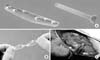
Fig. 2
(A) A 55-year-old woman had lateral malleolar fracture and deltoid ligament injury of the ankle joint.
(B) The lateral malleolar fracture was fixed with biodegradable plate and bicortical screw.
(C) At postoperative 12 weeks, the radiographs show the radiological bone union.
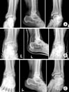
Fig. 3
(A) A 48-year-old woman had a trimalleolar fracture of the ankle joint.
(B) The lateral malleolar fracture was fixed with biodegradable plate and bicortical screws, and medial malleolar fragment with biodegradable screw and K-wire.
(C) At postoperative 15 weeks, the radiographs show the radiological bone union.
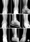
Fig. 4
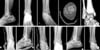
(A) Initial radiographs & CT of a 62-year-old woman show anterior tibio-fibular ligament avulsion fracture in lateral malleolar fracture.
(B) The lateral malleolar fracture was fixed with biodegradable plate and bicortical screws, and avulsion fragment of anterior tibio-fibular ligament with biodegradable screw.
(C) At postoperative 8 weeks, the radiographs show the minimal displacement less than 1 mm.
References
1. Blasier RD, Bucholz R, Cole W, Johnson LL, Mäklä EA. Bioresorbable implants: applications in orthopaedic surgery. Instr Course Lect. 1997; 174:531–546.
2. Böstman OM. Absorbable implants for the fixation of fractures. J Bone Joint Surg Am. 1991; 73:148–153.

3. Böstman OM. Osteolytic changes accompanying degradation of absorbable fracture fixation implants. J Bone Joint Surg Br. 1991; 73:679–682.
4. Böstman OM, Pihlajamäki HK. Late foreign-body reaction to an interosseous bioabsorbable polylactic acid screw. A case report. J Bone Joint Surg Am. 1998; 80:1791–1794.
5. Böstman OM, Pihlajamäki HK, Partio EK, Rokkanen PU. Clinical biocompatibility and degradation of polylevolactide screws in the ankle. Clin Orthop Relat Res. 1995; 320:101–109.
6. Bucholz RW, Henry S, Henley MB. Fixation with bioabsorbable screws for the treatment of fractures of the ankle. J Bone Joint Surg Am. 1994; 76:319–324.
7. Chung DW, Soh JH, Lim CT. Fixation with bioabsorbable polylactide screws for the treatment of the ankle fracture with syndesmotic injurues. J Korean Orthop Assoc. 2001; 36:395–401.
8. Daniels AU, Chang MK, Andriano KP. Mechanical properties of biodegradable polymers and composites proposed for internal fixation of bone. J Appl Biomater. 1990; 1:57–78.
9. Lee JI, Son MH, Lee JW, Namgung SH. Clinical evaluation for the ankle fractures by treatment. J Korean Soc Fract. 1997; 10:588–596.
10. Hovis WD, Bucholz RW. Polyglycolide bioabsorbable screws in the treatment of Ankle fractures. Foot Ankle Int. 1997; 18:128–131.
11. Hovis WD, Kaiser BW, Watson JT, Bucholz RW. Treatment of syndesmotic disruptions of the ankle with bioabsorbable screw fixation. J Bone Joint Surg Am. 2002; 84:26–31.
12. Meyer TL Jr, Kulmer KW. A.S.I.F. technique and ankle fractures. Clin Orthop Relat Res. 1980; 150:211–221.
13. Park MS, Lee JM, Chae SS. Management of the ankle fracture. J Korean Soc Fract. 1990; 3:238–246.
14. Rokkanen PU. Absorbable materials in orthopaedic surgery. Ann Med. 1991; 23:109–115.
15. Rokkanen P, Majola A, Vasenius J, Vainionpaa S. Strength retention of self-reinforced polyglycolide (SR-PGA) and SR-polylactic acid (PLA) composite rods in vitro and in vivo. Acta Ortho Scand. 1990; 235:Suppl. 51.
16. Simon JA, Ricci JL, Di Cesare PE. Bioreabsorbable fracture fixation in orthopedics: a comprehensive review. Part II. Clinical studies. Am J Orthop. 1997; 26:754–762.
17. Thordarson DB. Fixation of ankle syndesmosis with bioabsorbable screws. Tech Orthop. 1998; 13:187–191.
18. Yoon HK, Jeon KP, Kang KH, Kim KI. Biodegradable internal fixation for displaced non-communited malleolar fracture. J Korean Orthop Assoc. 1998; 33:309–313.
 XML Download