

 PDF
PDF ePub
ePub Citation
 Print
Print


Figures and Tables
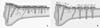 | Fig. 1Because the plate is not compressed on the bone, the blood supply will be preserved in LCP (A) than LC-DCP (B).
|
 | Fig. 2(A) Conical thread allows a secure fixation of the locking head screw.
(B) Dynamic compression can be achieved by eccentric insertion of standard screw at this hole.
|
 | Fig. 3(A) Once the metaphyseal fragment has been fixed with locking head screws, this portion can be compressed to the shaft with standard screws at compression hole.
(B) Arrangement of the combination holes.
|
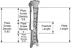 | Fig. 4Plate span ratio and plate screw density in bridge plate. Plate span ratio is the quotient of plate length and overall fracture length. Plate screw density is the quotient formed by the number of screws inserted and number of the plate holes (From Gautier E, Sommer C: Injury 34(Suppl 2), 2003).
|
 | Fig. 5(A) In normal bone, working length of monocortical screw is sufficient enough to withstand rotational displacement.
(B) In osteoporotic bone, working length of monocortical screw is insufficient due to thin cortex and under torque the bone thread soon will wear out and secondary displacement and instability will occur.
(C) In osteoporotic bone the bicortical screw is recommended because of the longer working length leading to a much higher torque resistance (From Gautier E, Sommer C: Injury 34(Suppl 2), 2003).
|
References
1. Aguila AZ, Manos JM, Orlansky AS, Todhunter RJ, Trotter EJ, Van der Meulen MC. In vitro biomechanical comparison of limited contat dynamic compression plate and locking compression plate. Vet Comp Orthop Traumatol. 2005; 18:220–226.

2. Ahmad M, Nanda R, Bajwa AS, Candal-Couto J, Green S, Hui AC. Biomechanical testing of the locking compression plate: when does the distance between bone and implant significantly reduce construct stability? Injury. 2007; 38:358–364.
3. Chang SA, Ahn HS, Byun YS, Kim JH, Bang HH, Kwon DY. Minimally invasive plate osteosynthesis in unstable fractures of the distal tibia. J Korean Fract Soc. 2005; 18:155–159.
4. Collinge CA, Sanders RW. Percutaneous plating in the lower extremity. J Am Acad Orthop Surg. 2000; 8:211–216.
5. Frigg R. Development of the locking compression plate. Injury. 2003; 34:Suppl 2. B6–B10.
6. Gautier E, Sommer C. Guidelines for the clinical application of the LCP. Injury. 2003; 34:Suppl 2. B63–B76.
7. Haidukewych GJ. Innovations in locking plate technology. J Am Acad Orthop Surg. 2004; 12:205–212.
8. Niemeyer P, Südkamp NP. Principles and clinical application of the locking compression plate (LCP). Acta Chir Orthop Traumatol Cech. 2006; 73:221–228.
9. Perren SM. Evolution and rationale of locked internal fixator technology. Introductory remarks. Injury. 2001; 32:Suppl 2. B3–B9.
10. Sim JC, Chung NS, Hong KD, Ha SS, Kang JH. Treatment of fractures of the distal radius using locking compression plate. J Korean Fract Soc. 2005; 18:100–104.
11. Sommer C, Gautier E, Müller M, Helfet DL, Wagner M. First clinical results of the Locking Compression Plate (LCP). Injury. 2003; 34:Suppl 2. B43–B54.
12. Wagner M. General principles for the clinical use of the LCP. Injury. 2003; 34:Suppl 2. B31–B42.
13. Wagner M, Frenk A, Frigg R. New concepts for bone fracture treatment and the Locking Compression Plate. Surg Technol Int. 2004; 12:271–277.
 XML Download