

 PDF
PDF ePub
ePub Citation
 Print
Print


INTRODUCTION
Intraductal papilloma (IDP) originates from both the large ducts of the subareolar region and the terminal duct lobular unit in the periphery, and it is histologically characterized by a fibrovascular core covered with epithelial and myoepithelial cells. It is a relatively common lesion found in breast biopsies. However, IDP often accompanies a variety of changes, including sclerosis, epithelial or myoepithelial hyperplasia, squamous or apocrine metaplasia, and even atypical proliferation [1].
A papilloma with atypia, which encompasses atypical ductal hyperplasia (ADH) or small foci of low-grade ductal carcinoma in situ (DCIS) within the papilloma, has an increased risk of developing malignancy and has been reported to show a high rate of upgrading on subsequent excision [2345]. Thus, surgical excision is usually recommended for IDP with atypia diagnosed by core needle biopsy (CNB) as standard management. On the contrary, no consensus has been met regarding the management of benign IDP without atypia diagnosed by CNB harboring no clinical symptoms. Some studies have shown that IDPs, even those without atypia, are significantly associated with higher-grade lesions [46], and surgical excision is recommended in all cases for accurate diagnosis. In contrast, other studies have shown low rates of upgrading for IDP without atypia, suggesting careful observation rather than surgical excision [7891011]. However, most of the previous studies were performed using small samples, and some studies included IDP with atypia or even malignancy in their analysis.
In the present study, we restricted our analysis to a large cohort of benign IDPs without atypia, and we evaluated the upgrade rate to malignancy, including DCIS and invasive carcinoma. In addition, evaluation of the presence of proliferative lesions with atypia, such as ADH, atypical lobular hyperplasia, and lobular carcinoma in situ, which are associated with a high risk for developing breast cancer, is also important in assessing breast cancer risk and determining patient management. Thus, we evaluated the upgrade rate to these high-risk lesions as well. Furthermore, we analyzed the clinicopathologic features associated with upgrading on excision.
Go to : 
METHODS
Case selection
We performed a retrospective search of the pathology database to identify IDPs without atypia diagnosed via CNB between January 2010 and December 2015 at the Department of Pathology, Seoul National University Bundang Hospital, Korea. All CNBs were performed using a 14-gauge automated biopsy gun (STERICUT®; TSK Laboratory, Tochigi, Japan) under ultrasound guidance by radiologists specialized in breast imaging. We selected cases that were excised with lumpectomy, excisional biopsy, or vacuum-assisted excision (VAE) at our institution. When patients presented with symptoms such as bloody nipple discharge or a palpable mass or when IDP was located near the skin or nipple, surgical management was recommended. When the lesion was single with a size less than 3 cm and located far from the skin or nipple, patients had an option of surgery or VAE. VAE was performed using an 8- or 11-gauge vacuum-assisted biopsy needle (Mammotome®; Devicor Medical Products, Cincinnati, USA).
Benign IDPs with benign proliferative lesions, such as usual ductal hyperplasia or adenosis, and those associated with sclerosis, radial scars, fibrocystic changes, and columnar cell changes, were included. IDPs coexisting with a malignant lesion, including invasive carcinoma and DCIS, or other highrisk lesions, such as ADH and lobular neoplasia (atypical lobular hyperplasia and lobular carcinoma in situ), in the same side of the breast were excluded. This study was approved by the Institutional Review Board of Seoul National University Bundang Hospital (protocol number: B-1708-414-107) and informed consent was waived.
Evaluation of clinical and radiologic features
Clinical variables, such as the age at the time of diagnosis; gender; cause of detection; clinical symptoms, including nipple discharge, palpable mass, and local pain; and history of breast cancer, were recorded. Radiologic findings, including the location of the lesion, size of the lesion defined as the largest dimension recorded on imaging, Breast Imaging Reporting and Data System (BI-RADS) classification of the lesion, multifocality, and the number of tissue cores in CNB, were retrieved from the medical records. Multifocality was defined as two or more lesions separated by normal breast tissue in imaging that were eventually proven to be benign IDPs on pathologic examination.
To analyze the radiologic-pathologic concordance, we reviewed all radiologic findings in each case and matched them with the pathologic diagnosis by the location of the CNB and the size of the lesion. Radiologic-pathologic discordance was considered to be present when the lesion was more than moderately suspicious for malignancy in radiologic findings, i.e., BI-RADS category 4b, 4c, or 5, but for which the histologic findings of IDP without atypia did not account for the imaging pattern.
Evaluation of excision specimens
We aszsessed accompanying lesions within the papilloma or in the adjacent breast tissue using excision specimens, and we classified them according to the World Health Organization classification of Tumours of the Breast, 4th edition. Upgrade to malignancy was defined as the presence of invasive carcinoma or DCIS in the excision specimen. The presence of atypical proliferative lesions such as ADH and lobular neoplasia, which are associated with a high risk of malignancy, was also assessed. Upgrade to high-risk lesions was defined as the presence of ADH or lobular neoplasia.
Statistical analysis
Statistical analysis was performed using SPSS version 21.0 software (IBM Corp., Armonk, USA). For patients with more than one IDP, analyses of the radiologic and pathologic features were based on each IDP. Chi-square tests or Fisher exact tests were used to compare the clinicopathologic variables between upgraded lesions and nonupgraded lesions. Statistical significance was defined as a p-value <0.05. All reported p-values were two-sided.
Go to :
RESULTS
Clinical, radiologic, and pathologic characteristics of IDP on CNB
A total of 511 benign IDPs without atypia diagnosed by CNB were identified, of which 398 cases were treated with surgical excision or VAE at our institution. After reviewing the medical records, pathologic reports, and hematoxylin and eosin stained slides of the CNBs, four cases that had high-risk lesions in the adjacent tissue, two cases that were re-diagnosed as papilloma with atypia, and nine cases of concurrent breast cancer in the same breast were excluded. Finally, 383 cases of benign IDP without atypia were included in the analyses.
The median age of the patients at the time of biopsy was 48 years (range, 23–82 years). In this cohort, the two major causes of initial detection were routine mammographic screening on medical checkup (n=224, 58.5%) and the presence of a symptom (n=108, 28.2%). All breast CNBs were performed under ultrasound guidance. The median number of core biopsy samples was 5 (range, 1–15). The other baseline characteristics of the patients are described in Table 1.
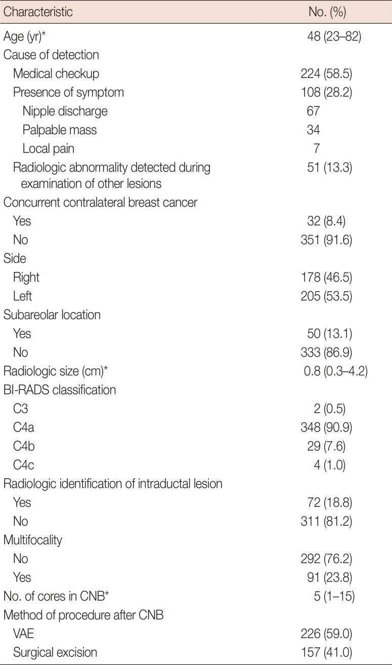
Table 1
Baseline characteristics (n=383)
![]()
Characteristics of IDPs with a likelihood of upgrading to malignancy or high-risk lesions on excision
Among the 383 IDPs without atypia on CNB, 20 cases (5.2%) were upgraded to malignancy or high-risk lesions, including ADH and lobular neoplasia, on excision; 10 cases were upgraded to ADH, five to atypical lobular hyperplasia, two to lobular carcinoma in situ, and three to DCIS. The rate of upgrading to malignancy and high-risk lesions after excision was 0.8% (n=3) and 4.4% (n=17), respectively. We also evaluated the characteristics of the lesions upgraded to malignancy on excision. Interestingly, we found that the presence of a symptom (p=0.022), the presence of concurrent contralateral breast cancer (p=0.019), and multifocality of the lesions (p=0.013) were predictive factors for upgrading to malignancy (Table 2). In addition, cases upgraded to malignancy or high-risk lesions were associated with surgical excision after CNB (p<0.001). The presence of concurrent contralateral breast cancer tended to have an association with upgrading to malignancy or high-risk lesions (p=0.075) (Table 3).
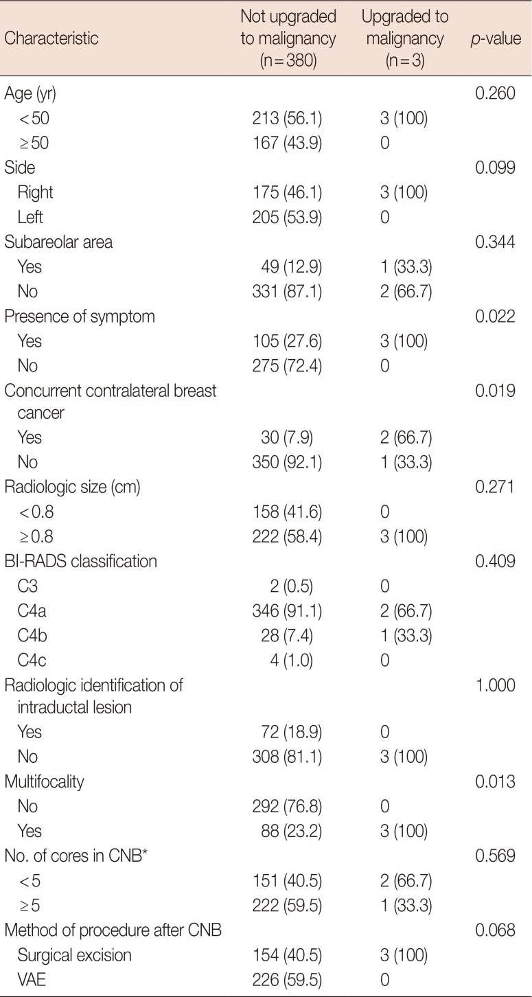
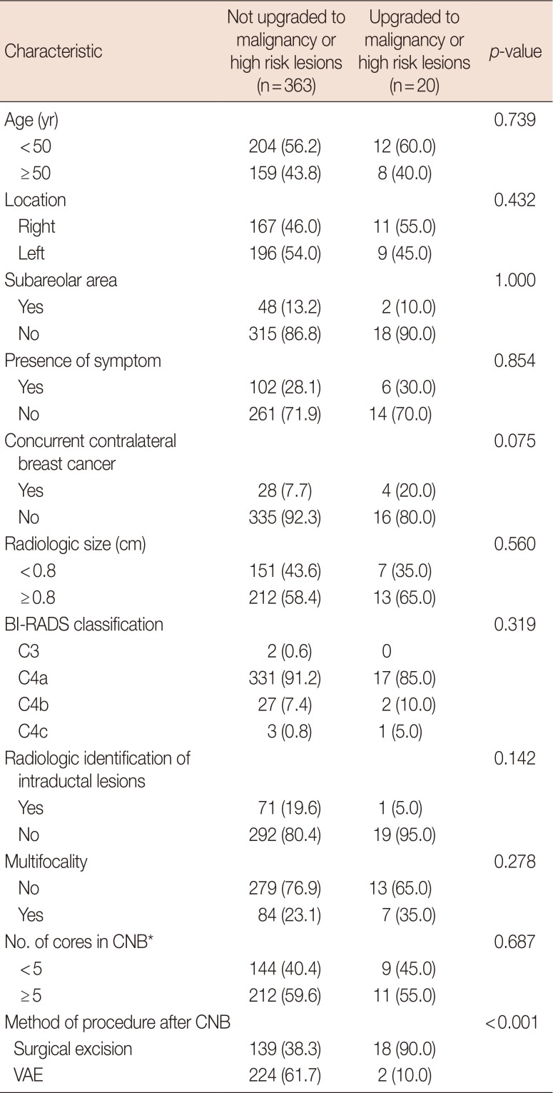
Table 2
Characteristics of intraductal papillomas without atypia upgraded to malignancy on subsequent excision
![]()
Table 3
Characteristics of intraductal papillomas without atypia upgraded to malignancy or other high-risk lesions on subsequent excision
![]()
Characteristics of the cases upgraded to malignancy on excision
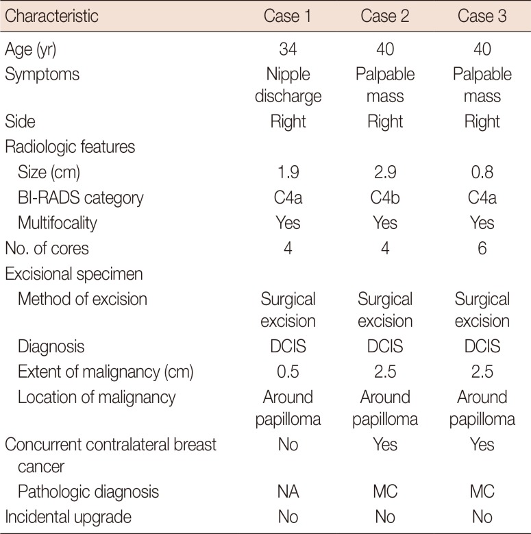
The clinical and pathologic characteristics of the cases that were upgraded to malignant lesions are summarized in Table 4. All of these cases were upgraded to DCIS, which was found around the papilloma (Figure 1). The extent of the DCIS was 0.5 cm, 2.5 cm, and 2.5 cm in each case. Of these three cases, two were from the same patient. All of the cases had clinical symptoms: one manifested as nipple discharge and the others as a palpable mass. Radiologic size of the lesions was 0.8 cm or greater and showed multifocality. Two cases had concurrent contralateral breast cancer.
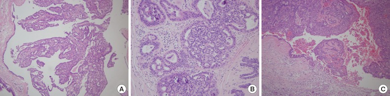 | Figure 1A representative case upgraded to ductal carcinoma in situ on excision. (A) Core needle biopsy shows typical benign intraductal papilloma without atypia (H&E stain, ×100). (B) Excision specimen reveals small foci of ductal carcinoma in situ with intermediate nuclear grade and a cribriform architectural pattern (H&E stain, ×200). (C) Residual papilloma in the excision specimen does not show any atypical proliferative change. Postbiopsy changes are seen in the lower portion of the papilloma (H&E stain, ×100).
|
Table 4
Summary of the characteristics of the lesions upgraded to malignancy on excision
![]()
Go to :
DISCUSSION
Advances in screening and imaging techniques for breast cancer have resulted in an increased number of suspicious lesions, which in turn, have led to an increased number of breast biopsies. The incidence of papillary lesions, including IDPs, has increased steadily over the past decade [12]. However, management of benign IDPs without atypia diagnosed by CNB remains controversial with some suggesting follow-up imaging and others excision. Moreover, in previous studies, the reported upgrading rates of benign IDP following excision vary widely [47891011131415]. This difference may have resulted from the study design, definition of upgrade lesions, and methodological details. Thus, we carefully selected our cohort to include only benign IDP cases diagnosed by CNB that had no malignant or high-risk lesions in the same breast. In this study, we showed that the rate of upgrading to malignancy was 0.8% at our institution. In addition, we found that the presence of a symptom, concurrent contralateral breast cancer, and multifocality were significant factors predictive of upgrading to malignancy. We also showed that upgrading to malignancy or high-risk lesions was associated with surgical excision rather than VAE. However, this may be caused by selection bias since patients with symptoms or multiple lesions were generally treated with surgical excision.
Several studies have attempted to identify factors that are associated with upgrading of benign IDP on subsequent excision [7916171819202122]. It was reported that old age at diagnosis was associated with upgrading to malignancy in benign IDPs [1622]. Regarding the size of the lesion, IDPs upgraded to malignancy have been reported to be larger [71722]. Previously, we also showed that large-sized IDP was a significant predictor of an upgrade by analyzing solitary IDPs diagnosed at different time points at our institution, excluding patients with breast cancer [9]. Although the size of the IDP was not a significant factor associated with upgrading to malignancy in the present study, all upgraded cases were above the median in size. Besides the size of the lesion, Kil et al. [18] have suggested that peripherally located IDPs require additional surgical excision. Holley et al. [19] reported that a reduced amount of tissue collected at biopsy (three cores vs. five cores; 14-gauge vs. 9-gauge needle) was associated with an upgraded lesion. Additionally, previous studies have reported varying results regarding the impact of the BI-RADS category on upgrade rates in benign IDP [2021].
Contrary to the previous studies, patient age, size, and location of the lesion, number of tissue cores, and BI-RADS score were not proven to be significant factors in our study. The most noticeable predictor of upgrading to malignancy was the presence of concurrent contralateral breast cancer, a finding that has never been reported in previous studies. While only 7.9% (30/380) of the nonupgraded cases had concurrent contralateral breast cancer, 66.7% (2/3) of upgraded cases had concurrent contralateral breast cancer. This association may be explained by the fact that the risk of developing new breast cancer is increased in patients with a history of breast cancer [23]. We also found that multifocality of the lesion was a significant predictor for upgrading to malignancy, similar to previous studies that showed that multiple papillomas were more likely to be associated with breast cancer than solitary papillomas [2425].
The mismatch between radiologic findings and pathologic diagnosis is an important issue that requires further evaluation. Some studies have documented that the discordance between radiographic and histologic findings was an important factor that should be evaluated by surgical excision. Thus, inclusion or exclusion of pathologic-radiologic discordant cases may be an important factor leading to variability in upgrade rates. After analyzing 234 benign IDPs, Rizzo et al. [26] reported the rate of upgrading to DCIS and invasive carcinoma as 8.9% and to ADH as 17.9%. Shin et al. [27] showed that an upgrade rate to DCIS and invasive carcinoma was 14% of 86 benign IDPs diagnosed by CNB. Mercado et al. [28] reported that 17% of 36 IDPs were upgraded to ADH. Other studies have reported similar high upgrade rates though these results may raise doubts about the definition of an upgrade [12930]. Collectively, these studies included pathologic-radiologic discordant cases in their analyses or failed to document pathologic-radiologic concordance [12627282930], leading to falsely high upgrade rates. Shin et al. [27] reported that the upgrade rate to malignancy was higher in imaging-pathologic discordant lesions than in concordant lesions. However, recent studies reported that the upgrade rate for benign IDP is low when the pathologic-radiologic discordant cases are excluded [1011].
Although physical findings such as a palpable mass and nipple discharge may not predict the risk of upgrading, lesions with such manifestations require careful evaluation and close observation and eventually need excision even if the pathologic diagnosis on CNB was benign. Nakhlis et al. [10] showed that the actual rate of upgrading in their study was 0% when they excluded both of the two patients from their cohort who presented with a clinically suspicious palpable mass. Other studies shared the same perspective, excluding symptomatic patients from their analyses of upgrading of benign IDP [819]. We agree that any patient with symptoms such as a palpable mass or nipple discharge should be treated by excision. Thus, of the 383 benign IDP diagnosed by CNB in our study, none of the cases were truly upgraded lesions when we excluded all three cases with clinical symptoms for which surgical treatment is indicated. Of the 17 cases upgraded to high-risk lesions, two cases had pathologic-radiologic discordance, and three cases had clinical symptoms. Therefore, the true upgrade rate to high-risk lesions was 3.1%. Given our results, close clinical and radiologic observation appear to be adequate for patients with benign IDP on CNB under proper clinical settings.
Although IDP has a relatively simple histologic definition, it encompasses a wide variety of lesions. Papillary lesions are often diagnostically challenging for pathologists. IDP may have a complex glandular architecture and epithelial hyperplasia; histologic distinctions between luminal epithelial and myoepithelial cells or benign hyperplasia and atypical hyperplasia can be subtle, leading to misinterpretations on CNB. Thus, accurate interpretation by an experienced pathologist and an adequate amount of an accurately-targeted specimen are essential for correct diagnosis. In our institution, all breast CNBs were diagnosed by an experienced breast pathologist (S.Y.P.), which can affect diagnostic accuracy. It was reported that one of the reasons for such diagnostic difficulties might be the limited amounts and fragmentation of the samples [16]. Renshaw et al. [3] pointed out that the incidence of upgrading after excision was associated with the adequacy of sampling in biopsy specimens. Several studies have shown different upgrading rates of benign IDP to malignancy depending on the needle gauge used during biopsy [1819]. In the current study, the number of tissue cores was sufficient with a median value of 5.0, which appears to have contributed to the low rate of upgrades.
In conclusion, the most significant finding of our study is that the rate of upgrading to malignancy of benign IDP without atypia diagnosed by CNB is very low. Moreover, when we excluded cases with suspicious clinical symptoms, the true rate of under-diagnosis in our cohort was 0%. For this reason, caution should be exercised in recommending surgical management for all benign IDPs. Our study suggests that patients with benign IDPs diagnosed by CNB who have solitary lesions without clinically suspicious symptoms and, in particular, no concurrent contralateral breast cancer may be candidates for close observation rather than prompt excision. However, due to the small number of cases upgraded to malignancy in this study, further large-scale studies are warranted to support this suggestion.
Go to :
 XML Download