

 PDF
PDF ePub
ePub Citation
 Print
Print


According to the mammography lexicon in the Breast Imaging Reporting and Data System (BI-RADS) Atlas 2013 (fifth edition), a "grouped" distribution of calcifications is defined as five or more calcifications within 1 cm of each other (lower limit) or larger numbers of calcifications grouped within 2 cm of each other (upper limit) [1]. The clinical significance of grouped microcalcifications varies widely, according to their morphology on mammography with a malignant potential of 16% to 36% [23].
Mammography is the gold standard for evaluation of microcalcifications, but has low specificity in terms of assessing grouped microcalcifications. Kim et al. [4] evaluated the utility of performing additional breast ultrasound (US) examinations in grouped microcalcifications for 61 pathologically verified breast lesions. They reported that additional breast US improves the specificity and accuracy of the diagnosis of breast carcinoma. However, microcalcifications are not easily detectable by US, particularly when they are not associated with a mass, but rather lie within normal breast tissue [5]. Because microcalcifications (at around 200 µm) are so small, normal fibroglandular tissues or US speckle artifacts interfere with their visualization. Therefore, mammography-guided tissue diagnoses should be performed for grouped microcalcifications with suspicious morphology.
Recently, an innovative imaging technology, MicroPure™ (Toshiba Medical Systems Corp., Tokyo, Japan), has been developed, which improves visualization of microcalcifications on US [6]. Improving the sensitivity of detection of microcalcifications on US can allow physicians to perform US-guided procedures without requiring radiation exposure, with reduced medical cost, and more accurate targeting in real-time imaging. In this brief communication, we introduce the early experiences with the use of the MicroPure™ US technique in 10 breast lesions of nine patients who had grouped microcalcifications not associated with masses on screening mammography and pathological verification, and demonstrate the utility of this advanced US technique for evaluation of microcalcifications.
This study was conducted with Institutional Review Board (Korea University Ansan Hospital) approval and the requirement for informed patient consent was waived (approval number: AS15212-001). All patients (mean age, 50 years; range, 40–70 years) had no clinical symptoms and no family history of breast cancer. We used the Aplio 500 (Toshiba Medical Systems Corp., Tokyo, Japan) US system with a 7- to 18-MHz linear transducer. Routine B-mode US examination was performed for both breasts in each patient as the first step, and then a targeted US examination with MicroPure™ mode was performed if a suspected region of grouped microcalcifications was found. During MicroPure™ examination, the two sections are displayed side-by-side (B-mode for the region of interest on the left side and MicroPure™ mode for the same region on the right side) on the screen. In MicroPure™ mode, microcalcifications are presented as bright white dots on a dark blue background. If a suspected microcalcification lesion was found on MicroPure™ imaging, an adhesive marker was placed over the region on the skin and additional mammography was then performed to precisely correlate mammography and US findings.
Two radiologists, with 3 and 16 years of experience in breast imaging, performed each evaluation and reached a consensus on the following radiologic findings. On mammography, the size, morphology, and number of grouped microcalcifications were evaluated. On breast US, the number of microcalcifications and the associated features were evaluated. The number of microcalcifications was counted on both B-mode and MicroPure™ images for each breast lesion. In addition, the image quality between B-mode and MicroPure™ imaging was compared in terms of the credibility of the presence of microcalcifications, and was graded into three types: -1, better on B-mode image; 0, the same with both imaging techniques; 1, better on MicroPure™ image.
US-guided 14-gauge automated core needle biopsy (n=3) or 11-gauge vacuum-assisted biopsy (n=7) was performed for all lesions. For four lesions, specimen mammography was conducted and confirmed that microcalcifications were well obtained. Five of nine patients underwent surgical excision after the biopsies as part of cancer treatment (n=4) or due to the patient's preference (n=1).
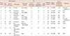
The radiological and pathologic findings of 10 breast lesions are summarized in Table 1. All lesions were classified as BI-RADS category 4 on mammography and the lesion size ranged from 3 to 18 mm (mean, 9.7 mm; median, 7 mm). Nine out of 10 lesions (90%) had associated US features: duct changes (n=4), masses (n=4), and duct changes and mass (n=1). The remaining lesion was observed as microcalcifications buried in the breast parenchyma on US (Figure 1). In six of the 10 lesions (60%), MicroPure™ images showed a greater number of microcalcifications than did B-mode images, and in the remaining four lesions, the number of microcalcifications observed was the same for the two imaging techniques. The mean number of microcalcifications on MicroPure™ and B-mode imaging was 6.8 and 4.7, respectively. In addition, image quality was better on MicroPure™ imaging in seven of the 10 lesions (70%); it was the same on both MicroPure™ and B-mode imaging in the remaining three lesions (30%). The pathological diagnoses were fibrocystic changes in six of the lesions and ductal carcinoma in situ in the other four lesions.
The MicroPure™ imaging is an innovative US technique, which adopts high-end image processing, including the "ApliPure" technique and the "constant false-alarm rate (CFAR)" filter [6]. ApliPure combines spatial and frequency compounding and leads to high-contrast resolution and high tissue uniformity. CFAR is a special interpolation filter that extracts only isolated high-brightness echoes against heterogeneous background clutter. These two image-processing techniques allow reduction in speckling and can separate true microcalcifications from artifacts in normal breast tissue. Lastly, the filtered image that presents only high-brightness dots overlap on the B-mode image and are layered with dark blue or purple color. This process further improves the ease of detection of the filtered microcalcifications.
Few published reports on the clinical utility of the MicroPure™ technique in breast evaluations are available [78]. In a study by Machado et al. [7], four readers evaluated 20 patients with diffuse microcalcifications identified on mammography using MicroPure™ and B-mode US imaging. They concluded that MicroPure™ showed more calcifications (mean number, 0.7±1.1 vs. 1.9±1.7) and fewer artifacts than did B-mode US. A recent study by Tan et al. [8] evaluated 70 pathologically proven breast lesions (0.2–9.6 cm in size) with suspected microcalcifications; 100% (70/70) of these microcalcifications could be seen using MicroPure™ US, and 71.4% (50/70) could be observed in B-mode US. The current study also revealed that MicroPure™ images show more microcalcifications in 60% (6/10) of lesions and that lesions are more conspicuous in 70% (7/10) than in B-mode images. Based on these studies, it can be concluded that MicroPure™ imaging is more sensitive for visualization of breast microcalcifications than B-mode US. A point of difference of this study is that we focused on the evaluation of grouped microcalcifications not associated with mass on mammography. This study revealed that the MicroPure™ technique could be useful for detection of suspicious grouped microcalcifications, unassociated with a mass, and conveniently facilitate tissue confirmation with US guidance.
In conclusion, MicroPure™ imaging is a promising US technique that could improve the sensitivity for detecting grouped microcalcifications that are not associated with mass in the breast on mammography. Further large-scale studies are recommended for assessing the potential future contribution of this new technique to diagnostic performance and the objective clinical benefits associated with detection of grouped microcalcifications.

 XML Download