

 PDF
PDF ePub
ePub Citation
 Print
Print


INTRODUCTION
Breast-conserving surgery (BCS) followed by adjuvant radiotherapy has been reported to be as effective as mastectomy in localized breast cancer [123]. Adjuvant radiotherapy conventionally consists of whole breast irradiation with a dose of 45 to 50.4 Gy and a boost with 10 to 20 Gy. As ipsilateral breast tumor recurrence usually occurs in the area of excision cavity, the cavity is the main target of boost radiotherapy. A boost on the excision cavity significantly reduces the risk of ipsilateral breast recurrence [4567]. However, when radiotherapy is administered after complete resection of a breast tumor, excision cavity is sometimes not obviously defined on planning computed tomography (CT) scan. Since the surgical scar does not always correspond to the tumor bed, surgical clips can increase the accuracy to define the tumor bed [89]. Target volume for a boost radiation is usually contoured using surgical clips, surgical scar, postoperative change, and seroma, if available.
During adjuvant radiotherapy for breast cancer treatment, replanning CT scan for a boost radiation is optional because of cost, convenience, and unnecessary radiation exposure. Without CT for boost plan, boost target contour is defined on the initial planning CT. However, the breast is an organ consisting of fatty parenchyma without external wall or internal septum. Surgical clips can be displaced inside breast fat tissue. Contours of the breast also can be changed depending on position, postoperative change, and radiation induced reaction. Whole breast irradiation usually takes more than 4 weeks using conventional-fraction schedule. During irradiation, postoperative change and radiation-induced inflammation can occur.
Consequently, the coordinates of the clips in the breast may change during radiotherapy. In order to investigate the displacement of surgical clips during whole breast irradiation and to determine the proper margin to compensate the displacement, we compared the coordinates of the surgical clips in patients with breast cancer who received BCS and adjuvant radiotherapy.
METHODS
Patients
A total of 178 patients who received BCS and adjuvant radiotherapy between September 2011 and October 2014 were analyzed. All patients had pathologically proven breast cancer. The tumor was completely removed with a margin of the surrounding normal breast parenchyma including the retromammary fat layer. Surgeons marked the excision cavity with four surgical clips, which were placed at the superior, inferior, lateral, and medial sides (Surgiclip™ Clip Applier; Covidien, Dublin, Ireland). Residual breast parenchyma and fat tissue were mobilized to fill in the excision cavity. Patients who were suspicious of microscopic or gross residual disease after resection were excluded. Adjuvant chemotherapy was performed in 136 patients (76.4%). Doxorubicin regimen was delivered to 122 patients (68.5%) and cyclophosphamide/methotrexate/fluorouracil to 14 patients (7.9%). Radiotherapy was performed following adjuvant chemotherapy or within 6 weeks after surgery in patients who did not receive adjuvant chemotherapy. All patients were measured for their bust and underbust circumferences before the start of radiotherapy. The breasts of patients with a difference of ≤5 cm, >5 cm, and ≤10 cm, and >10 cm between these circumferences were classified as small breast, medium breast, and large breast [10]. Breast seroma ≥15 mL observed in simulation CT scan was considered statistically significant [11]. The data including the clinical characteristics, radiological findings, operation findings, pathological reports, radiation dose, and planning CT images were reviewed retrospectively from medical records. Institutional Review Board approval (VC14RISI0064) was obtained for the present study.
Computed tomography scan and radiotherapy
All patients undertook an initial planning CT before whole breast irradiation of 50.4 Gy and a second planning CT before a boost irradiation. The planning CT was scanned in the supine position with both arms raised over the head. The position was immobilized using a wing board. The thickness of the CT slice was 3 mm. A dose of 50.4 Gy to the whole breast and a dose of 9 Gy to the excision cavity were delivered. The superior border of the whole breast was the sternal notch, the inferior border was 2 cm below the breast fold, the lateral border was the midaxillary line, and the medial border was the midline. An opposite tangential photon beam was used to cover the whole breast. Boost volume was defined as 1 cm expansion from the clips. Boost dose of 9 Gy was delivered using an electron beam of 6 to 15 MeV.
Surgical clip evaluation
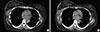
Both initial planning CT and second planning CT image sets were transferred to a three-dimensional (3D) treatment-planning software system Eclipse version 7.3.10 (Varian Medical Systems, Palo Alto, USA). In 3D planning software, image fusion of two CT sets was performed. Clips were delineated on both CT images. The reference point was placed at the midline of the sternal notch. The 3D planning software presented a relative coordinate of the clips from the reference point. The superior clip was selected as a representative. Coordinates of the surgical clips on both CT scans were compared to measure the displacement of the clips in a 3D direction including lateromedial (X), anteroposterior (Y), and superoinferior (Z). The 3D distance was calculated as the root of (X2+Y2+Z2) (Figure 1).
Statistical analyses
Chi-square method was used to perform the univariate analysis evaluating the association between the displacement of the surgical clips and clinicopathological factors. Logistic regression was used to perform the multivariate analysis. All p-values were two-sided, and p<0.05 was considered as statistically significant. All statistical analyses were performed using R software version 3.1.2 (R Foundation for Statistical Computing, Vienna, Austria; http://www.r-project.org).
RESULTS
Patients
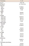
Baseline characteristics of 178 patients are detailed in Table 1. Eighty-six patients (48.3%) had right-side lesions and 92 patients (51.7%) had left-side lesions. Seroma was observed in 38 patients (21.3%) and seroma ≥15 mL in 14 patients (7.9%). The median interval between surgery and initiation of radiotherapy was 21 weeks (range, 3.1–36.0 weeks), and the median interval between the initiation of radiotherapy and boost irradiation was 5 weeks (range, 4.4–8.1 weeks). For 42 patients (23.6%) who did not receive chemotherapy, the median interval between surgery and radiotherapy and between radiotherapy and boost irradiation were median 34.5 and 35 days, respectively.
Displacement of surgical clip
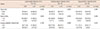
Median displacement of the surgical clips was 2.6 mm (range, 0.0–22.2 mm) in the lateromedial direction, 1.8 mm (range, 0.0–14.2 mm) in the anteroposterior direction, and 2.0 mm (range, 0.0–10.0 mm) in the superoinferior direction. Median displacement of the 3D distance was 5.3 mm (range, 0.0–28.2 mm). The 90th percentile of displacement was 5.3, 7.1, and 6.0 mm in the lateromedial, anteroposterior, and superoinferior directions, respectively, and the 3D distance was 9.8 mm. The 3D displacements of the surgical clips are detailed in Table 2.
Association with clinicopathological factors
Clinicopathological factors including age, body mass index, breast size, side, quadrant, axillary dissection, the number of dissected lymph node, pathological T stage, pathological N stage, adjuvant chemotherapy, seroma, and the time interval between surgery and radiotherapy were analyzed. Displacements of the surgical clips were grouped into less than the third quartile and more than the third quartile. The third quartile was 3.5, 4.5, and 5.0 mm in the lateromedial, anteroposterior, and superoinferior directions, respectively (Table 3). Univariate analysis revealed significant associations of outer quadrant lesion (p=0.040) and seroma ≥15 mL (p=0.004) with displacement in the lateromedial direction. Seroma (p=0.005) and the time interval between surgery and radiotherapy (p=0.040) were significantly associated with displacement in the superoinferior direction. Multivariate analyses revealed that seroma ≥15 mL was the only significant factor associated with displacement in the lateromedial (p=0.002) and superoinferior (p=0.003) directions. Outer quadrant lesion and time interval between surgery and radiotherapy showed a trend associated with displacement in the lateromedial (p=0.063) and superoinferior (p=0.057) directions (Supplementary Table 1).
Subgroup analyses of patients with seroma ≥15 mL were performed. The 90th percentile of displacement of the surgical clips was 15.1 mm in the lateromedial direction, 12.7 mm in the anteroposterior direction, 10.0 mm in the superoinferior direction, and 21.75 mm in the 3D distance (Table 4).
DISCUSSION
BCS has become a standard treatment for early stage breast cancer, allowing breast preservation in patients with breast cancer. Radiotherapy is an essential treatment after BCS to achieve an oncological outcome equivalent to mastectomy [12312]. Because tumor recurrences reportedly appear adjacent to the primary tumor, the surgical bed should be covered with a high radiation dose. Radiation delivery to exact surgical bed on CT scan is a major concern for radiation oncologists. Radiotherapy to patients with breast cancer takes 5 to 6 weeks using a conventional fractionation schedule. Radiation should be delivered to the intended target site accurately from the beginning to the end of treatment. Geometric uncertainties exist during radiotherapy because of setup error, intrafractional motion, and interfractional target change. An adequate margin to the radiation target can ensure that the target is not missed. The proper margin differs according to the treatment sites and modalities.
The lumpectomy cavity may change after breast cancer surgery. Patients who do not receive adjuvant chemotherapy start postoperative radiotherapy within 6 weeks after surgery. Postoperative healing may continue during radiotherapy. Fibrosis, contraction, and absorption of fluid can influence the size and shape of the lumpectomy cavity. Disturbance of the lymphatic flow may induce an edematous change in breast tissue of patients who undergo lymph node dissection. The breast may be influenced by postoperative and radiation-induced change more than other solid organs, because the breast is not a fixed organ; it consists of fat and lacks an external wall, so is easily movable. With this background, we investigated the possible factors influencing the surgical cavity in patients with breast cancer including breast size, quadrant, lymph node dissection, adjuvant chemotherapy, the time interval between surgery and radiotherapy, and seroma.
The 90th percentile of displacement of the surgical clips between initial simulation CT before the start of radiotherapy and re-simulation CT after 50.4 Gy of radiotherapy was within 10 mm in all patients, but more than 10 mm in patients with seroma. Conventional radiotherapy consists of whole breast irradiation and surgical bed boost. Simulation CT scan for surgical bed boost after whole breast irradiation allows adaptive planning for change of surgical cavity. However, this takes time and involves extra cost and extra radiation exposure. Presently, displacements of surgical clips were within 10 mm in all directions. Given a 10 mm margin to the surgical cavity, a plan based on initial simulation CT scan would be sufficient to cover the primary tumor bed. Recently, intensity-modulated radiotherapy (IMRT) has been adopted to treat patients with breast cancer. The heart, coronary arteries, and lungs can be saved from a high dose radiation using IMRT [1314]. External beam-partial breast irradiation using IMRT also can be considered for selected patients. IMRT preserves the normal organs but has a higher risk of missing the target. Interfractional change in the target volume can cause unintended dose delivery [15]. Our results suggest that a 10 mm margin may be sufficient for IMRT planning to compensate for the alteration of the surgical cavity during radiotherapy.
Presently, seroma ≥15 mL on initial simulation CT was associated with >10 mm displacement of the surgical clips. Seroma has been reported as a significant factor associated with tumor bed volumetric change of 5% or greater during radiotherapy [16]. Tumor volume was observed to increase in 30% of patients and to decrease in 70% of patients. In another study, the presence of seroma was significantly associated with a change in the lumpectomy cavity volume of more than 40% (p=0.021). A significant seroma volume change was also reported elsewhere [17]. Mean seroma volume was 65.7 mL before whole breast irradiation and 35.6 mL at boost planning. Two of 24 patients showed an increase in the volume of seroma by 9.7% and 10.7%, respectively. Our data suggest that for patients who have seroma ≥15 mL, re-simulation CT scan should be considered before boost planning. Boost margin of 20 mm from the clinical target volume may be sufficient to cover the displacement of lumpectomy cavity in patients with seroma, but could unnecessarily irradiate normal tissue. According to previous reports, the lumpectomy cavity undergoes volume reduction rather than volume expansion during radiotherapy in most cases. CT scans for boost plan and adjustment of target volume may be a suitable option to cover the lumpectomy cavity adequately and to minimize the irradiation of normal breast tissue.
Patients who received radiotherapy within 23 weeks after surgery showed a trend to have a larger displacement of the surgical clips in the superoinferior direction. When the time interval between surgery and radiotherapy is shorter, there is a greater possibility that postoperative healing changes the lumpectomy cavity. An analysis of the changes in the breast during whole breast irradiation reported that the mean volume reduction in the excision cavity was 22.5% (p<0.0001) and was inversely correlated with the time between surgery and radiotherapy (p<0.01) [18]. Another study compared the volume of the lumpectomy cavity from postoperative CT scan to planning CT scan. Change in the tumor bed volume between the two CT scans was 2.1% per day in patients who received immediate postoperative radiotherapy after surgery and 0.4% per day in patients who had a delay for adjuvant chemotherapy between surgery and radiotherapy [19]. These findings suggested that the volumetric change is inversely proportional to the interval between surgery and radiotherapy. However, the study was limited by its retrospective nature and relatively small sample size [20]. The results from the retrospective data should be interpreted with caution. We showed the displacement of selected single clip for each patient and analyzed it using the 90th percentile as a reference value [21]. Variation between all clips in the same patient was not analyzed in this study.
In conclusion, displacements of the surgical clips during whole breast irradiation typically range under 10 mm. Given a margin of 10 mm, a target volume based on initial simulation CT scan would sufficiently cover the lumpectomy cavity. However, in patients who have seroma ≥15 mL after BCS, resimulation before a boost treatment may be necessary to make exact planning adaptive to volumetric changes in the lumpectomy cavity.


 XML Download