

 PDF
PDF ePub
ePub Citation
 Print
Print


INTRODUCTION
Globally, breast cancer is the most frequently diagnosed cancer and the leading cause of cancer death in women. In Korea, breast cancer is the second most common newly diagnosed malignancy in women (more than 15,000 new cases annually) [1]. Therefore, it is important to identify factors predictive of prognosis and therapeutic significance.
Recently, somatic mutations of the AT-rich interactive domain 1A (SWI-like) gene (ARID1A) located in chromosome 1p36 were identified in many human cancers, including breast cancer [234]. BAF250a, the protein encoded by ARID1A, is a key component of the multiprotein SWI/SNF chromatin remodeling complex, which is critical for differentiation, proliferation, DNA repair, and tumor suppression [45]. ARID1A has recently been the subject of intense investigation because it has been found to be lost or mutated in various types of cancer, including ovarian clear cell carcinoma [6], endometrial carcinoma [78], cervical cancer [9], clear cell renal cell carcinoma [10], small intestinal carcinoma [11], malignant rhabdoid tumors [12], gastric carcinoma [13], non-small cell lung cancer [14], and urothelial bladder tumors [15]. Loss of ARID1A protein expression correlates closely with ARID1A mutations and can be used as a surrogate marker of ARID1A mutation [1617].
Previous studies have implicated that loss of ARID1A expression is associated with an unfavorable outcome of breast cancer [21819]. The relationship between ARID1A protein expression and clinicopathological variables, including prognostic significance, in breast cancer has been investigated only in a limited way, and details remain largely unknown. The purpose of this study was to evaluate the nuclear expression of ARID1A in 476 cases of Korean breast cancer by immunohistochemistry (IHC) and to correlate the findings to molecular subtype and clinicopathologic variables, including prognostic significance.
METHODS
Patients
Formalin-fixed and paraffin-embedded tissues from 476 consecutively resected primary breast cancers from patients treated at Soonchunhyang University Cheonan Hospital from 2001 to 2013 were retrospectively examined. The inclusion criteria for these samples were as follows: patients underwent curative surgeries, resected specimens were pathologically examined, and complete medical records were available. All patients received standardized comprehensive treatment. Two pathologists (H.D.C. and H.J.L.) reviewed hematoxylin and eosinstained slides of all cases, according to the 2012 World Health Organization classification [20]. Data regarding patient age at initial diagnosis, tumor size, histological type, histological tumor grade [21], lymph node status, and surgery type were also collected. Pathologic TNM classification and staging were performed for the 476 cases using the current TNM international staging system (seventh edition of the American Joint Committee on Cancer criteria). This study was approved by the institutional review boards at the Soonchunhyang University Cheonan Hospital (SCHCA 2015-04-009-002).
Construction of the tissue microarrays
For uniform and simultaneous protein expression analysis of multiple tissue samples, tissue microarrays (TMAs) were prepared. Representative core tissue sections 2 mm in diameter were taken from paraffin blocks and arranged in new TMA blocks using a manual TMA device (Superbiochips Laboratories, Seoul, Korea). In cases with variable histological features, the most representative area was selected for TMA construction. Six cores were sampled and included in the TMA block. Using a standard microtome, 4 µm-thick sections were cut from TMA blocks and were used to perform IHC.
Immunohistochemistry
ARID1A expression was analyzed by IHC. Four micrometer-thick sections from the TMA blocks were deparaffinized in xylene and rehydrated through gradually decreasing concentrations of ethanol in distilled water. IHC staining of the TMA samples was performed using a Benchmark® automatic immunostaining device (Ventana Medical Systems, Tucson, USA) and an UltraView™ Universal DAB detection kit (Ventana Medical Systems) according to the manufacturer's recommendations. The primary anti-ARID1A mouse monoclonal antibody (PSG3, SC-32761; Santa Cruz, Dallas, USA) was used at a dilution of 1:150. For negative controls, sections were treated omitting the primary antibody. For positive controls, normal breast tissue section staining was positive. Cells positive for ARID1A protein were defined as those with distinct brown granules located in cell nuclei. Two independent observers (H.D.C. and H.J.L.) read the slides in a blinded manner. Only epithelial cells were evaluated, and the result for each core was recorded separately. At the time of review, neither of the investigators was aware of the clinicopathologic data of the breast cancers, since all of the slides had been coded. The average maximal staining intensity (no staining [0], weak [1+], moderate [2+], or strong [3+]) for each of the two cores per sample was recorded. The extent of staining was also initially assessed on a three-point scale: 0, ≤10% positive cells; 1, 11%-50% positive cells; and 2, ≥51% positive cells. Subsequently, the total score was calculated by multiplying each score. According to these assessment criteria, the immunostaining results were classified as follows: scores of 0-2 indicated low or no expression of ARID1A protein, and scores of 3-6 indicated high expression of ARID1A protein [19].
IHC staining for estrogen receptor (ER; 1:50; Dako Co., Carpinteria, USA), progesterone receptor (PR; 1:50; Dako Co.), human epidermal growth factor receptor 2 (HER2; 1:200; Novocastra Laboratories Ltd., Newcastle, UK), Ki-67 (1:800; Dako Co.), cytokeratin 5/6 (CK5/6; 1:50; Dako Co.), epidermal growth factor receptor (EGFR; 1:100; Dako Co.), and p53 (1:1,200; Dako Co.) was also performed on 4 µm-sections of the TMA blocks. The IHC staining for ER and PR was evaluated using the Allred method [22]. An Allred score of 3 or higher was considered positive. HER2 expression was analyzed according to the general guidelines set by the American Society of Clinical Oncology/College of American Pathologists. When the IHC yielded equivocal results, HER2 status was determined using fluorescent in situ hybridization. The expression of Ki-67 was counted in 1,000 tumor cells, and the percentage of positive cells was categorized as ≥14%. For CK5/6 and EGFR expression, the cells were considered positive when the cytoplasmic and/or membranous reaction was ≥10%. The expression of p53 was counted in 1,000 tumor cells, and the percentage of positive cells was categorized as >10%. The phenotypes were classified as follows: luminal A type: ER and/or PR positive, HER2 negative, and Ki-67 index <14%; luminal B HER2 negative type: ER and/or PR positive, HER2 negative, and Ki-67 index ≥14%; luminal B HER2 positive type: ER and/or PR positive, HER2 positive, and any Ki-67 index; HER2 type: ER and PR negative and HER2 positive; triple-negative breast cancer (TNBC) basal type: ER, PR, and HER2 negative and CK5/6 and/or EGFR positive; and TNBC nonbasal type: ER, PR, HER2, CK5/6, and EGFR negative.
Statistical analyses
The analyses were performed using the software package SPSS version 19.0 for Windows (IBM Corp., Armonk, USA). Associations between ARID1A expression and the clinicopathologic characteristics were analyzed using Pearson chi-square test, Fisher exact test, or an independent t-test, according to test conditions. Survival curves were plotted using the Kaplan-Meier method, and statistical significance was assessed using the log-rank test. Disease-free survival (DFS) was defined as the interval between primary surgery and the last follow-up visit without disease or evidence of recurrence or metastasis of breast cancers (locoregional relapse, distant metastasis). Overall survival (OS) was defined as the interval between primary surgery and the last follow-up visit or death from any cause. The Cox proportional hazards model was used for multivariate analysis. A p-value <0.05 was considered statistically significant.
RESULTS
Patient characteristics and ARID1A immunoreactivity
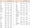
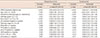
The clinicopathological characteristics of the patients with primary breast cancer (n=476) are listed in Table 1. Patient age ranged from 24 to 81 years (median, 50.0 years; mean, 52.3 years). There were 470 (98.7%) female and six (1.3%) male patients. Of the 476 included samples, 241 patients (50.6%) underwent breast-conserving surgery, and 235 patients (49.4%) underwent mastectomy. The histological types included invasive ductal carcinoma not otherwise specified (432 samples, 90.8%), invasive lobular carcinoma (20 samples, 4.2%), and others (24 samples, 5.0%). The histological grade was available for 476 samples; 59 (12.4%) were grade 1, 243 (51.1%) were grade 2, and 174 (36.6%) were grade 3. Tumor sizes varied from 0.3 to 12 cm (mean, 2.42 cm). Among 476 patients for whom primary tumor size data were available, 230 (48.3%), 219 (46.0%), and 23 (4.8%) tumors were categorized as pT1, pT2, and pT3, respectively. Of the 476 patients, 173 (36.3%) had lymph node positivity at the time of surgery. The 476 patients were classified using the TNM classification system as follows: stage I, 169 patients (35.5%); stage II, 220 patients (46.2%); and stage III, 87 patients (18.3%). The proportions of patients positive for ER and PR expression were 66.8% and 36.1%, respectively. Upon analysis of HER2 expression, 17.4% of all patients were positive. The percentage of cases with high Ki-67 expression was 43.9%. CK5/6 and EGFR expression was found in 9.5% and 20.6% of cases, respectively. For p53 expression, 17.4% of patients were positive.
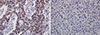
ARID1A protein expression in breast cancer appeared mainly in the nuclei of tumor cells (Figure 1). After evaluation of the 476 immunostained breast cancer specimens, 150 (31.5%) showed no positivity, 75 (15.8%) had score 1 positivity, 114 (23.9%) had score 2 positivity, 44 (9.2%) had score 3 positivity, 45 (9.5%) had score 4 positivity, and 48 (10.1%) had score 6 positivity. For the statistical analysis, the cases were subdivided into an ARID1A high expression group (scores 3, 4, and 6; n=137, 28.8%) and an ARID1A low expression group (scores 0, 1, and 2; n=339, 71.2%).
Correlations between ARID1A expression and clinicopathologic parameters
Low expression of ARID1A was significantly correlated with mastectomy (p=0.023), positive lymph node metastasis (p=0.027), advanced pathologic stage (p-stage, p=0.001), low Ki-67 labeling index (p=0.003), negative p53 expression (p=0.017), and neoadjuvant chemotherapy status (p=0.002) (Table 1). Weak correlations between low ARID1A expression level and low histologic grade (p=0.056) were also found without reaching formal statistical significance. Other clinicopathologic variables, including age, sex, histologic type, tumor size, ER positivity, PR positivity, HER2 positivity, CK5/6 positivity, EGFR positivity, molecular subtype, chemotherapy, and radiotherapy did not correlate with ARID1A expression.
Survival analysis
All patients were closely followed after surgery, with a median follow-up period of 39 months (range, 1-158 months). During follow-up, 88 patients (18.5%) relapsed, and 51 patients (10.7%) died. Patterns of relapse were reviewed, and we found that most patients had distant metastasis (n=79, 16.6%) rather than locoregional relapse (n=9, 1.9%) (Table 1). The OS rates for breast cancer patients with high and low ARID1A expression were 98.5% and 85.5%, respectively.
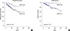
On univariate survival analysis, conventional prognostic parameters, including age, operation methods, tumor size, lymph node metastasis, and p-stage, reached significance for DFS and OS (p<0.05 for all) (Table 2). In addition, hormonal expression (ER or PR) and HER2 expression were factors affecting DFS or OS of breast cancer patients. Patients with low ARID1A expression had significantly shorter DFS and OS than patients with high ARID1A expression (p<0.001 and p<0.001, respectively) (Figure 2).
To evaluate whether ARID1A positivity in breast cancer was an independent predictor of DFS and OS, a multivariate analysis using the Cox proportional hazard model was performed with the following variables: age, operation methods, tumor size, lymph node metastasis, p-stage, hormonal expression (ER or PR), HER2 expression, and ARID1A expression. All variables with a p-value<0.05 in the univariate analysis were included in the multivariate Cox model. Age (p=0.038 and p<0.001 for DFS and OS, respectively), operation methods (p=0.024, only for DFS), lymph node metastasis (p=0.008, only for DFS), ER positivity (p=0.037, only for OS), and HER2 positivity (p=0.043, only for DFS) were significant prognostic factors for breast cancer patients (Table 2). Multivariate analysis identified low ARID1A expression as significant independent factor for poor DFS and OS in patients with breast cancer (DFS: hazard ratio, 0.38, 95% confidence interval [CI], 0.20-0.73, p=0.004; OS: hazard ratio, 0.11, 95% CI, 0.03-0.46, p=0.003, respectively).
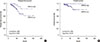
The DFS and OS of the ARID1A high and low expression groups, stratified according to molecular subtype, are shown in Figure 3 and Supplementary Figure 1 (available online). In patients with luminal A type disease, low ARID1A expression was associated with significantly shorter DFS and OS than high ARID1A expression (p=0.022 and p=0.018, respectively) (Figure 3). In patients with luminal B, HER2 negative type disease, both DFS and OS did not show any statistically significant differences according to ARID1A expression (p=0.874 and p=0.313, respectively) (Supplementary Figure 1A, B). In patients with luminal B, HER2 positive type disease, both DFS and OS did not show any statistically significant differences according to ARID1A expression (p=0.238 and p=0.067, respectively) (Supplementary Figure 1C, D). In patients with HER2 type disease, low ARID1A expression was associated with significantly shorter DFS than high ARID1A expression (p=0.016) (Supplementary Figure 1E). In contrast, OS was not significantly different between the ARID1A high and low expression groups (p=0.087) (Supplementary Figure 1F). In patients with TNBC basal type disease, both DFS and OS did not show any statistically significant differences according to ARID1A expression (p=0.144 and p= 0.114, respectively) (Supplementary Figure 1G, H). In patients with TNBC nonbasal type disease, both DFS and OS did not show any statistically significant differences according to ARID1A expression (p=0.258 and p=0.408, respectively) (Supplementary Figure 1I, J). In multivariate analysis using the Cox proportional hazard model, low ARID1A expression was not a significant independent prognostic factor for DFS and OS, according to molecular subtype.
DISCUSSION
ARID1A functions as a tumor suppressor and may participate in both tumor initiation and progression in human cancers [23]. ARID1A is most frequently mutated in endometrium-derived tumors (about 50% of ovarian clear cell carcinomas and 30% of ovarian endometrioid carcinomas) [817]. Comprehensive molecular studies indicate that the ARID1A gene mutation rate is about 4% in breast cancers, but copy number loss occurs in 13%-35% of cases [224]. Low expression of ARID1A mRNA has been reported to be strongly associated with promoter hypermethylation of the ARID1A gene in invasive ductal carcinomas (86.4%) [25]. The authors identified an association of low ARID1A RNA or nuclear protein expression with more aggressive breast cancer phenotypes [2].
In our study, we used a large number of breast cancer samples (n=476) to detect the expression of ARID1A protein by IHC. The majority (339/476, 71.2%) of the breast cancer tissues exhibited low ARID1A expression. This percentage is consistent, but somewhat higher, than previous reports (Mamo et al. [2]: 64% [151/236]; Zhang et al. [18]: 56% [63/112]; and Zhao et al. [19]: 65.3% [324/496]). Differences in patient race and IHC methods may account for this discrepancy.
In this study, clinicopathological analysis revealed that low ARID1A expression in breast cancer was associated with mastectomy, lymph node metastasis, advanced p-stage, low Ki-67 labeling index, and negative p53 expression. Weak correlations between low ARID1A expression and low histologic grade were also found. With regard to lymph node metastasis and advanced p-stage, these results are similar to those reported in previous studies of breast cancer [21819]. Our findings indicated that loss of ARID1A expression was related to low Ki-67 labeling index, negative p53 expression, and low histologic grade, which have been identified as good prognostic factors. Some studies reported the loss of ARID1A expression may be related to less invasive clinicopathologic features in colorectal cancer and gastric cancer [26]. However, one study reported that tumors with low ARID1A expression were associated with ER negativity, higher p53(+) percentage, higher Ki-67 labeling index, and TNBC subtype in breast cancer [18], with other authors reporting that low ARID1A expression was associated with ER negativity, high histologic grade, and higher p53(+) percentage [19]. A statistically significant inverse correlation between the mutational statuses of the ARID1A and TP53 genes in tumor samples of ovarian clear cell carcinoma and endometrial endometrioid carcinoma was reported [27]. The authors suggested that ARID1A and p53 collaborate to prevent tumorigenesis. In contrast, other researchers reported no significant relationship between loss of ARID1A expression and p53 overexpression in endometrial clear cell carcinoma [28]. These differences may be attributed to differences in the organ and carcinoma types, patient races or sample sizes, variations in antibodies, laboratory IHC methods, as well as other cofactors that affect tumor behavior. Therefore, additional studies on a larger cohort will be needed to confirm our findings.
In agreement with previous studies [21819], patients with low ARID1A expression had worse DFS and OS than those with high ARID1A expression. The multivariate analysis also revealed that low ARID1A expression was a significant independent prognostic factor for shorter DFS and OS in patients with breast cancer. Thus, low ARID1A expression might be applied as a valuable prognostic marker for relapse and disease-related death in patients with breast cancer. In this study, we were also able to demonstrate an association between ARID1A expression and clinical outcomes according to the molecular subtype. In luminal A type disease, there was a significant trend toward shorter DFS and OS in the group with low ARID1A expression compared with the group with high ARID1A expression. With this result, we could speculate that luminal A type disease with loss of ARID1A expression might be correlated with poor clinical outcomes.
There are some limitations to the generalization of these results. First, the retrospective design and the small sample size drawn from a single institution resulted in selection bias. Second, the survival analysis was limited as the follow-up period was too short to determine 5-year survival rates. Third, the significant association between ARID1A expression and p53 and Ki-67 might be affected by several confounders. These need to be evaluated in subsequent studies. Despite these limitations, this is the first study to examine ARID1A expression in a large number of breast cancer patients in Korea.
Recent studies have focused on the possible mechanism of ARID1A mutation and protein expression loss in tumorigenesis. A relationship between ARID1A mutations and enhancer of zeste homologue 2 (EZH2) has been suggested. Inhibition of EZH2 methyltransferase acts in a synthetic lethal manner in ARID1A-mutated ovarian cancer cells, with ARID1A mutational status correlated with response to the EZH2 inhibitor, making it a potential target for targeted therapy [29].
In summary, we assessed clinicopathological correlations, molecular subtype, and prognostic significance of ARID1A expression by IHC in primary breast cancer. This is the first study to reveal the prognostic significance of ARID1A expression in a large number of breast cancer patients in Korea. Low expression of ARID1A is an independent predictive factor for poor DFS and OS in breast cancer patients and may be associated with luminal A type disease. The exact role of the ARID1A pathway in breast cancer is not clear. Additional functional studies using breast cancer cell lines and further validation with in vivo experiments are needed to elucidate the role of the ARID1A pathway in the tumorigenesis and progression of breast cancer.
 XML Download