

 PDF
PDF ePub
ePub Citation
 Print
Print


INTRODUCTION
The breast is composed of adipose tissue and glandular tissue. A terminal ductal-lobular unit is the basic unit of the glandular tissue and is postulated as the site of origin of most breast cancers [1]. Invasive lobular carcinoma (ILC) arises in the lobule and is microscopically characterized by linear infiltration of small uniform cells. It is the second most common invasive breast cancer after invasive ductal carcinoma (IDC), accounting for 2% to 3% of breast cancers in Korean women [23456]. Multicentric, bilateral, and estrogen receptor (ER)-positive cancers are frequent in patients with ILC compared to those with IDC [789]. Although some studies have claimed that the local recurrence rate after partial mastectomy in patients with ILC is high [1011], the treatment outcomes of breast conserving surgery (BCS) followed by radiation ther apy are comparable to those of mastectomy [12].
The purpose of the current study was to assess the incidence of ILC among patients who underwent BCS followed by radio therapy and to compare the clinicopathological features and treatment results between ILC and IDC.
METHODS
Patients
This was a retrospective study of 1,071 patients with invasive ductal or lobular carcinomas of the breast who underwent BCS followed by radiotherapy in our institution between 1994 and 2007. All patients were newly diagnosed, and those with a prior history of breast cancer, as well as those diagnosed with mixed ductal-lobular types or types other than IDC or ILC, were excluded from the study.
Treatment
All patients underwent upfront BCS. Patients who received preoperative chemotherapy were not included in the study. If the surgical margins were involved by ductal carcinoma in situ or invasive tumor, a re-excision was performed. Sentinel lymph node biopsies were performed in clinically node-negative patients, and axillary lymph node dissections were performed in clinically node-positive or sentinel lymph node-positive patients.
Adjuvant chemotherapy was recommended for node-positive patients as well as those with tumors larger than 1 cm or basal-like subtypes. The chemotherapy regimens consisted of cyclophosphamide, methotrexate, and 5-fluorouracil (CMF); doxorubicin and cyclophosphamide (AC); 5-fluorouracil, doxorubicin, and cyclophosphamide (FAC); and AC followed by paclitaxel. Anthracycline-based chemotherapy was adopted in 2001 and replaced CMF chemotherapy beginning in 2004. Hormone therapy was recommended for patients with hormone receptor-positive tumors.
Radiation therapy was started 4 to 6 weeks after surgery or completion of adjuvant chemotherapy or was delivered between AC and paclitaxel. The radiation field was matched to the tangential field covering the whole breast and the lower part of the level I and II axillary lymph nodes. The field-in-field technique or the wedge was used to improve the dose homogeneity. Supraclavicular fossa irradiation was performed in patients with pathological N2 or high-risk N1 disease. A median dose of 50.4 Gy (range, 50.0-50.4 Gy) at 1.8 to 2.0 Gy per fraction was delivered with 4 or 6 MV photon beams. An electron boost to the tumor bed with a median dose of 10.0 Gy (range, 6.0-12.0 Gy) was delivered to all patients except those with microinvasive carcinomas.
Clinicopathological features
Medical records and pathological reports were retrospectively reviewed to assess clinicopathological features including age, laterality, pathologic stage, nuclear grade, ER status, progesterone receptor (PR) status, human epidermal growth factor receptor 2 (HER2) status, extensive intraductal carcinoma (EIC), and lymphovascular invasion (LVI). Pathologic stage was classified according to the seventh edition of the American Joint Committee on Cancer Staging Manual [13]. The histologic grade was scored according to the Bloom-Richardson grading system and the Elston-Ellis modification of the Scarff-Bloom-Richardson grading system (Nottingham histologic score system) [141516]. The hormone receptor status, HER2 status, and p53 protein expression were determined by immunohistochemical (IHC) staining. The tumors were classified into three IHC subtypes: luminal (ER- or PR-positive), basal-like (ER-, PR-, and HER2-negative), and erbB-2 overexpressing (ER-, PR-negative, and HER2-positive) [17]. EIC was defined as an intraductal carcinoma occupying more than 25% of the primary tumor with intraductal foci separate from the main tumor mass.
Statistical methods
The clinicopathological features of ILC and IDC were compared using Pearson chi-square test. Disease-specific survival (DSS) was measured from the date of surgery to the date of death from breast cancer, and deaths from other cancers or diseases were censored. Disease-free survival (DFS) was measured from the date of surgery to the date of any recurrence or to the date the patient was last known to be recurrence-free. Metachronous contralateral breast cancer was not considered recurrence. Kaplan-Meier analysis and log-rank tests were used to estimate and compare the DSS and DFS. Multivariate analysis was performed using the Cox proportional hazards model. A Bonferroni correction was applied for multiple testing. The SPSS statistical software version 18.0 (SPSS Inc., Chicago, USA) was used for statistical analyses. A p-value of less than 0.05 was considered statistically significant.
RESULTS
Clinicopathological features
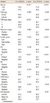
Among 1,071 patients with invasive breast cancer, 56 patients (5.2%) were diagnosed with ILC. Table 1 shows the comparison of clinicopathological features between ILC and IDC. There were many cases in which EIC and LVI data were not reported, because it was not obligatory to report these until the development of a unified format for pathology reports in 2005. However, the proportions of cases in which the hormone receptor status and IHC subtype were unreported were less than 10%. The statistical analyses were performed excluding cases with unreported data. There were no statistically significant differences in age, pathologic stage, resection margins, EIC, or HER2 status. Statistically significant differences were found in laterality, nuclear grade, LVI, hormone receptor status, p53 status, and IHC subtype. Bilateral breast cancer was more frequent in patients with ILC than in those with IDC, at 7.1% vs. 1.5%, respectively. ILC was found to have a lower nuclear grade than IDC. There were no cases of ILC with LVI. The proportion of hormone receptor-positive breast cancers was higher in patients with ILC than in those with IDC. With respect to IHC subtypes, the erbB-2 overexpressing subtype was less frequent in ILC, and there were no instances of the basal-like subtype found among patients with ILC.
Treatment results
A total of 825 patients (77.0%) received chemotherapy after BCS. There was no statistically significant difference in regimens between patients with ILC and those with IDC (p=0.494). A total of 699 patients (65.3%) received hormone therapy including tamoxifen or aromatase inhibitors. Among 722 patients (67.4%) with ER- or PR-positive cancers, 676 patients (63.1%) received hormone therapy.
The median follow-up duration, calculated from the date of surgery, was 114 months (range, 5-238 months). During the follow-up period, 105 patients died of breast cancer and 15 patients died of other causes, including other cancers, myocardial infarction, intracranial hemorrhage, and pneumonia. The 10-year DSS and DFS rates were 89.4% and 84.0%, respectively.
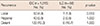
Recurrence occurred in 163 patients (15.2%). There were no statistically significant differences in the patterns of recurrence between ILC and IDC (Table 2). Twenty-eight patients (2.6%) developed contralateral breast cancer, which is not defined as recurrence, and all but one of these patients were initially diagnosed with IDC.
Statistical analyses
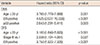
On univariate analyses, age, stage, nuclear grade, EIC, LVI, ER status, PR status, HER2 status, p53 status, and IHC subtypes were statistically significant prognostic factors for DSS. Age, stage, nuclear grade, LVI, PR status, HER2 status, and p53 status were statistically significant prognostic factors for DFS (Table 3).
On multivariate analyses, age, LVI, and p53 status were statistically significant prognostic factors for DSS. Age, stage, and LVI were statistically significant prognostic factors for DFS (Table 4).
DISCUSSION
ILC is the second most common type of invasive breast cancer after IDC. In 2000, Li et al. [18] analyzed the data of the Surveillance, Epidemiology, and End Results (SEER) Program, including 240,018 patients with invasive breast cancer, and reported that 16,476 patients (6.9%) were diagnosed with ILC. In addition, they reported that the incidence rate of ILC increased steadily from 1987 to 1995 in women older than 50 years. Eheman et al. [19] reported on the changing incidences of IDC and ILC in the United States between 1999 and 2004. The incidence of ILC decreased from 11.7% to 9.3%. Interestingly, the authors noted differences in incidence rates according to race. The age-adjusted incidences of ILC were 11.2, 6.6, 4.4, and 3.6 per 100,000 in Caucasians, African-Americans, Asians, and American Indians, respectively. In Korean women, the incidence of ILC was reported as 2% to 3% [23456]. In our study, it was 5.2%, slightly higher than in other Korean studies. However, even considering selection bias, the figure reported in our study was still lower than that in Western women.
Many studies had reported several differences in clinicopathological features between ILC and IDC [78920212223]. Tumor size, tumor grade, hormone receptor status, and incidence of contralateral breast cancer were the most commonly reported differences in various studies. ILCs were larger and had a lower tumor grade than IDCs. Approximately 76% to 93% of ILCs were hormone receptor-positive. Contralateral breast cancer was more common in patients with ILC, with a reported incidence of 14% to 21%. Although several studies reported statistically significant differences in average age between patients with ILC and those with other invasive carcinomas, the age gap was small (less than 3 years). In our study, there were no statistically significant differences in age, pathologic stage, resection margins, EIC, or HER2 status. Statistically significant differences were found in laterality, nuclear grade, LVI, hormone receptor status, p53 status, and IHC subtype. However, these results should be evaluated carefully, because our study included patients who were suitable for BCS, and EIC and LVI were unreported in more than 50% of cases in the patients with ILC.
Several studies reported higher rates of local recurrence after BCS than after mastectomy and suggested mastectomy for patients with ILC [1011]. In a recently published study by Fodor [24], DSS was not affected by the surgical extent, but the 15-year local recurrence-free survival rates were 77% and 89% after BCS and mastectomy, respectively (p=0.005). However, among the 72 patients who underwent BCS, 19 patients (26.0%) did not receive adjuvant radiotherapy. Add itional analysis revealed that the 15-year local recurrence rates for the BCS groups with or without adjuvant radiotherapy were 10% and 53%, respectively (p<0.001). As adjuvant radiotherapy after BCS has been proven in the National Surgical Adjuvant Breast and Bowel Project clinical trial to decrease local recurrence [25], previous results of BCS should be carefully investigated, with this factor taken into consideration.
Many studies have found that there are no differences in survival or local recurrence between patients with ILC and those with IDC after BCS followed by radiotherapy [212627282930]. The 10-year local recurrence rates were found to be 9%-18% and 7%-12% for ILC and IDC, respectively. In our study, while the differences were not statistically significant, there was a trend toward better DSS and DFS in patients with ILC. The 10-year DSS rates were 98.0% and 89.0% in ILC and IDC, respectively (p=0.262), while the 10-year local recurrence rates were 3.6% and 8.8% in ILC and IDC, respectively (p=0.457). The better outcomes might be due to a higher proportion of hormone-positive breast cancers in patients with ILC. The number of patients with ILC was too small to show statistically significant differences.
Our study does have some limitations. First, we included only patients who underwent BCS, and this population does not represent the entire spectrum of invasive breast cancer patients. Second, although LVI was an independent prognostic factor in both univariate and multivariate analyses, it was not reported in 695 cases (64.9%). Depending on the LVI status in the cases in which it was unreported, the results of the statistical analyses could change. Finally, there was an evolution in the chemotherapy regimens over time, from CMF to anthracycline-based chemotherapy, and this change could have affected regional or distant recurrences.
In conclusion, the incidence of ILC in our study was 5.2%, slightly higher than that observed in other Korean studies, but still lower than those reported in Western studies. Bilateral breast cancer, lower nuclear grade, and hormone receptor-positive breast cancer were more frequent in patients with ILC than in those with IDC. There were no cases of LVI or the basal-like subtype among the patients with ILC. There were no statistically significant differences in the patterns of treatment failure. The development of metachronous contralateral breast cancer was more frequent in patients with IDC (n=27). Only one patient with ILC developed contralateral breast cancer, with ductal carcinoma in situ. Although the difference was not statistically significant, there was a trend toward better DSS and DFS rates in patients with ILC.
 XML Download