

 PDF
PDF ePub
ePub Citation
 Print
Print


INTRODUCTION
Multiple symmetric lipomatosis (MSL) is a very rare lipid metabolism disorder characterized by the accumulation of multiple, symmetric, nonencapsulated lipomatous masses on the face, neck, shoulders, and upper arms [1].
The disorder was first described by Brodie in 1846. Madelung then reported the cases of 33 patients in 1888, and Launois and Bensaude provided the standard description of MSL in 1898. Thus, MSL is also known as Madelung's disease or Launois-Bensaude syndrome [2].
The cause of the condition is unknown, but a number of theories have been proposed. The incidence rate is higher in men than in women, and the average age of incidence is approximately 60 years; the condition is known to be linked to alcoholism [1,2].
Herein, we report a case of MSL in a 66-year-old man and review the current literature on the condition.
CASE REPORT
A 66-year-old man presented with bilateral breast bulging. His breasts had slowly become enlarged over a number of years and this had led to huge breast bulging; the patient reported that he was ashamed of his breast disfigurement when in public baths and swimming pools. He had undergone an excisional biopsy of a mass in the central upper back region 1 year earlier, which revealed a lipoma.
He reported more than 25 years of alcohol abuse, and confirmed that he consumed alcohol 5 times a week. Furthermore, he had a history of cigarette use, but had stopped smoking 4 years previously. He also had a 10-year history of hypertension and diabetes mellitus. The patient's father had a history of multiple large disfiguring masses in the neck and upper body.
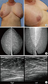
Physical examination of the patient revealed multiple large, disfiguring masses in the occipital region as well as on the breasts, upper arms, shoulders, neck, and upper trunk. These appeared to be symmetrical on the breasts, upper arms, and shoulders. Palpation revealed that the masses had an elastic consistency, could be moved without causing the patient any pain, and had no definite margins (Figure 1A).
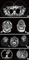
Laboratory examinations revealed normal liver and thyroid function, and a hormone assay confirmed that the results were within the normal ranges. A mammography was performed which revealed that the patient's breasts were comprised almost entirely of fat. Breast ultrasonography revealed excessively fatty breasts without any definite glandular tissue (Figure 1B). Computed tomography and magnetic resonance imaging showed diffuse and marked fatty deposits in the superficial soft tissue layers of the neck, chest, abdomen, and shoulder wall with marked bulging contours of both breasts (Figure 2).
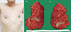
The patient underwent bilateral nipple-sparing mastectomy with nipple interposition. Pathology revealed soft tissue lipomas measuring 30×17×5 cm and 27×16×5 cm, and both lipomas were homogenous without hemorrhage and necrosis (Figures 3, 4). No hidden malignancy was found. The patient did well postoperatively and was satisfied with the cosmetic result.
DISCUSSION
MSL is a rare disease with an incidence of approximately in 1/25,000 men. The condition is more common in men, especially those aged 30 to 60 years; the male to female ratio is approximately 15:1 to 30:1. Its incidence is known to be associated with a history of alcoholic abuse. The etiology of MSL remains unknown, however, several theories have been suggested, such as mitochondrial disorder related to genetic dysfunction, defective embryogenic brown adipose tissue, and a lack of adrenergic-induced lipolysis [1,2,3,4,5]. MSL presents as large painless growing adipose masses with little circumscription that are usually located in the neck region, on the shoulders and upper extremities, and in the upper trunk region. Pathologically, MSL shows characteristics of normal adipose tissue and accumulation of fat in the brown adipose tissue [1,2,3]. MSL may occur sporadically or be inherited (autosomal dominant transmittance). Sporadic cases are more frequently observed and are strongly related to alcoholic abuse and tobacco smoking. In the case reported here, the family history supports a hereditary origin of the disease [1,2].
Enzi [6] classified MSL into two types according to its distribution site. Type I is horse collar lipoma, which presents as circumscribed protruding masses in the neck area as well as the clavicular and deltoid regions. In severe cases, this type can cause mediastinal extension and tracheal obstruction. Type II is characterized by a pseudoathletic appearance with diffuse involvements of the trunk and the proximal end of the extremities, resembling obesity [1,3]. Donhauser et al. [4] added gynecoid type to the categories of the disease and classified MSL as follows: type I, horse collar lipoma; type II, pseudoathletic appearance; and type III, gynecoid type.
There was one interesting difference in the case presented here compared with previous reports. The presenting symptom of MSL is somewhat different according to gender; men who presented with condition usually present with a submental mass [2,5,7,8]. However, the patient in our case presented mainly with large bilateral breast masses. Due to this extraordinary clinical presentation, we first had to exclude simple gynecomastia by taking a through medical history and performing a physical examination.
MSL is a benign disease and its malignant transformation is very rare. Malignant degeneration of fatty tissue into liposarcoma has only been reported in two cases [9,10]. Despite the benign nature of MSL, it can present a serious clinical scenario. In the early period of the disease, it grows rapidly and thereafter, shows slow progression or remains stable for a long period of time. The long-term deposition of lipomatous tissue often results in a cosmetic problem, and more seriously it can cause obstructive symptoms such as dysphagia, dysphonia, or dyspnea [2,3,5]. In some cases, neurological symptoms, which are related to mitochondrial defect, maybe present [11].
Differential diagnoses of MSL are simple obesity, Cushing syndrome, angiolipoma, neurofibromatosis, salivary gland diseases, conditions with a neck mass such as thyroid goiter, Frohlich's syndrome, Dercum's disease, and lipomatosis in human immunodeficiency virus [1,2]. For the accurate diagnosis of MSL, taking the clinical history of patients and performing physical examinations are very important. In the case presented here, the patient presented with large bilateral breast masses, which had slowly become enlarged over a number of years. He also had a history of a lipomatous mass on his central upper back and 25 years of alcohol abuse. Physical examination revealed multiple large, disfiguring masses in the occipital region, upper arms, shoulders, and upper trunk. Of note, the masses on the arms and, shoulders were located symmetrically. We were able to exclude the possibility of simple gynecomastia and diagnose MSL through careful history taking and physical examination.
Surgery is known to be the most effective treatment for MSL [7]. Alcohol withdrawal and weight reduction are recommended, however, these cannot reverse or stop the course of the disease [12]. Ultrasound-guided liposuction can also achieve good effects. However, liposuction is of limited effectiveness with large masses [13]. The overall recurrence rate following surgery is approximately 63%, open surgery results in a recurrence rate of 51% compared to a recurrence rate of 95% with liposuction [7].
In conclusion, MSL can present as gynecomastia. Increasing awareness of the clinical characteristics of MSL, especially among breast surgeons, could facilitate its recognition. Surgery is known to be the most effective treatment for MSL. In addition, because MSL is a genetic disorder, family members should be followed up to allow for early detection and appropriate surgical treatment, thus avoiding a delayed diagnosis and subsequent unsatisfactory treatment outcome accompanying a series of further operations.

 XML Download