

 PDF
PDF ePub
ePub Citation
 Print
Print


INTRODUCTION
Sentinel lymph node biopsy (SLNB) has been validated in early breast cancer patients and is the accepted standard of care for axillary staging in patients with clinically node-negative axilla [1,2,3]. Although patients undergoing axillary lymph node dissection (ALND) have higher morbidities including seroma, lymphedema, and arm weakness than SLNB [4], it is still a valid procedure for patients with a positive SLN. Nevertheless, 40% to 70% of patients with positive SLNs on the final histology have no metastases in the nonsentinel lymph nodes (NSLN) [5,6,7]. For this reason, most investigators beleive that ALND has minimal therapeutic effect for patients with only SLN involvement [8,9].
Currently, several risk factors are defined for the probability of metastasis in NSLNs, such as the histological primary tumor size, size of the SLN metastasis, number of positive SLNs, the ratio of positive SLNs to all removed SLNs, lymphovascular invasion, and extracapsular extension of the positive SLN [8,10,11]. None of these parameters alone is sufficient to identify which patients need ALND. Furthermore, although some mathematical models have been developed to predict the NSLN status in these patients [11,12,13,14], the predictive value of these models does not always reach a sufficiently high accuracy.
In the last decade, breast cancer has been classified into distinct molecular subtypes based on the prognostic significance of gene expression profiles [15,16]. For practical use in hospital setting, it has been suggested that immunohistochemical surrogate panels, composed of the estrogen receptor (ER), progesterone receptor (PR), and oncogene erbB-2/human epidermal growth factor receptor 2 (HER2), be used to define breast cancer subtypes rather than gene expression profiling [17,18,19]. It has been reported that different breast cancer subtypes have variable responses to treatments and oncologic outcomes [20,21]. Additionally, it has been demonstrated that there is a close relationship between subtype and risk of metastasis to one or more axillary SLNs [22,23]. However, data on which subtype has a higher risk of NSLN metastasis are limited. Here, we determined if the breast cancer subtype influences NSLN metastasis independently compared to other clinicopathological factors in 104 patients with a positive SLN.
METHODS
We examined the records of 480 patients with invasive breast cancer who underwent SLNB between April 2009 and September 2013 at Ankara Oncology Training and Research Hospital. Institutional Review Board approval was obtained with approval number 2014/352, for the study. Of these patients, 104 women with a positive SLN who underwent subsequent ALND were included in the study. The inclusion criteria were early infiltrating breast carcinoma based on clinical and radiological findings, normal physical examination of the axilla, and initial treatment of mastectomy or lumpectomy plus SLNB. Patients who received neoadjuvant chemotherapy and/or radiotherapy were excluded from the study.
The SLNB procedure was performed using a blue dye and radioisotope combination. SLN imaging was done 2 to 18 hours before the operation by superficial injection of a radioisotope followed by gamma camera imaging. On the day of surgery, blue dye was administered and manual massage was applied. All blue and/or radioactive lymph nodes were considered as SLNs and examined by frozen section intraoperatively. Any firm or enlarged node encountered during the SLNB procedure was also considered as an SLN [24]. Standard ALND was performed during the same procedure when metastases were detected in SLNs. If the definitive diagnosis with either hematoxylin and eosin staining or immunohistochemistry (IHC) revealed metastasis in an SLN postoperatively, when the frozen section had been negative, a second operation for ALND was performed.
Pathological examinations were performed by the same team for all patients. SLN materials were reviewed by the pathologist following serial sectioning and staining with hematoxylin and eosin. Positive SLNs were classified into two groups according to the size of the metastasis: micrometastasis (≤2 mm) and macrometastasis (>2 mm). Patients who had isolated tumor cells in NSLN were not included in this study. For analysis of NSLNs, routine hematoxylin and eosin staining was used. For hormone receptor status determination, tumor cells that showed at least 1% immunohistochemical staining in paraffin blocks were accepted as receptor positive [25]. HER2 overexpression status was determined according to the American Society of Clinical Oncology guidelines [26]. Patients were classified into four distinct subtypes based on the different possible IHC combinations of ER, PR, and HER2 status: luminal A (ER-positive and/or PR-positive, HER2-negative), luminal/HER2+, HER2 overexpression (ER-negative, PR-negative, HER2-positive), and triple-negative (ER-negative, PR-negative, HER2-negative) [17,18,19].
Clinicopathological factors including age, menopausal status, histological primary tumor size, histological grade, lymphovascular invasion (LVI), number of SLNs removed, size of the largest SLN metastasis, extracapsular extension status of the positive SLNs, the ratio of positive SLNs to all removed SLNs, and subtype of the tumor were assessed as predictors of NSLN metastasis using chi-square or Fisher exact tests in the univariate analysis. Statistically significant risk factors for NSLN metastasis in the univariate analysis were included in a multivariate analysis using the logistic regression model. All tests of significance were two-sided and a p-value of <0.05 was considered statistically significant. Statistical tests were carried out using the SPSS version 15.0 for Windows (SPSS Inc., Chicago, USA).
RESULTS
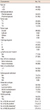
Clinicopathological characteristics of 104 early-stage breast cancer patients with a positive SLN who underwent ALND are summarized in Table 1. The median age of these patients was 49 years (range, 26-75 years) and the median pathological tumor size was 3 cm (range, 0.5-6.5 cm). The mean number of SLNs removed was 2.4±1.3 (mean±SD) and the mean number of positive SLNs was 1.5±0.8. Moreover, the mean number of ALNs dissected was 18.2±7.1. The median number of NSLNs dissected in luminal A, luminal/HER2+, HER2 overexpression, and triple-negative cases was 18, 16, 19, and 19, respectively. The mean number of positive NSLNs was 2.0±0.5 and the rate of the patients with a positive NSLN was 48%. In this retrospective cohort, the proportion of the patients who had the luminal A subtype was 48%, while the proportion of patients with the luminal/HER2+, HER2 overexpression and triple-negative subtypes was 21%, 15%, and 16%, respectively.
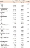
Univariate analysis results are presented in Table 2. Age, menopausal status, number of SLNs removed, size of SLN metastasis, and extracapsular extension were not significant risk factors for NSLN metastasis (p>0.166). However, tumor size, histological grade, LVI, ratio of positive SLNs, and subtype classification were significant risk factors for NSLN metastsis (p<0.012). With respect to the breast cancer subtypes, patients with the luminal/HER2+ and HER2 overexpression subtypes had a significantly higher rate of NSLN metastasis than patients with the luminal A and triple negatif subtypes (p<0.001). The rate of NSLN metastasis in both patients with luminal/HER2+ and HER2 overexpression subtypes was 73%.
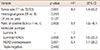
In the multivariate analysis (Table 3), a large tumor size and a high ratio of positive SLN were significant predictors of NSLN metastasis (p<0.001 and p<0.002, respectively). Furthermore, a significant relationship was revealed between subtype classification and NSLN metastasis in the multivariate model. Among patients with a positive SLN, both patients with the luminal/HER2+ and HER2 overexpression subtypes had a higher risk of NSLN metastasis than patients with the luminal A subtype (p<0.006; hazard ratio [HR], 6.1; 95% confidence interval [95% CI], 1.6-22.2; and p<0.03; HR, 5.4; 95% CI, 1.1-26.0, respectively). However, patients with the triple-negative subtype did not have a higher risk than patients with the luminal A subtype (p<0.442).
DISCUSSION
SLNB has become the standard of care to assess axillary status, particularly in patients with early-stage invasive breast cancer. Generally, ALND is still the standard surgical intervention for patients with a positive SLN. However, the therapeutic efficacy of this procedure is not clear.
Various risk factors for likelihood of NSLN metastasis, such as the primary tumor size, grade of primary tumor, size of the SLN metastasis, number of positive SLNs, ratio of positive SLNs, and LVI have been identified [8,10]. In addition, although some nomograms using various combinations of these factors have been developed to predict NSLN status [11,12,13,14], the predictive value of these models does not always reach the intended high accuracy level.
Our study shows that clinicopathological factors including the primary tumor size and the ratio of positive SLNs predict the presence of NSLN metastasis in patients with a positive SLN. Patients with a tumor size >2 cm had a significantly higher risk of NSLN metastasis than those with tumors ≤2 cm (60% vs. 13%). Similarly, patients with the ratio of positive SLNs to all removed SLNs equal to 1, that is, patients with metastases in all SLNs, had a higher risk of NSLN metastasis than those with a ratio <1 (68% vs. 35%). All groups with a ratio of positive SLN lower than 1 (e.g., values of 0.5, values from 0.5 to <1) had a significantly lower risk of NSLN metastasis than patients with a ratio of 1. Thus, we divided the ratio of positive SLN into two groups (<1 and 1) in this study. The size of the SLN metastasis was not a risk factor for NSLN metastasis. However, the rate of NSLN metastasis in patients with macrometastatic SLN (51%) was higher than that in patients with micrometastatic SLN (27%). Sample size and the small number of patients with a micrometastatic SLN may have contributed to this result. Similarly, although histological grade and LVI were significant risk factors for NSLN metastasis in the univariate analysis, they were not of significant predictive value in the multivariable models. Although the effect of extracapsular extension of the positive SLN on the risk of NSLN metastasis did not reach statistical significance, patients with extracapsular extension had a higher rate of NSLN metastasis (68%) than the patients without extracapsular extension (47%). This difference might reach statistical significance in a larger study population.
ER, PR, and HER2 status have been investigated in several studies in terms of the effects on NSLN metastasis in patients with a positive SLN, but they have not been identified as risk factors [7,27,28]. Conversely, a limited number of studies have been published regarding the effect of breast cancer subtypes on NSLN metastasis. We tested whether the subtype of breast cancer, classified based on different IHC combinations of ER, PR, and HER2 status had a significant influence on NSLN metastasis in patients with a positive SLN.
Certain luminal subtypes of breast cancer have been reported to have a greater risk of metastasis to axillary lymph nodes than other subtypes [22,23]. One study suggested that subtype classification was a predictor of SLN positivity and found that patients with the ER-negative and HER2-negative subtype had a lower risk of SLN metastasis than patients with other subtypes [23]. Conversely, the association between breast cancer subtype and NSLN metastasis in patients with a positive SLN is not well defined. A recent study analyzed how breast cancer subtypes interact with the NSLN status of patients with an SLN metastasis [29]. In order to validate their hypothesis the authors also tested the performance of the Tenon score and Memorial Sloan-Kettering Cancer Center nomogram designed to predict NSLN status in patients with a positive SLN. Significant differences between the four molecular subtypes in terms of percentage of NSLN metastasis were identified. Although the performance of the two predictors was high in patients with ER-positive and HER2-negative subtype, at least 200 samples in each breast cancer molecular subgroup were required to obtain a definitive result. In another recent study consisting of 130 patients with a positive SLN, Zhou et al. [30] evaluated the relationship between subtype and NSLN metastasis, as well as, if breast cancer subtype increased the predictive accuracy of the Cambridge model. This study reported that patients with the luminal subtypes had a higher risk of NSLN metastasis than those with triple-negative subtype breast cancer. Patients with the HER2 overexpression subtype also had a higher risk of NSLN metastasis, but this difference was not significant. Additionally, the authors emphasized that their new version of the Cambridge model had a more accurate predictive performance when the molecular subtype was added to the model. Our study showed a statistically significant association between the subtype classification and NSLN metastasis in patients with a positive SLN. In this retrospective series, the luminal A subtype of breast cancer had the lowest risk of NSLN metastasis. Patients with the luminal/HER2+ subtype had a higher risk of NSLN metastasis than those with the luminal A subtype. In addition, the HER2 overexpression subtype was significantly associated with NSLN metastasis, in contrast to the study findings of Zhou et al. [30]. However, we found no relationship between the triple-negative breast cancer and NSLN metastasis. Although triple-negative breast cancer is generally accepted as a more aggressive subtype of breast cancer, both the present study and those mentioned above have observed that it has a smaller effect on NSLN metastasis.
The limitations of this study include a small sample size, the retrospective nature of the study, and the limited number of patients with an SLN micrometastatis. However, the conclusions of this study are quite interesting and are useful when planning additional studies. Future prospective studies with a large sample size are needed to validate our findings.
In conclusion, the results of this study suggest that, in addition to a large tumor size and a high ratio of positive SLNs, the subtype of breast cancer should be considered as an independent predictor of NSLN involvement in patients with a positive SLN. These results support the development of new nomograms including breast cancer subtypes to increase predictive accuracy
 XML Download