

 PDF
PDF ePub
ePub Citation
 Print
Print


INTRODUCTION
With a highly polar domain and two nonpolar methyl groups, dimethyl sulfoxide (DMSO; a simple amphipathic molecule) is able to dissolve many insoluble compounds. While DMSO produces cytotoxicity because of its effect on apical membrane permeability or on cell-to-cell tight junctional complexes, it also exhibits strong effectiveness at inducing differentiation [1,2,3,4,5]. DMSO has the potential to induce human leukemia cell maturity. DMSO could also promote differentiation into granulocyte-like or monocyte-like cells and terminate their proliferative ability, especially in the human promyelocytic leukemia cell line HL-60 [5]. In addition to inducing cancer cell differentiation, DMSO can also promote phagocytic abilities in mouse macrophage-like cells [6]. However, the metastatic potential of low-metastatic Lewis lung carcinoma cells (P-20) was enhanced by DMSO [7]. Accordingly, understanding the different effects of DMSO on tumors may be of great exploratory significance.
Tumor-associated macrophages (TAMs) are important immune cells that exist in tumor microenvironments and play a role in modulating tumor-associated immune reaction. TAMs could be induced into different types, including from M1-type to M2-type TAMs. Typically, the M1-type (also called the classically activated macrophage) originates from macrophages stimulated with granulocyte-macrophage colony-stimulating factor (GM-CSF), interferon γ, and lipopolysaccharides (LPS) and highly expresses interleukin (IL)-12. Additionally, the M2-type (also called the alternatively activated macrophage) always originates from macrophages stimulated with M-CSF and IL-4, and the M2-type highly expresses IL-10. At the same time, the M2-type can promote its own maturation by producing an autocrine factor, IL-10. According to a previous study, the M1-type is characterized by the surface marker CD11b+F4/80lowLy-6C+CD206-, whereas the M2-type is characterized by the surface marker CD11b+F4/80highLy-6C-/lowLy-6G-CD206+ [8]. Moreover, migration stimulating factor, a whole new marker for M2-type differentiation, was identified in recent research [9]. Previous studies have also shown that the M2-type is the dominant form of TAM [10,11]. This dominant position could be attributed to the lack of stimulation factors, which M1-type polarization requires. However, the M2-type usually plays a negative role in anticancer therapy by releasing cytokines that can promote cancer progression, such as vascular endothelial growth factor, IL-10, transforming growth factor β [10], and chemokine ligand 18 [12]. Thus, depletion or impairment of the M2-type could be a good approach to treating cancer.
In the current study, we focused on the effects of DMSO on 4T1 tumors. It has been found that DMSO leads to tumor retardation when injected into mouse peritoneal cavities in a certain concentration range (0.5-1.0 mg/g). Furthermore, when exploring the associated mechanisms, TAMs were found to be selectively (M1-type) induced. To further clarify this phenomenon, we mimicked a tumor microenvironment in vitro by administrating DMSO to mouse peritoneal macrophages cultured in 4T1 tumor cell conditioned medium (TC-medium). Furthermore, the M1-type was also found to be induced, and the M2-type was suppressed in TC-medium compared with the control. Our work provides evidence that DMSO may influence mouse breast cancer growth by modulating TAM differentiation.
METHODS
Cell line and reagents
Mouse breast cancer cell line 4T1 was purchased from American Type Culture Collection (Manassas, USA). The cells were cultured using traditional methods in Roswell Park Memorial Institute 1640 (RPMI-1640) culture medium (Sigma Aldrich, St. Louis, USA) containing 10% fetal bovine serum (FBS) under 5% CO2 and incubated at 37℃. DMSO was purchased from Sigma Aldrich. Antibodies for flow cytometry (FCM) were purchased from Becton Dickinson (Oxford, UK).
Animal model
A total of 25 Balb/c female mice (5-7 weeks old) were purchased from HFK Bioscience Co., Ltd. (Beijing, China). The mice were raised according to institutional guidelines approved by Sichuan University that are in accordance with the current regulations and standards of Ministry, Labor, and Welfare. Logarithmic phase 4T1 cells (1×10⁶) in 100 µL serum-free medium were inoculated subcutaneously into the backs of mice. Mice were randomly divided into five groups including four experimental groups and one control group when the tumor was observably large enough. DMSO was diluted by 0.9% normal saline (NS) for each concentration (0.25, 0.5, 0.75, and 1.0 mg/g) tested, and 200 µL of solution was injected into four experimental mice peritoneal cavities once per day for a total of 10 days. In accordance with a previous study, the maximum concentration was limited to 1 mg/g body weight [13]. The control group was injected with NS. Tumor size was tested every 3 days from the first injection. After the fifth measurement on day 19, the mice were killed to remove tumors, and the tumor weight was recorded. The experiment was repeated twice.
Flow cytometry in vivo
Fresh tumor tissue was digested in 1 mg/mL collagenase-1 (Gibco Ltd., Grand Island, USA) and diluted in RPMI-1640 culture without serum and antibiotics for 1.5 to 2 hours. Then, tissue homogenate was centrifuged (352×g) for 3 minutes, supernatant was removed, and the homogenate was washed with phosphate buffer saline (PBS) 3 times. Sediment was resuspended with 5 to 8 mL PBS (pH, 7.4). Single-cell suspension was achieved after the tissue mass was filtered. After calculation with the blood cell counters, cell concentration was modulated to 1×10⁵/100 µL. Then, a 100-µL cell suspension was extracted to incubate with FCM antibodies. The surface markers, CD11b-FITC and F4/80-PE (BD Biosciences Pharmingen, Franklin Lakes, USA), were applied and incubated with the specimen for 30 minutes at 4℃. Moreover, isotype controls were set as the negative controls. Cells were resuspended with 200 µL PBS to be tested by a Flow Cytometer (Becton Dickinson). Total cells to be harvested were set to 1×10⁴, and collection speed was controlled at 200 to 300 cells/sec. Additionally, FCM tests were repeated twice. Data analysis was completed using CELL Quest software (Becton Dickinson ,Oxford, UK).
Cell culture
The 4T1 TC-medium was prepared as described previously [14]. Briefly, 4T1 cells were regularly cultured in RPMI-1640. The media was discarded when cells grew to 80% to 90% confluence. Petri dishes were rinsed with sterile saline 3 times. Then, cells were added to serum-free RPMI-1640 medium and incubated for another 24 hours. The TC-medium was collected and filtered with a 0.2-µm plastic filter. Finally, TC-medium was stored at -20℃ for preservation. Furthermore, Balb/c mice peritoneal macrophages were extracted and purified using the adherence method. Primary macrophages were then subcultured in RPMI-1640 with 10% FBS overnight. Subsequently, old medium was discarded and new medium was prepared as 30% v/v TC-medium and 70% v/v (RPMI-1640+DMSO of the given concentration). The control was prepared as 30% v/v TC-medium and 70% v/v RPMI-1640 without DMSO. Cell morphology was photographed after 24 and 48 hours.
3-(4,5-Dimethylthiazol-2-yl)-2,5-diphenyltetrazolium bromide assays
After macrophages were cultured in TC-medium for 48 hours, 3-(4,5-dimethylthiazol-2-yl)-2,5-diphenyltetrazolium bromide (MTT) assays were performed to evaluate cell viability. A total of 20 µL of MTT solution (5 mg/mL) was added, and the mixture was incubated for 1 to 4 hours. Supernatant was discarded carefully, and the plates were washed with PBS 3 times. A total of 150 µL of DMSO was added to every well, and the plates were put on a shaker for 15 to 20 minutes to make the formazan crystal violet completely dissolve. The absorbance of macrophages was tested at 570 nm with the enzyme-linked immunosorbent assay (ELISA) reader (Thermo Fisher Scientific China Co., Ltd., Beijing, China). The suppression ratio was considered as follows: relative suppression ratio=(Ae-Ab)×100/(Ac-Ab). Ac represents the absorbance of the control. Ae and Ab indicate mean absorbance of experimental groups and background absorbance, respectively.
Enzyme-linked immunosorbent assay
TC-medium was collected by centrifuge to detect the relevant cytokine secretions of TAMs after 48 hours. The IL-10 and IL-12p40 cytokine secretions in the culture medium were measured using Mouse Cytokine ELISA Kits (R&D Systems China Co., Ltd., Shanghai, China). This assay was performed using the general ELISA protocol with a sandwich design.
Flow cytometry in vitro
The triple stain technique was applied to observe the macrophage surface markers in vitro. After being cultured for 48 hours, peritoneal macrophages were harvested and washed twice with PBS. Then, the cells were resuspended in 100 µL PBS. One group sample was stained with CD206-PERCP, CD11b-FITC, and F4/80-PE. Another group was stained with Ly6c-FITC, CD11b-PERCP, and F4/80-PE. All samples were incubated on ice for 30 minutes. Similar to the in vivo test, three corresponding color isotype controls were also set as the negative controls. Detection and data analysis were completed using a Flow Cytometer and CELL Quest software (Becton Dickinson ,Oxford, UK).
RESULTS
DMSO inhibited tumor growth
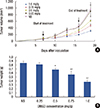
Tumor growth was initially inhibited on day 13 except for in the 0.25 mg/g group. Furthermore, the inhibition effect lasted for 3 days after the last injection and displayed a dose-dependent effect (Figure 1A). The most prominent growth suppression was observed when DMSO was administered at 1.0 mg/g, where a statistically significant retardation of tumor growth was observed on days 13 to 19 as compared with the control (p<0.01). Similarly, except for the 0.25 mg/g group, tumor weight significantly reduced compared with the control group at the time of the last injection (p<0.03) (Figure 1B).
TAM polarization was selectively induced or inhibited
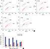
Although alteration of other immune cells was not observed, TAMs were selectively induced or inhibited. Results showed that the percentage of CD11b+ F4/80high cells decreased after treatment (37.54%±2.34%, 41.62%±3.10%, 29.28%±4.42%, and 19.92%±4.80% for 0.25, 0.5, 0.75, and 1.0 mg/g, respectively). The percentage in the control group was 45.00%±3.43%. At the same time, percentage of CD11b+ F4/80low cells in the treatment groups variably increased (9.27%±1.21%, 18.74%±2.55%, 12.84%±1.04%, and 22.26%±1.38% for 0.25, 0.5, 0.75, and 1.0 mg/g, respectively). Alternatively, the control group value was 7.29%±1.59% (Figure 2A). Significant CD11b+ F4/80low cell increase occurred at the 0.5, 0.75, and 1.0 mg/g groups (p<0.03), and the most remarkable reduction of CD11b+ F4/80high cells was observed in the 1.0 mg/g group (p<0.01) (Figure 2B).
DMSO changed macrophage morphology and downregulated relevant cytokines in TC-medium
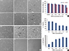
To further test the effect of reversing polarization direction by DMSO, we mimicked a 4T1 tumor microenvironment by using 4T1 cultured cell supernatant and placed the primary peritoneal macrophage into this microenvironment. We found that after culture with DMSO of given concentrations for 24 hours (Figure 3A) and 48 hours (Figure 3B), the refractive index of macrophages decreased. In particular, the macrophages appeared more round and smooth compared with the control macrophages, which were rough and branched. ELISA revealed that IL-10 expression was downregulated and IL-12 expression was upregulated compared with the control group (Figure 3D). DMSO exerted significant cytotoxicity to TAMs after 48 hours at a 2% concentration (Figure 3C).
DMSO reversed polarization direction of TAMs to M1-type
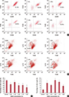
Similar to the effect of in vivo experiment, the macrophages in TC-medium were also selectively induced or inhibited. It showed that the percentage of CD11b+ F4/80high CD206+ cells decreased after cultured in TC-medium (47.42%±4.25%, 59.13%±5.03%, 41.32%±5.43%, 42.58%±4.27%, and 30.51%±7.23% for 1.0%, 1.25%, 1.50%, 1.75%, and 2.0%, respectively). And the percentage in the control group was 68.03%±3.52%. The significant concentration dependent CD11b+ F4/80high CD206+ cells decrease occurred at the 1.50%, 1.75%, and 2.0% groups compared with 1.25% (Figure 4A, C). Also, percentage of CD11b+ F4/80low Ly-6c+ cells in the treatment groups variably increased (10.32%±1.83%, 8.25%±1.16%, 13.73%±0.74%, 11.76%±1.03%, and 7.45%±1.92% for 1.0%, 1.25%, 1.50%, 1.75%, and 2.0%, respectively). And this value was 3.42%±0.85% in control. Furthermore, the significant CD11b+ F4/80low Ly-6c+ cells increase occurred at 1.0%, 1.5%, and 1.75% compared with the control (Figure 4B, D).
DISCUSSION
DMSO has been shown to be a potential anticancer drug in treating lung adenocarcinoma by stimulating tumor suppressor proteins [15]. Additionally, the combination of antineoplastic agents and DMSO showed significant synergistic cytotoxicity in ovarian cancer [16]. Its antitumor effects were also verified by in vivo experiments of lymphoma, which could be attributed to the induction of the tumor necrosis factor α-p53-mitochondrial pathway [17]. In addition to slowing cancer growth, DMSO intravenous injection has been proven to be an effective way of treating refractory cancer pain [18]. In vivo experiment results indicated that a low dose of DMSO (0.5-1.0 mg/g) could delay the growth of mouse breast cancer (Figure 1A, B). Considering that serious cytotoxicity from DMSO can occur, higher DMSO doses were not employed during in vivo tests. Similar to the finding that human melanoma cells showed no dendrite-like structures when exposed to DMSO [1], we observed that macrophages also appeared round and less branched (Figure 3A, B). However, there is no research to suggest that morphological differences exist between M1- and M2-type TAMs. Thus, this morphological transformation could result from an inhibitory effect resulting from attachment by DMSO.
The effects of DMSO on tumor cells have been universally researched. However, studies regarding the impact of DMSO on cells in a tumor microenvironment are limited. The direct proapoptosis effects of DMSO on tumor cells were evaluated. However, possible indirect bypass effects may have been overlooked. In this study, we surprisingly found that DMSO could suppress mouse breast cancer growth by indirect immunoregulation, which induces M1-type polarization while inhibiting M2-type polarization.
The exact mechanisms behind DMSO-induced differentiation are unclear. According to previous research, DMSO could induce maturation of human promyelocytic leukemia cell line HL-60 by increasing phospholipid- and Ca2+-independent protein kinase activity [19]. Ca2+ elevation played an important role in the differentiation of various cell types induced by DMSO [20]. Additionally, the effect of DMSO-induced differentiation differentiation in human rectal adenocarcinoma cell may be attributed to reducing alkaline phosphatase activity [21]. Furthermore, decrease of DNA ligase activity could be related to differentiation of mouse erythroleukemia cells induced by DMSO [22].
DMSO exerted anti-inflammatory effects by inhibiting the expression of many inflammation factors, such as IL-6, monocyte chemotactic protein 1, and prostaglandin E2 [23]. Also, in this study, some inflammation factors and/or cytokines could be downregulated after DMSO was added to the 4T1 tumor microenvironment and the inflammation factor (or factors) and/or cytokines could facilitate TAMs polarizing toward the M2-type. Hence, this inhibiting effect directly impaired the M2-type oriented polarizing ability of TAMs. The possibility that DMSO upregulates cytokines that direct M1-type polarization cannot be excluded, and these factors are being investigated. However, it has been considered that DMSO could suppress inflammatory response through the nuclear factor-κB pathway and impair the function of LPS-orientated macrophages [24]. On the other hand, on an intracellular level, DMSO may penetrate into the cell membrane and influence transcription and/or translation of surface proteins by modulating nuclear factors. This results in downregulation of CD206 and F4/80 mRNA levels and upregulation of Ly-6C mRNA levels.
Recently, the Notch signal was considered the determinant in mediating polarization of M1- and M2-type TAMs [25]. Therefore, the Notch signal may be one mechanism that mediates DMSO-induced differentiation. However, the precise pathway needs to be studied further. More investigations are needed to illustrate the exact mechanisms behind TAM differentiation regulation. Overall, our findings suggest that a low dose of DMSO could exert antitumor effects in 4T1 cancer-bearing mice. This antitumor effect could be attributed to reversing orientation of TAM polarization from pro- to antitumor type. In turn, these results may provide potential guidance for further research regarding breast cancer immunotherapy.
 XML Download