

 PDF
PDF ePub
ePub Citation
 Print
Print


INTRODUCTION
Precise preoperative marking of a discharging breast duct and intraductal lesions facilitates a minimal-volume microdochectomy [1,2]. Ultrasound (US) provides valuable guidance for localizing intraductal abnormalities in patients with nipple discharge [3,4,5], and several studies have reported the use of preoperative US-guided wire localization for cases involving problems with cannulation of the discharging duct [4,6]. We herein report an alternative method of preoperative US-guided indigo carmine dye staining in a patient with no discharge on the day of surgery.
CASE REPORT
A 35-year-old woman presented with spontaneously intermittent bloody discharge from her right nipple. The patient incidentally detected the discharge 4 months prior to visiting our clinic. She had no history of breast abnormalities, previous breast surgery, or radiation therapy. A physical examination revealed bloody discharge from one orifice of the right nipple. There was no overlying skin abnormality or palpable mass in her right breast.
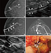
Mammography showed an asymmetric tubular structure in the subareolar area and a 3.2 cm tortuous tubular structure in the upper outer quadrant of the right breast (Figure 1A). US showed a solitary dilated duct containing two isoechoic intraductal lesions 2 cm from the nipple and an isoechoic lesion 5 cm from the nipple in the 10 o'clock region of the right breast (Figure 1B). Galactography demonstrated a dilated duct with several polypoid filling defects and complete obstruction at the site of the tortuous tubular structure (Figure 1C).
We performed a US-guided core needle biopsy for the intraductal lesions 2 cm from the nipple and the tubular lesion 5 cm from the nipple in the 10 o'clock region of the right breast, and all specimens were placed into one specimen bottle. Histopathological examination of the biopsy specimens indicated invasive ductal carcinoma, including intraductal proliferation with a papillary growth pattern. Magnetic resonance imaging for preoperative staging demonstrated a 4.4 cm area of linear and clumped nonmass enhancement that extended from the upper outer quadrant to the subareolar portion of the right breast (Figure 1D). The surgeon recommended right mastectomy, but the patient desired to undergo breast-conserving surgery. The surgeon requested localization of the dilated duct and intraductal lesions for microdochectomy, and preoperative wire localization under galactographic guidance was planned.
On the day of surgery, no discharge was stimulated, and we could not perform conventional preoperative ductal cannulation. Thus, we considered an alternative method using US-guidance. We inserted a needle under US guidance into the dilated duct in the 10 o'clock region of the right breast. Under US guidance, a short bevel 25-gauge needle was used to avoid counterpuncture and contrast extravasation and a small amount of indigo carmine (0.8% indigotindisulfonate sodium; Korea United Pharm., Seoul, Korea) was injected at low pressure (Figure 1E). After injection, the targeted duct became more dilated, and dye was discharged from the right nipple. We performed US-guided hook wire localization for the mass at 5 cm from the nipple. Surgery was performed using a radial skin incision. During the microdochectomy, the indigo carmine-stained duct was identified in the operative field (Figure 1F). For the mass 5 cm from the nipple, lumpectomy was performed with a sufficient resection margin. Histopathology of the excised specimen confirmed invasive ductal carcinoma with a negative margin. On follow-up mammography and US performed 8 months after the operation, tumor recurrence was not detected.
DISCUSSION
The discharging duct and intraductal lesions can be identified before surgical excision through several methods, such as reviewing the preoperative galactogram [7], preoperative marking with blue dye staining [1,4], or wire localization under galactography guidance [8]. In addition to these postoper-ative procedures, intraoperative intraductal blue dye injection [9], ductoscopic wire marking [2], and insertion of a lacrimal probe into the discharging duct [10] can be performed.
It is sometimes difficult to insert a needle or catheter into the secreting duct. Moreover, lesions causing intermittent discharge are difficult to localize; the less discharge, the more difficult the cannulation [5]. If no discharge can be provoked, duct cannulation is not possible [4], and it may not provide adequate guidance for surgery, leading to unnecessary wide excision of breast tissue or failure to remove the cause of nipple discharge. Rissanen et al. [4] and Hussain and Lui [6] reported several successful cases of an alternative method, namely preoperative US-guided wire localization, which was useful, when problems in cannulation of the discharging duct were encountered.
We designed a convenient alternative method under US guidance for preoperative marking of the dilated duct and intraductal lesions in a patient with no nipple discharge. When a dilated duct is detected on US, a needle should be placed into the pathologic duct and US-guided indigo carmine injection should be followed. The oblique needle approach along the long axis of the dilated duct can facilitate the procedure by providing an elongated target. Subtle adjustments with both hands, maintenance of proper needle alignment, exact placement of the needle into the pathologic duct, and careful dye injection with low pressure are factors for the success of this procedure.
Rissanen et al. [5] reported contrast medium extravasation during US-guided percutaneous galactography although the needle tip was seen inside the duct during contrast injection. It is assumed that the needle punctured the posterior duct wall when the syringe was pushed. Similarly, it is important that the needle tip does not puncture the posterior duct wall during US-guided percutaneous indigo carmine staining. Another important factor is careful monitoring of the patient and preparation of a vasopressor before using indigo carmine. Although the procedure has clinically been considered safe, Jo et al. [11] recently reported two cases of hypotension after intradermal injection of indigo carmine into the periareolar area for sentinel node mapping. To date, no adverse reactions have been reported after intraductal injection of indigo carmine.
In conclusion, US-guided percutaneous indigo carmine staining of the dilated duct and intraductal lesions can facilitate minimal-volume microdochectomy in patients with no nipple discharge.
 XML Download