Journal List > J Breast Cancer > v.17(2) > 1036470

Kim, Jung, Ryu, and Lee: Clinicopathologic Characteristics of Breast Cancer Stem Cells Identified on the Basis of Aldehyde Dehydrogenase 1 Expression

Abstract

Purpose

Breast cancer displays varying molecular and clinical features. The ability to form breast tumors has been shown by several studies with aldehyde dehydrogenase 1 (ALDH1) positive cells. The aim of this study is to investigate the association between ALDH1 expression and clinicopathologic characteristics of invasive ductal carcinoma.

Methods

We investigated breast cancer tissues for the prevalence of ALDH1+ tumor cells and their prognostic value. The present study included paraffin-embedded tissues of 70 patients with or without recurrences. We applied immunohistochemical staining for the detection of ALDH1+ cells. Analysis of the association of clinical outcomes and molecular subtype with marker status was conducted.

Results

ALDH1+ and ALDH1- tumors were more frequent in triple-negative breast cancers and in luminal A breast cancers, respectively (p<0.01). ALDH1 expression was found to exert significant impact on disease free survival (DFS) (ALDH1+ vs. ALDH1-, 53.1±6.7 months vs. 79.2±4.7 months; p=0.03) and overall survival (OS) (ALDH1+ vs. ALDH1-, 68.5±4.7 months vs. 95.3±1.1 months; p<0.01). In triple-negative breast cancer (TNBC) patients, DFS and OS showed no statistical differences according to ALDH1 expression (ALDH1+ vs. ALDH1-, 45.3±9.4 months vs. 81.3±7.4 months, p=0.52; 69.0±7.5 months vs. 91.3±6.3 months, p=0.67). However, non-TNBC patients showed significant OS difference between ALDH1+ and ALDH1- tumors (ALDH1+ vs. ALDH1-, 77.6±3.6 months vs. 98.0±1.0 months; p=0.04) with no statistical difference of DFS (ALDH1+ vs. ALDH1-, 60.5±8.0 months vs. 81.8±4.6 months; p=0.27).

Conclusion

Our findings suggest that the expression of ALDH1 in breast cancer may be associated with TNBC and poor clinical outcomes. On the basis of our findings, we propose that ALDH1 expression in breast cancer could be correlated with poor prognosis, and may contribute to a more aggressive cancer phenotype.

INTRODUCTION

Breast cancer is a highly heterogeneous disease that often acquires treatment resistance [1]. In spite of many advances in the therapy for breast cancer, many patients still die owing to recurrence. One theory that could explain the treatment resistance is the cancer stem cell (CSC) theory [2]. CSCs are a subset of tumor cells that have been thought to contribute to the heterogeneous nature of cancers. These cells have the capacity for indefinite self-renewal and differentiation like normal stem cells [3]. These CSCs have the capacity to give rise to all cell types within the tumor, and could constitute drug-resistant cells that induce recurrence or metastasis after anticancer therapy. [4]. While these CSCs have been associated with poor prognosis in some patients with breast cancer, the relationship between CSCs and tumor recurrence remains unclear. Several previous studies have reported that disease-specific survival is poorer in CSC-positive patients. However, other reports have shown contradicting results [1,4,5,6,7,8,9].
A number of markers such as cluster of differentiation (CD) 44, CD24, and aldehyde dehydrogenase 1 (ALDH1) have been proposed for the identification and enrichment of breast CSCs [10,11,12,13,14,15]. Through the study of cell surface markers, Al Hajj et al. [16] verified that cells with a CD44+/CD24- phenotype have the capacity to form breast cancers. In addition, on the basis of ALDH1 activity, Ginestier et al. [17] reported that ALDH1+ tumor cells have the ability to produce tumors in nude mice. Additionally, several studies indicated that CD44+/CD24- and ALDH1+ breast cancer cells have tumor-initiating properties, and are associated with triple-negative breast cancers [11,12,13,14].
These findings led us to hypothesize that the expression of ALDH1 might be associated with clinicopathologic outcomes of patients with breast cancer. In this study, we investigated the association of ALDH1+ breast cancer cells with disease-free survival (DFS), overall survival (OS), and with the breast cancer molecular subtypes. We evaluated the expression of ALDH1 to determine its clinical utility in predicting tumor recurrence and patients' survival.

METHODS

Patients

This study was approved by the Kosin University Gospel Hospital Institutional Review Board (approval number, 13-100). Patients who underwent surgery for invasive ductal carcinoma between July 2005 and March 2007 were evaluated. Patients were excluded if they had received palliative surgery, neoadjuvant chemotherapy, or neoadjuvant radiotherapy for preoperative metastasis of breast cancer at any site (stage IV). In addition, patients with <5 years of follow up and incomplete immunohistochemistry (IHC) data were excluded. We identified 428 patients diagnosed with breast cancer between July 2005 and March 2007. Of them, 70 patients (16.4%) were included in the current analysis (Figure 1). The patients were observed from the date of diagnosis until death resulting from any cause before September 2013. Data on age at diagnosis, tumor size, axillary nodal status, American Joint Committee on Cancer (AJCC) stage, hormonal receptor status, human epidermal growth factor receptor 2 (HER2) status, p53 mutation, histological grade, and lymphovascular invasion status were available.

Definitions of terms and follow-up evaluations

DFS was defined as the period a patient with disease lived without known recurrence after surgery. OS was defined as the time from the primary operation to the date of death. When the patient showed recurrence of breast cancer at any site, we regarded it as a recurrent event. Patients still alive without an event at the last follow-up were considered censored.
Baseline assessments including breast ultrasonography, mammography, chest radiography, abdominal ultrasonography, and bone scintigraphy were performed every 6 months, and positive emission tomography was performed annually. In case of suspicious lesions at baseline, additional radiologic and interventional evaluations were performed.

Immunohistochemical Staining

IHC was performed by using 4-µm thick, formalin-fixed, paraffin-embedded tissues. The tissues were dried overnight in an oven at 60℃, and placed in a Bond™ polymer refine detection system (Leica Biosystems Newcastle Ltd., Newcastle upon Tyne, UK). After deparaffinizing with Bond™ dewax solution (Leica Biosystems Newcastle Ltd.), pretreatment was performed by using Bond™ epitope retrieval solution 1 (Leica Biosystems Newcastle Ltd.) for 20 minutes at 98℃. Following this, the endogenous peroxidase was quenched by incubation with hydrogen peroxide for 15 minutes. Sections were incubated for 15 minutes at room temperature with the monoclonal antibody for ALDH1 (EP1933Y, 1:100; Abcam, Cambridge, UK) using biotin-free polymeric horseradish peroxidase-linker antibody conjugate system, and developed with 3,3 diaminobenzidine chromogen, in a Bond-maX™ automated slide stainer (Leica Biosystems Melbourne Pty. Ltd., Melbourne, Australia).

Evaluation of Immunohistochemical Staining

ALDH1 staining was evaluated in tumor cells and stromal cells in desmoplastic peritumoral connective tissue. Regardless of the extent or intensity, ALDH1 staining was considered positive when the cytoplasm of each cellular component showed a positive reaction (Figure 2). IHC results for estrogen receptor (ER), progesterone receptor (PR), HER2, and p53 were obtained from the patients' pathology reports. Hormone receptors such as ER and PR were evaluated by using the Allred scoring system, as follows: the proportion of positive cells and staining intensity were scored on a scale of 0 to 5 and 0 to 3, respectively. Subsequently, the total score was calculated by adding each score. When the total score was >3, it was regarded as positive [18]. The cells were considered positive for p53 expression when the nuclear reaction was ≥1%. HER2 was analyzed according to the general guidelines set by the American Society of Clinical Oncology/College of American Pathologists. When the IHC yielded equivocal results, the HER2 status was determined by using fluorescent in situ hybridization.
On the basis of IHC, every cancer was divided into the following four kinds of molecular subtypes: luminal A (positive for ER and/or PR), luminal B (positive for ER and/or PR and HER2), HER2 positive (positive for HER2 only), and triple-negative breast cancer (TNBC; negative for ER, PR, and HER2).

Statistical analysis

Statistical analysis was performed by using PASW version 18.0 (SPSS Inc., Chicago, USA). Evaluation of pathologic differences and molecular subtypes according to ALDH1 expression were assessed by using the Pearson chi-square test. Kaplan-Meier estimates and curves were prepared for the survival outcomes. Multivariate analysis by using the Cox proportional hazards model was performed to assess the independent significance of pathologic factors and survival probabilities. Hazard ratios and their corresponding 95% confidence intervals were computed to provide quantitative information about the relevance of the statistical results. All p-values were obtained by performing two-sided testing, and p<0.05 was considered statistically significant.

RESULTS

Pathologic characteristics of patients and pathologic differences associated with a recurrent event

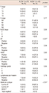
The pathologic characteristics of all patients and pathologic differences associated with events are given in Table 1. All patients were women, with a mean age of 58.1±10.1 years. Out of 70 patients, ALDH1+ and ALDH1- tumors were reported in 27 patients (38.6%) and 43 patients (61.4%), respectively. The number of patients with a recurrent event was 38.6% (27/70). In patients with recurrent events, the T stage, N stage, AJCC stage, ER status, PR status, and molecular subtypes were correlated with the recurrence.

Comparison of pathologic differences and molecular subtypes according to ALDH1 expression

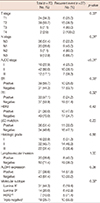
The comparison of pathologic differences and molecular subtype according to ALDH1 expression is shown in Table 2. In patients with ALDH1+ tumors, ER negativity was significantly associated with the expression of the stem cell marker ALDH1 (p<0.01). No other markers correlated with ALDH1+ tumors. Among patients with ALDH1+ tumors, TNBC subtype was observed in 15 patients (55.6%). In addition, luminal A subtype was observed in 26 patients (60.5%) with ALDH1- tumors (p<0.01). No association was detected between ALDH1 expression and other pathological markers.

Association between ALDH1 expression and survival outcomes

The median observation time was 80.7 months (range, 60-99 months) for patients who were alive until the cutoff date for follow-up. The DFS and OS according to ALDH1 expression are shown in Figure 3. In total, 38.6% (27/70) of the patients had tumors positive for ALDH1 expression. The mean DFS for all of the 70 patients was 71.4±4.3 months. When the patients were stratified according to the presence or absence of ALDH1 positivity, the mean DFS was 53.1±6.7 and 79.2±4.7 months for the ALDH1+ and ALDH1- groups, respectively (p=0.03). The estimated mean OS was 68.5±4.7 and 95.3±1.1 months for the ALDH1+ and ALDH1- groups, respectively (p<0.01).

Survival analysis in patients with ALDH1+ and ALDH1- according to molecular subtypes

The DFS and OS between ALDH1+ and ALDH1- patients stratified according to molecular subtypes are shown in Figure 4. We dichotomized the molecular subtypes into non-TNBC and TNBC. In patients with non-TNBC, the mean DFS was 60.5±8.0 months and 81.8±4.6 months for the ALDH1+ and ALDH1- groups, respectively. In patients with TNBC, the mean DFS was 45.3±9.4 months and 81.3±7.4 months for the ALDH1+ and ALDH1- groups, respectively. Both patients with non-TNBC and TNBC did not show any statistically significant DFS differences according to ALDH1 expression (p=0.27; p=0.52). In patients with non-TNBC, the ALDH1+ and ALDH1- groups showed 77.6±3.6 and 98.0±1.0 months of mean OS, respectively. These results indicated a statistical difference between each group (p=0.04). In contrast, in patients with TNBC, the mean OS was not significantly different between those with ALDH1+ and ALDH1- tumors (69.0±7.5 months vs. 91.3±6.3 months, p=0.67) (Figure 4).

DISCUSSION

As CSCs are a minor population of cells associated with recurrence or distant metastasis after anticancer therapy, it is very important to understand these cells in order to improve the outcome of conventional cancer therapy [4,19,20].
We therefore investigated the importance of CSCs in the relapse of invasive ductal carcinoma cells. In this study, we used IHC expression of ALDH1 as a surrogate marker for breast CSCs. We hypothesized that the molecular characteristics and the clinical outcomes of breast cancer might be associated with ALDH1 expression. We analyzed the IHC expression of ALDH1 and sought to correlate these data with the molecular characteristics and survival outcomes such as DFS and OS in a cohort of 70 patients with breast cancers (invasive ductal carcinoma).
Firstly, we evaluated the association between ALDH1 expression and pathologic characteristics known to be important for the clinical outcome, such as tumor size, nodal status, hormonal receptor status, HER2 status, p53 mutation, histologic grade, and lymphovascular invasion. The correlation between the expression of ALDH1 expression and the pathologic characteristics is controversial. Neumeister et al. [5] reported that ALDH1 expression was not associated with pathologic characteristics. However, in two other studies, the expression of ALDH1 was found to be correlated with poor prognostic features such as high histologic grade, HER2 overexpression, and the absence of ER and PR expression [17,21]. In the present study, we were able to demonstrate an association between ALDH1 expression and ER negativity.
Secondly, we investigated whether a correlation exists between ALDH1 expression and the molecular subtypes of breast cancer. As several reports have demonstrated the relationship between the breast cancer subgroups defined according to tumor markers and the molecular subtypes, we evaluated the expression of ER, PR, and HER2 to classify the breast cancers into four subtypes [22,23]. Many previous studies documented that CD44+/CD24- and ALDH1+ cells are associated with HER2+ type breast cancer and TNBC [2,3,6,21,24]. Similarly, in this study, we discovered an association between ALDH1 expression and the molecular subtypes. We found that the ALDH1+ tumors were significantly correlated with TNBCs, and that luminal A type breast cancers occurred in a high proportion of patients with ALDH1- tumors. It is well documented that TNBCs are associated with a worse prognosis than luminal A type breast cancers. Therefore, our results indicate that ALDH1 expression could play a role in the biological heterogeneity and aggressiveness of breast cancer.
Lastly, we evaluated the association between ALDH1 expression and clinical outcomes in patients with invasive ductal carcinoma. Several previous studies have documented that ALDH1 expression was associated with a poor clinical outcome, and it could be an independent prognostic factor [5,17,25]. In this study, we were also able to demonstrate an association between ALDH1 expression and clinical outcomes. There was a significant trend toward shorter DFS and OS in the group with ALDH1+ tumors than in the group with ALDH1- tumors. With this result, we could speculate that breast cancer cells with ALDH1 expression might be correlated with poor clinical outcomes.
In the entire patient cohort, as described above, those with ALDH1+ tumors showed shorter DFS than those with ALDH1- tumors. However, after correcting the frequency of molecular subtype, there were no significant DFS differences between patients with ALDH1+ and ALDH1- tumors regardless of the occurrence of TNBC. Therefore, when the tumor expressed ALDH1, the shorter DFS may result from the high incidence of TNBC rather than ALDH1 expression itself. This result was further verified by analyzing DFS probability with the Cox proportional hazard regression model. In the analysis of OS, the expression of ALDH1 was also associated with shorter OS. In terms of the molecular subtype, the OS did not significantly differ between patients with ALDH1+ or ALDH1- TNBC. In patients with non-TNBC, however, ALDH1+ tumors led to shorter OS than ALDH1- tumors. This result may mean that patients with TNBC had poorer OS than patients with non-TNBC when ALDH1 was expressed. Thus, ALDH1 rather than the molecular subtype could be a contributing factor to determine the OS. This result could be ascertained in the analysis of OS probability by using the Cox proportional hazard regression model. Additionally, in this study, AJCC stage and PR seemed to be factors that influenced recurrence. By using the Cox proportional hazard regression model, these factors were found to be associated with the DFS, but not with the OS. Although these two factors had no influence on the OS, ALDH1 expression was shown as a factor that affected the OS. This result indicates that ALDH1 expression was a significant factor that affected the OS (Table 3). Three possible reasons could be postulated to explain such a result. Firstly, ALDH1+ tumors themselves might have a tendency for rapid progression after recurrence. Secondly, ALDH1+ tumors might be beneficial for the progression of recurrent cancer cells as these tumors have the capacity to be resistant to anticancer therapy. Lastly, for the treatment of recurrent tumors, additional anticancer therapy could be detrimental to the survival of normal cells, consequently providing a growth advantage to the ALDH1+ tumor cells. However, as these theories have not been demonstrated experimentally, additional studies may be required to determine the exact reason(s) for the short OS in patients with ALDH1+ tumors in cases of non-TNBC.
The limitation of this study was that only 70 of the 428 patients (16.7%) were enrolled in this study. Therefore, additional studies on a larger cohort will be needed to confirm our findings.
In conclusion, the expression of ALDH1 is associated with poor clinical outcomes and TNBC. The significant correlation of this marker with the breast cancer molecular subtypes and patient clinical outcomes may suggest that the presence of ALDH1+ cells is associated with poor prognostic features and could contribute to an aggressive breast cancer phenotype.

Figures and Tables

Figure 1
Consolidated Standards of Reporting Trials diagram.
IHC=immunohistochemistry.
jbc-17-121-g001
Figure 2
Immunohistochemical staining of aldehyde dehydrogenase 1 (ALDH1) in tumor cells. (A) Diffuse staining was observed in cytoplasm and nucleus of cancer cells (positive reaction). (B) No ALDH1 staining was shown in cancer cells (negative reaction) (×400).
jbc-17-121-g002
Figure 3
Survival curves between aldehyde dehydrogenase 1 (ALDH1)+ and ALDH1- patients. (A) Disease-free survival and (B) overall survival.
jbc-17-121-g003
Figure 4
Survival curves between aldehyde dehydrogenase 1 (ALDH1)+ and ALDH1- patients according to molecular subtype. Disease-free survival in patients with (A) non-triple-negative breast cancer (TNBC) and (B) TNBC. Overall survival in patients with (C) non-TNBC and (D) TNBC.
jbc-17-121-g004
Table 1
Correlation between pathologic characteristics and recurrent event
jbc-17-121-i001

AJCC=American Joint Committee on Cancer; ER=estrogen receptor; PR=progesterone receptor; HER2=human epidermal growth factor receptor 2; ALDH1=aldehyde dehydrogenase 1.

*Statistically correlated with cancer recurrence; ER or PR(+); ER or PR(+) and HER2(+); §HER2(+) only; ER, PR and HER2(-).

Table 2
Comparison of pathologic differences and molecular subtype according to aldehyde dehydrogenase 1 expression
jbc-17-121-i002

ALDH1=aldehyde dehydrogenase 1; AJCC=American Joint Committee on Cancer stage; ER=estrogen receptor; PR=progesterone receptor; HER2=human epidermal growth factor receptor 2.

*Statistically correlated with aldehyde dehydrogenase 1 expression; ER or PR(+); ER or PR(+) and HER2(+); §HER2(+) only; ER, PR and HER2(-).

Table 3
Disease-free survival and overall survival probabilities* as calculated by Cox proportional hazards regression model
jbc-17-121-i003

DFS=disease-free survival; OS=overall survival; HR=hazard ratio; CI=confidence interval; AJCC=American Joint Committee on Cancer stage; ER=estrogen receptor; PR=progesterone receptor; TNBC=triple-negative breast cancer; ALDH1=aldehyde dehydrogenase 1.

*With an event defined as development of recurrence and death by disease; Risk of recurrence; Risk of death; §Statistically associated with survival outcomes.

Notes

The authors declare that they have no competing interests.

References

1. Sung JY, Kim GY, Park YK, Lee J, Kim YW, Lim SJ. Clinicopathological significance of invasive ductal carcinoma with high prevalence of CD44(+)/CD24(-/low) tumor cells in breast cancer. Korean J Pathol. 2010; 44:390–396.
crossref
2. de Beça FF, Caetano P, Gerhard R, Alvarenga CA, Gomes M, Paredes J, et al. Cancer stem cells markers CD44, CD24 and ALDH1 in breast cancer special histological types. J Clin Pathol. 2013; 66:187–191.
crossref
3. Park SY, Lee HE, Li H, Shipitsin M, Gelman R, Polyak K. Heterogeneity for stem cell-related markers according to tumor subtype and histologic stage in breast cancer. Clin Cancer Res. 2010; 16:876–887.
crossref
4. Jordan CT, Guzman ML, Noble M. Cancer stem cells. N Engl J Med. 2006; 355:1253–1261.
crossref
5. Neumeister V, Agarwal S, Bordeaux J, Camp RL, Rimm DL. In situ identification of putative cancer stem cells by multiplexing ALDH1, CD44, and cytokeratin identifies breast cancer patients with poor prognosis. Am J Pathol. 2010; 176:2131–2138.
crossref
6. Idowu MO, Kmieciak M, Dumur C, Burton RS, Grimes MM, Powers CN, et al. CD44(+)/CD24(-/low) cancer stem/progenitor cells are more abundant in triple-negative invasive breast carcinoma phenotype and are associated with poor outcome. Hum Pathol. 2012; 43:364–373.
crossref
7. Lin Y, Zhong Y, Guan H, Zhang X, Sun Q. CD44+/CD24- phenotype contributes to malignant relapse following surgical resection and chemotherapy in patients with invasive ductal carcinoma. J Exp Clin Cancer Res. 2012; 31:59.
crossref
8. Currie MJ, Beardsley BE, Harris GC, Gunningham SP, Dachs GU, Dijkstra B, et al. Immunohistochemical analysis of cancer stem cell markers in invasive breast carcinoma and associated ductal carcinoma in situ: relationships with markers of tumor hypoxia and microvascularity. Hum Pathol. 2013; 44:402–411.
crossref
9. Mylona E, Giannopoulou I, Fasomytakis E, Nomikos A, Magkou C, Bakarakos P, et al. The clinicopathologic and prognostic significance of CD44+/CD24(-/low) and CD44-/CD24+ tumor cells in invasive breast carcinomas. Hum Pathol. 2008; 39:1096–1102.
crossref
10. Bane A, Viloria-Petit A, Pinnaduwage D, Mulligan AM, O'Malley FP, Andrulis IL. Clinical-pathologic significance of cancer stem cell marker expression in familial breast cancers. Breast Cancer Res Treat. 2013; 140:195–205.
crossref
11. Perrone G, Gaeta LM, Zagami M, Nasorri F, Coppola R, Borzomati D, et al. In situ identification of CD44+/CD24- cancer cells in primary human breast carcinomas. PLoS One. 2012; 7:e43110.
crossref
12. Sheridan C, Kishimoto H, Fuchs RK, Mehrotra S, Bhat-Nakshatri P, Turner CH, et al. CD44+/CD24- breast cancer cells exhibit enhanced invasive properties: an early step necessary for metastasis. Breast Cancer Res. 2006; 8:R59.
13. Sun H, Jia J, Wang X, Ma B, Di L, Song G, et al. CD44+/CD24- breast cancer cells isolated from MCF-7 cultures exhibit enhanced angiogenic properties. Clin Transl Oncol. 2013; 15:46–54.
crossref
14. Guler G, Balci S, Costinean S, Ussakli CH, Irkkan C, Suren D, et al. Stem cell-related markers in primary breast cancers and associated metastatic lesions. Mod Pathol. 2012; 25:949–955.
crossref
15. Douville J, Beaulieu R, Balicki D. ALDH1 as a functional marker of cancer stem and progenitor cells. Stem Cells Dev. 2009; 18:17–25.
crossref
16. Al-Hajj M, Wicha MS, Benito-Hernandez A, Morrison SJ, Clarke MF. Prospective identification of tumorigenic breast cancer cells. Proc Natl Acad Sci U S A. 2003; 100:3983–3988.
crossref
17. Ginestier C, Hur MH, Charafe-Jauffret E, Monville F, Dutcher J, Brown M, et al. ALDH1 is a marker of normal and malignant human mammary stem cells and a predictor of poor clinical outcome. Cell Stem Cell. 2007; 1:555–567.
crossref
18. Allred DC, Harvey JM, Berardo M, Clark GM. Prognostic and predictive factors in breast cancer by immunohistochemical analysis. Mod Pathol. 1998; 11:155–168.
19. Reya T, Morrison SJ, Clarke MF, Weissman IL. Stem cells, cancer, and cancer stem cells. Nature. 2001; 414:105–111.
crossref
20. Takahashi RU, Takeshita F, Fujiwara T, Ono M, Ochiya T. Cancer stem cells in breast cancer. Cancers (Basel). 2011; 3:1311–1328.
crossref
21. Ricardo S, Vieira AF, Gerhard R, Leitão D, Pinto R, Cameselle-Teijeiro JF, et al. Breast cancer stem cell markers CD44, CD24 and ALDH1: expression distribution within intrinsic molecular subtype. J Clin Pathol. 2011; 64:937–946.
crossref
22. Sorlie T, Perou CM, Tibshirani R, Aas T, Geisler S, Johnsen H, et al. Gene expression patterns of breast carcinomas distinguish tumor subclasses with clinical implications. Proc Natl Acad Sci U S A. 2001; 98:10869–10874.
crossref
23. Sorlie T, Tibshirani R, Parker J, Hastie T, Marron JS, Nobel A, et al. Repeated observation of breast tumor subtypes in independent gene expression data sets. Proc Natl Acad Sci U S A. 2003; 100:8418–8423.
crossref
24. Honeth G, Bendahl PO, Ringnér M, Saal LH, Gruvberger-Saal SK, Lövgren K, et al. The CD44+/CD24- phenotype is enriched in basal-like breast tumors. Breast Cancer Res. 2008; 10:R53.
25. Tanei T, Morimoto K, Shimazu K, Kim SJ, Tanji Y, Taguchi T, et al. Association of breast cancer stem cells identified by aldehyde dehydrogenase 1 expression with resistance to sequential paclitaxel and epirubicin-based chemotherapy for breast cancers. Clin Cancer Res. 2009; 15:4234–4241.
crossref
TOOLS
Similar articles