

 PDF
PDF ePub
ePub Citation
 Print
Print


INTRODUCTION
Association of different cancers is classified depending on the timing of their detection. According to Gluckman's definition "synchronous malignancy" includes carcinomas that present either simultaneously or within a 6-month period of identification of the original malignancy. Beyond the 6-month interval they are referred to as "metachronous malignancy" [1]. The presence of multiple synchronous malignancies in the head and neck area and the upper aerodigestive tract is well established [2] and has been explained by the concept of "field cancerization" [3]. Few cases of association of synchronous breast malignancy in males with other malignancies have been reported but they are far and few in between. Yaman et al. [4] and Sun et al. [5] have reported synchronous bilateral breast cancer in males who had no family history of breast cancer. Another case of synchronous chronic lymphocytic leukemia and breast malignancy in a male was reported by Dubashi et al. [6] in a 69-year-old man who had a family history breast cancer. Another case of synchronous breast malignancy and Large B cell Non-Hodgkin's lymphoma was reported by Sordi et al. [7]. This patient too had a family history of breast cancer. However, association of synchronous breast malignancy with head and neck cancer is relatively rare. Strobel et al. [8] in a systematic detection of synchronous primaries to squamous head and neck carcinomas on 589 patients noted only one case of second cancer in breast. Synchronous cancers were predominantly located in the aerodigestive tract, primarily in the lung, head and neck, and oesophagus. A case of synchronous undifferentiated nasopharyngeal carcinoma and infiltrating ductal breast carcinoma in a 47-year-old Moroccan female is on records by Mesmoudi et al. [9]. Our case is probably the first report of a random synchronous male breast carcinoma in a known case of head and neck malignancy which was detected on whole body 18-fluorine deoxyglucose positron emission tomography and computed tomography (18F-FDG PET/CT) study.
CASE REPORT
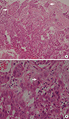
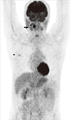
This 58-year-old male Indian male presented with an indurated lesion of size 2×2 cm in the posterior 1/3rd of right side of tongue, not crossing midline with normal tongue movements. Biopsy confirmed a poorly differentiated squamous cell carcinoma of base of tongue (Figure 1). Subsequently he underwent a contrast enhanced whole body 18F-FDG PET/CT (head to mid thigh) on our General Electric Discovery PET 8 slice CT scanner. Standardized uptake value (SUVmax) of the lesions were calculated for body weight and expressed in gram per milliliter (g/mL). Apart from the known base of tongue lesion (Figures 2 and 3) in the right posterior 1/3rd (SUVmax, 8.2), a right level II cervical lymph nodal metastases (SUVmax, 5.3). Incidental FDG avid enhancing subcutaneous soft tissue lesion with spiculated margins of size 15×14 mm, in retro areolar region of right breast was noted (SUVmax, 2.1) (Figures 2 and 4). On subsequent physical examination, a right breast lump of size 2×2 cm at 9 o'clock position with surrounding periareolar nodularity was noted with no palpable axillary lymph nodes. Left breast and axilla were normal. Excision biopsy revealed an invasive ductal carcinoma, not otherwise specified, modified Bloom Richardson grade 1 (tubule 2, atypia 1, mitosis 1), tumor size of 1.5×1.3×1 cm. Immunohistochemistry revealed estrogen and progesterone receptor positive (Figure 5). He underwent a right modified radical mastectomy. Histopathology showed no residual tumor while one lymph node was positive for metastases. After percutaneous endoscopic gastrostomy tube placement, concurrent chemoradiation for poorly differentiated squamous cell carcinoma of base of tongue (T1N1M0) was started. Patient received intensity modulated radiation therapy with 69 Gy to the tumor in 30 fractions, 60 Gy to level II-IV lymphnodes and 54 Gy to low anterior neck and weekly chemotherapy with Cisplatin 50 mg. Patient tolerated the concurrent chemoradiation well. On follow-up after 1 month of completion of concurrent chemoradiation, there was no visible base of tongue growth on endoscopy or on palpapation. Subsequently, patient received six cycles of adriamycin, cyclophosphamide, and 5-flurouracil for carcinoma breast (pT1cN1M0). Patient was subsequently on tamoxifen. Apart from a positive family history, patient does not have any known risk factors like alcoholism, liver cirrhosis, etc. One of his brother is known to have breast carcinoma. Genetic mutation testing was not done as this is locally not available to us in our hospital and outsourced testing for the same was not insisted upon taking in consideration the poor financial status of the patient.
DISCUSSION
The overall occurrence rate of multiple primary malignancies reported in literature is between 0.73% and 11.7% [10]. Three diagnostic criteria have been proposed for multiple primary malignancy: 1) each tumor must present with definite features of malignancy; 2) each must be distinct; and 3) the chance of one being a metastasis of the other must be excluded [11]. Our patient was detected with the second malignancy within a month of detection of the first malignancy, fulfilling the criteria laid down by Gluckman's definition of synchronous malignancy. Based on the location, multiple primary malignancies are classified into four types: 1) multicentric, if the two distinct carcinoma arise in the same organ or tissue; 2) systemic, if they arise on anatomically or functionally allied organs of the same system (colon and rectum cancers); 3) paired organs, as in the breasts; and 4) random, if they occur as a coincidental or accidental association in unrelated sites [12]. Our case can be categorized into random synchronous malignancy as base of tongue and breast are unrelated sites. Mechanisms involved are yet to be clarified. However, some factors such as heredity, constitution, environmental, immunological, carcinogenic, viral, radiological, and chemical factors have been implicated [13]. Genetic predisposition (e.g., higher racial prevalence in Ashkenazi Jews and African-Americans), exposure to female hormones (in cases of sex change operations), cirrhosis, other testicular dysfunction such as orchitis or undescended testis, soap and perfume workers, alcohol, etc. have been implicated for development of male breast cancer. While there was no family history of breast cancer in cases reported by Yaman et al. [4] and Sun et al. [5], family history was positive in cases reported by Dubashi et al. [6] and Sordi et al. [7]. A family history is known to be a definite risk factor for breast malignancy-5% to 30% of male breast cancer patients report a family history of the disease. In view of this, the role of genetic testing for BRCA1 and BRCA2 and other molecular genetic markers assumes importance [14]. All histologic types of breast cancer have been identified in men, the most common type being infiltrating ductal adenocarcinoma (70%-95% of cases). Ductal carcinoma in situ is infrequent. The rate of receptor positivity is high, up to 85% (higher than in females) as in our patient [15]. This case report highlights the incremental value of whole body PET/CT in the oncological screening of patients. This case merits attention as it is relatively uncommon to have a synchronous male breast malignancy especially in a case of head and neck cancer. This case also corroborates the utility of 18F-FDG PET/CT in detecting known as well as unknown malignant lesions and projects the need for a thorough screening with a whole body modality like 18F-FDG PET/CT in patients with family history of those malignancies which are seen in family members like breast carcinomas.


 XML Download