Journal List > J Breast Cancer > v.16(4) > 1036451

Min, Bae, Lee, Lee, Kim, Kim, Lee, Kil, Kim, Lee, and Nam: Tubular Carcinoma of the Breast: Clinicopathologic Features and Survival Outcome Compared with Ductal Carcinoma In Situ

Abstract

Purpose

Tubular carcinoma (TC) of the breast is an uncommon histological subtype of invasive breast cancer with an excellent prognosis compared with standard invasive ductal carcinoma. Recent studies suggested a possible precursor role for low grade ductal carcinoma in situ (DCIS) in the development of TC. The goal of this analysis was to understand the clinicopathologic features and outcomes of TC by comparing TC with DCIS.

Methods

A retrospective review identified 70 patients with TC and 1,106 patients with DCIS between 1995 and 2011. Student t-test and Fisher exact test were used to compare the clinicopathologic characteristics of TC patients with those of DCIS patients. The Kaplan-Meier method and Cox regression analysis were used to determine disease-free survival (DFS) rates.

Results

Compared to DCIS, TC exhibited favorable clinicopathologic characteristics such as a lower nuclear grade (92.3%), higher expression of hormonal receptors (estrogen receptor-positive, 92.9%; progesterone receptor-positive, 87.0%), and less frequent overexpression of human epidermal growth receptor 2 (12.9%). DFS did not differ significantly between the TC and DCIS groups (5-year DFS, 100% vs. 96.7%; 10-year DFS, 92.3% vs. 93.3%; p=0.324), and cancer-specific deaths were not noted in either group. However, axillary lymph node involvement was observed in six (8.6%) of the 70 patients with TC. Three of these patients had small tumors (≤1 cm).

Conclusion

In our study cohort, TC was associated with an excellent prognosis and a low rate of lymph node metastasis. However, lymph nodes metastases were found even in patients with small tumors (≤1 cm). Axillary staging must be considered for all patients with TC of the breast.

INTRODUCTION

Tubular carcinoma (TC) of the breast is an uncommon histological subtype of invasive breast cancer that accounts for approximately 1% to 5% of invasive breast carcinomas [1-3]. TC is defined as a well-differentiated invasive carcinoma with regular cells arranged in well-defined tubules (typically one layer thick) surrounded by an abundant fibrohyaline stroma, classified as pure TC or mixed TC [1-3]. The term pure TC is assigned to tumors with a tubular composition of ≥90%, a low nuclear grade, and no mitoses [4], whereas mixed TC has a tubular composition of ≥75% [5,6].
TC is generally associated with an excellent prognosis, manifesting in a low incidence of lymph node (LN) metastases (approximately 2%-11% [1,3,7,8]), a low rate of local recurrence, and a high overall survival rate [3,5,7,9]. The 5-year disease free survival (DFS) rate is generally more than 90% [1,7,9], and the 10-year overall survival (OS) rate is comparable to that of an age-matched general population [2,9].
It has often been suggested that axillary staging may be unnecessary in patients with small TC [2,10,11]. However, there is currently no consensus on the omission of surgical axillary node staging according to the type of TC. Moreover, recent studies have documented an association between TC and micropapillary and cribriform ductal carcinoma in situ (DCIS) and suggested a possible precursor role of low grade DCIS in the development of tubular breast cancer [12-16].
The goals of this analysis were to compare TC with DCIS in order to identify the clinicopathologic features of TC, including the frequency of LN metastasis, and to determine the most efficacious treatment and whether LN evaluation can be safely omitted.

METHODS

Patients diagnosed with breast cancer between January 1, 1995, and July 31, 2011, were selected from the database of the Breast Cancer Center at Samsung Medical Center, Korea. Of the 10,323 recorded breast carcinomas, 70 were TC (0.7%) and 1,106 were DCIS (10.7%). Patients diagnosed with a concurrent cancer of a different histological type were not included.
We analyzed the clinicopathologic factors, including age; tumor size; axillary LN status; nuclear grade (NG); and expression of estrogen receptor (ER), progesterone receptor (PR), and human epidermal growth receptor 2 (HER2). We also examined the type of treatment received (surgical treatment or adjuvant treatment [use of chemotherapy, radiotherapy, and/or hormone therapy]).
The pathological tumor stage was assessed according to the American Joint Committee on Cancer's 7th Staging System. ER and PR staining data were acquired from the pathology reports. Staining was scored using the Allred score (AS), a semiquantitative method that calculates the proportion of positive cells (scored on a scale of 0-5) and staining intensity (scored on a scale of 0-3), with a maximum score of 8; an AS >2 was considered positive.
Differences in the frequencies of clinicopathologic factors were analyzed using the chi-square test, Fisher exact test, and binary logistic-regression analysis. DFS was defined as the time from the date of diagnosis to the date of a documented recurrence, including locoregional recurrence and/or distant metastasis. OS was expressed as the number of months from the operation to the date of death. Survival curves were constructed using the Kaplan-Meier method. Statistical significance was defined as p<0.05. All statistical analyses were performed using SPSS Statistics version 21.0 (IBM Corp., Armonk, USA).

RESULTS

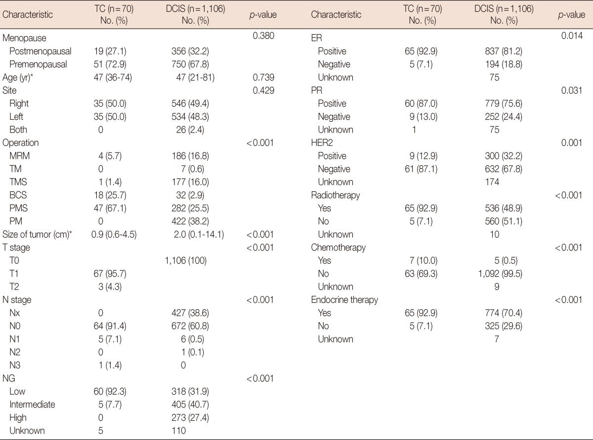
Between 1995 and 2011, 70 patients with TC (0.7%) and 1,106 patients with DCIS (10.7%) were identified. The clinicopathologic characteristics of the two study groups are shown in Table 1. The median age of the TC group was 47 years (range, 36-74 years), and the median tumor size was 0.9 cm (range, 0.3-4.5 cm). Partial mastectomy was performed in 65 cases (92.8%), whereas five patients (6.1%) underwent total mastectomy. All patients with TC underwent surgical evaluation for axillary LN staging, and six patients (8.5%) had LN metastases. Sixty patients (92.3%) had tumors with a low NG, and five patients (7.7%) had tumors with an intermediate grade. Almost all tumors were ER positive (65 tumors, 92.9%), PR positive (60 tumors, 87%), and HER2 negative (61 tumors, 87.1%). In 22 patients (31.4%), TC was associated with DCIS, and of these patients, 15 (68.1%) had the cribriform type.
Seven patients (10%) received chemotherapy, including five of six patients with LN metastases, one patient with isolated tumor cells, and one patient with multiple tumors. Radiotherapy was administered to 65 patients (92.9%), 64 (98.5%) of whom underwent breast conserving surgery. Adjuvant endocrine therapy was administered to 65 patients (92.9%), 60 (92.3%) of whom received tamoxifen.
Of the patients who underwent surgery for axillary staging, 48 (68.6%) underwent sentinel lymph node (SLN) surgery and 22 patients (31.4%) underwent axillary lymph node dissection (ALND). Four patients with SLN positivity underwent ALND. Axillary nodal metastases were identified in six cases (8.5%) (Table 1). Five patients had macrometastasis, and one patient had micrometastasis. The characteristics of the patients with LN metastasis are compared with patients without LN metastasis (Supplementary Table 1), the median pathological tumor size was 1.6 cm (range, 0.3-4.5 cm) for patients with LN metastasis and 0.9 cm (range, 0.3-2.0 cm) for patients without LN metastasis (p<0.001).
Multivariate analysis was performed on six parameters identified by prior univariate analysis: patient age, pathological tumor size, coexistence of DCIS, NG, hormonal receptor status, and overexpression of HER2. This analysis identified pathological tumor size (p=0.040) and NG (p=0.013) as the parameters significantly associated with the risk of LN involvement (Table 2). However, of the 51 patients with a pathological tumor size <10 mm, LN metastases were found in three patients, and of these, two patients had LN macrometastasis and one patient had sentinel node micrometastasis.
The median follow-up time for these patients was 51.8 months (range, 0.5-202.4 months). One patient (1.4%) in the TC group had a recurrence of invasive ductal carcinoma 8.6 years (103.4 months) after the primary operation. The primary tumor was 0.8 cm in size without LN metastasis in the upper center of the left breast, with a low nuclear grade, ER positivity, PR positivity, and HER2 negativity. The tumor that recurred was a 1.5 cm high grade, ER-positive, PR-positive, and HER2-positive invasive ductal carcinoma in the mid-outer region of the left breast.
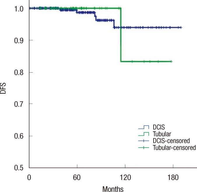
Thirty-two patients (2.9%) in the DCIS group had recurrence. DFS did not differ significantly between the TC and DCIS groups (5-year DFS, 100% vs. 96.7%; 10-year, 92.3% vs. 93.3%; p=0.324) (Figure 1). No cancer specific deaths were noted in either group. Twenty-two patients (31.4%) with TC had an associated DCIS. However, the presence of DCIS was not associated with DFS.
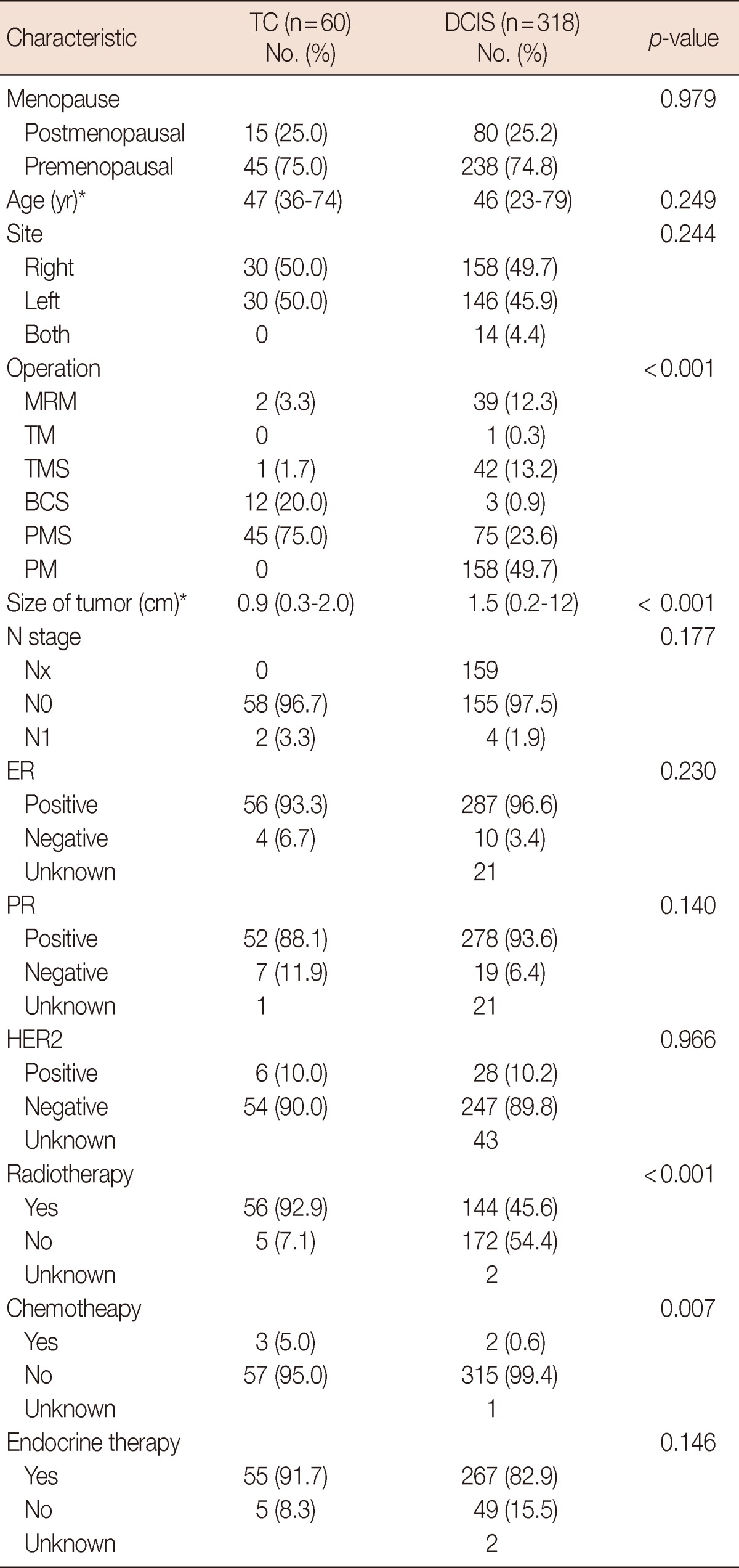
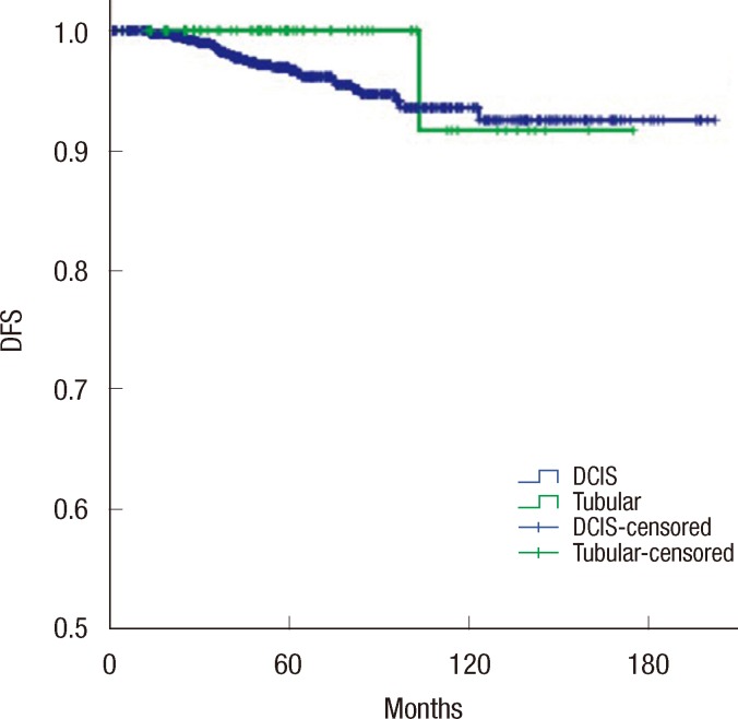
Additionally, 60 patients (92.3%) with low grade TC were compared with 318 patients (31.9%) with low grade DCIS (Table 3). Low grade TC exhibited similar clinicopathologic features as low grade DCIS, excluding tumor size (0.9 cm vs. 1.5 cm, p<0.001) and associated factors (operation and radiotherapy). In patients with low grade tumors, the recurrence rate was 1.7% (1/60 patients) in the TC group and 1.6% (5/318 patients) in the DCIS group (p=0.957). There was no significant difference in DFS between the TC and DCIS groups (5-year DFS, 100% vs. 98.7%; 10-year DFS, 83.3% vs. 94.0%; p=0.918) (Figure 2).

DISCUSSION

Pure TC of the breast is a well-differentiated and distinct histologic subtype of invasive carcinoma accounting for less than 2% of all breast carcinomas and is associated with an excellent prognosis; the survival of patients with TC is similar to that of the general population [2,3,5,7,15]. Other studies have also reported low recurrence rates among patients with TC [3,5,10,17]. In one study, tumors only recurred in the group of patients who were treated with wide local excision [18]. Even in patients who did not receive adjuvant therapy, life expectancy is close to that of the general population after complete surgical excision and radiotherapy [3,9,19]. In our study, only one patient who received radiotherapy showed recurrence of invasive ductal carcinoma 8.6 years (103.4 months) after the primary operation. However, because some tumors could represent a phenotypic alteration in the recurrent tumor tissue and others may be new primary tumors [3,20], it is difficult to distinguish the exact mechanism of recurrence.
In our study, there were no cases of distant metastasis or cancer-related death. This result was consistent with previous studies revealing that TC has a better prognosis than grade 1 ductal carcinomas [3]. Morphologic and molecular studies have also reported similarities between TC and other low grade, luminal type breast carcinomas [12,13,21-23]. Moreover, recent studies have suggested a possible precursor role of low grade DCIS in the development of tubular breast cancer by documenting the association between TC and micropapillary and cribriform DCIS [12-16,22,24]. In our study, 22 patients (31.4%) with TC also showed DCIS, and of these, 15 patients (68.1%) had a cribriform type. Our study compared the clinicopathologic features and outcomes of patients with TC with those of patients with DCIS. There was no difference in DFS between the two groups. However, patients with TC had a greater number of favorable prognostic factors than those with DCIS, such as a lower NG, higher ER/PR expression, and lower HER2 overexpression. Specifically, high rates of hormone receptor positivity, HER2 negativity, and lower NG were also reported in several other studies [3,25]. However, patients with TC showed a significantly higher rate of LN metastasis than those with DCIS. Axillary LN metastases are uncommon in patients with TC; in our study, six patients (8.5%) had LN metastases. This result is similar to the incidence (6%-11%) in other large series [3,5,10,11]. Of 679 patients with DCIS who underwent axillary staging, only seven (1%) had LN metastases (p<0.001). Pathological tumor size is considered the main risk factor for breast cancer axillary LN metastasis. In TC, a pathological tumor size of less than 1 cm is associated with a low risk of LN involvement [17,26]. Although several previous studies suggested that axillary staging may be unnecessary in patients with TC with a tumor size of less than 1 cm [2,8,10,11], current guideline do not consider the omission of surgical axillary node staging [27]. Tumor size was the only significant risk factor associated with LN metastases in our multivariate analysis, but we found LN metastases in three patients with a primary TC ≤1 cm in size. In addition, LN macrometastasis were found in two patients. Axillary macrometastasis is considered the main risk factor for distant metastatic diffusion and is the main parameter used to indicate systemic adjuvant therapy. Moreover, large clinical trials demonstrated that the morbidity of SLN surgery is low [28,29], and axillary staging or SLN biopsy may be needed in patients with TC of the breast.
This study has some limitations. These patients were identified from a cancer registry database, and the retrospective nature of the study introduces significant bias with respect to patient selection and intrinsic, retrospective data collection. Treatment of TC was not dictated by protocol, therefore, this may not have been homogenous due to variations in treatment patterns over the study period.
In conclusion, TC has a favorable biological behavior including a low rate of LN metastasis and an excellent prognosis, and the survival of patients with TC is similar to that of patients with DCIS. However, despite the low incidence of positive nodes, we found LN metastases in patients with small tumors (≤1 cm). Although well-designed and controlled randomized studies are needed, axillary staging must be considered for all patients with TC of the breast.

Notes

This work was supported by a grant of the Korea Healthcare Technology R&D Project, Ministry for Health and Welfare Affairs, Republic of Korea (A092255) and in part by Samsung Biomedical Research Institute grant (GL1B32711).

The authors declare that they have no competing interests.

References

1. Sullivan T, Raad RA, Goldberg S, Assaad SI, Gadd M, Smith BL, et al. Tubular carcinoma of the breast: a retrospective analysis and review of the literature. Breast Cancer Res Treat. 2005; 93:199–205. PMID: 16142444.
crossref
2. Cabral AH, Recine M, Paramo JC, McPhee MM, Poppiti R, Mesko TW. Tubular carcinoma of the breast: an institutional experience and review of the literature. Breast J. 2003; 9:298–301. PMID: 12846864.
crossref
3. Rakha EA, Lee AH, Evans AJ, Menon S, Assad NY, Hodi Z, et al. Tubular carcinoma of the breast: further evidence to support its excellent prognosis. J Clin Oncol. 2010; 28:99–104. PMID: 19917872.
crossref
4. Cooper HS, Patchefsky AS, Krall RA. Tubular carcinoma of the breast. Cancer. 1978; 42:2334–2342. PMID: 214219.
crossref
5. McDivitt RW, Boyce W, Gersell D. Tubular carcinoma of the breast: clinical and pathological observations concerning 135 cases. Am J Surg Pathol. 1982; 6:401–411. PMID: 6289683.
6. Leibman AJ, Lewis M, Kruse B. Tubular carcinoma of the breast: mammographic appearance. AJR Am J Roentgenol. 1993; 160:263–265. PMID: 8424330.
crossref
7. Livi L, Paiar F, Meldolesi E, Talamonti C, Simontacchi G, Detti B, et al. Tubular carcinoma of the breast: outcome and loco-regional recurrence in 307 patients. Eur J Surg Oncol. 2005; 31:9–12. PMID: 15642419.
crossref
8. Fedko MG, Scow JS, Shah SS, Reynolds C, Degnim AC, Jakub JW, et al. Pure tubular carcinoma and axillary nodal metastases. Ann Surg Oncol. 2010; 17(Suppl 3):338–342. PMID: 20853056.
crossref
9. Diab SG, Clark GM, Osborne CK, Libby A, Allred DC, Elledge RM. Tumor characteristics and clinical outcome of tubular and mucinous breast carcinomas. J Clin Oncol. 1999; 17:1442–1448. PMID: 10334529.
crossref
10. Javid SH, Smith BL, Mayer E, Bellon J, Murphy CD, Lipsitz S, et al. Tubular carcinoma of the breast: results of a large contemporary series. Am J Surg. 2009; 197:674–677. PMID: 18789411.
crossref
11. Kader HA, Jackson J, Mates D, Andersen S, Hayes M, Olivotto IA. Tubular carcinoma of the breast: a population-based study of nodal metastases at presentation and of patterns of relapse. Breast J. 2001; 7:8–13. PMID: 11348409.
crossref
12. Fernandez-Aguilar S, Noël JC. Expression of cathepsin D and galectin 3 in tubular carcinomas of the breast. APMIS. 2008; 116:33–40. PMID: 18254778.
crossref
13. Abdel-Fatah TM, Powe DG, Hodi Z, Lee AH, Reis-Filho JS, Ellis IO. High frequency of coexistence of columnar cell lesions, lobular neoplasia, and low grade ductal carcinoma in situ with invasive tubular carcinoma and invasive lobular carcinoma. Am J Surg Pathol. 2007; 31:417–426. PMID: 17325484.
crossref
14. Aulmann S, Elsawaf Z, Penzel R, Schirmacher P, Sinn HP. Invasive tubular carcinoma of the breast frequently is clonally related to flat epithelial atypia and low-grade ductal carcinoma in situ. Am J Surg Pathol. 2009; 33:1646–1653. PMID: 19675453.
crossref
15. Kunju LP, Ding Y, Kleer CG. Tubular carcinoma and grade 1 (well-differentiated) invasive ductal carcinoma: comparison of flat epithelial atypia and other intra-epithelial lesions. Pathol Int. 2008; 58:620–625. PMID: 18801081.
crossref
16. Man S, Ellis IO, Sibbering M, Blamey RW, Brook JD. High levels of allele loss at the FHIT and ATM genes in non-comedo ductal carcinoma in situ and grade I tubular invasive breast cancers. Cancer Res. 1996; 56:5484–5489. PMID: 8968105.
17. Winchester DJ, Sahin AA, Tucker SL, Singletary SE. Tubular carcinoma of the breast: predicting axillary nodal metastases and recurrence. Ann Surg. 1996; 223:342–347. PMID: 8604915.
18. Stalsberg H, Hartmann WH. The delimitation of tubular carcinoma of the breast. Hum Pathol. 2000; 31:601–607. PMID: 10836300.
19. Hansen CJ, Kenny L, Lakhani SR, Ung O, Keller J, Tripcony L, et al. Tubular breast carcinoma: an argument against treatment de-escalation. J Med Imaging Radiat Oncol. 2012; 56:116–122. PMID: 22339755.
crossref
20. McBoyle MF, Razek HA, Carter JL, Helmer SD. Tubular carcinoma of the breast: an institutional review. Am Surg. 1997; 63:639–644. PMID: 9202540.
21. Shin HJ, Kim HH, Kim SM, Kim DB, Lee YR, Kim MJ, et al. Pure and mixed tubular carcinoma of the breast: mammographic and sonographic differential features. Korean J Radiol. 2007; 8:103–110. PMID: 17420627.
crossref
22. Abdel-Fatah TM, Powe DG, Hodi Z, Reis-Filho JS, Lee AH, Ellis IO. Morphologic and molecular evolutionary pathways of low nuclear grade invasive breast cancers and their putative precursor lesions: further evidence to support the concept of low nuclear grade breast neoplasia family. Am J Surg Pathol. 2008; 32:513–523. PMID: 18223478.
crossref
23. Fernandez-Aguilar S, Jondet M, Simonart T, Nöel JC. Microvessel and lymphatic density in tubular carcinoma of the breast: comparative study with invasive low-grade ductal carcinoma. Breast. 2006; 15:782–785. PMID: 16931017.
crossref
24. Fernández-Aguilar S, Simon P, Buxant F, Simonart T, Noël JC. Tubular carcinoma of the breast and associated intra-epithelial lesions: a comparative study with invasive low-grade ductal carcinomas. Virchows Arch. 2005; 447:683–687. PMID: 16091953.
crossref
25. Fasano M, Vamvakas E, Delgado Y, Inghirami G, Mitnick J, Roses D, et al. Tubular carcinoma of the breast: immunohistochemical and DNA flow cytometric profile. Breast J. 1999; 5:252–255. PMID: 11348296.
crossref
26. Dejode M, Sagan C, Campion L, Houvenaeghel G, Giard S, Rodier JF, et al. Pure tubular carcinoma of the breast and sentinel lymph node biopsy: a retrospective multi-institutional study of 234 cases. Eur J Surg Oncol. 2013; 39:248–254. PMID: 23273874.
crossref
27. Clinical practice guidelines in oncology - v.3.2013. National Comprehensive Cancer Network. Accessed August 12th, 2013. http://www.nccn.org/professionals/physician_gls/pdf/breast.pdf.
28. Lucci A, McCall LM, Beitsch PD, Whitworth PW, Reintgen DS, Blumencranz PW, et al. Surgical complications associated with sentinel lymph node dissection (SLND) plus axillary lymph node dissection compared with SLND alone in the American College of Surgeons Oncology Group Trial Z0011. J Clin Oncol. 2007; 25:3657–3663. PMID: 17485711.
crossref
29. Mansel RE, Fallowfield L, Kissin M, Goyal A, Newcombe RG, Dixon JM, et al. Randomized multicenter trial of sentinel node biopsy versus standard axillary treatment in operable breast cancer: the ALMANAC Trial. J Natl Cancer Inst. 2006; 98:599–609. PMID: 16670385.
crossref

Supplementary Material

Supplementary Table 1

The clinicopathologic characteristics of tubular carcinoma by lymph node metastasis
jbc-16-404-s001.pdf
Figure 1
Disease-free survival (DFS) of tubular carcinoma and ductal carcinoma in situ (DCIS).
jbc-16-404-g001
Figure 2
Disease-free survival (DFS) of low grade tubular carcinoma and low grade ductal carcinoma in situ (DCIS).
jbc-16-404-g002
Table 1
The clinicopathologic characteristics of tubular carcinoma and ductal carcinoma in situ
jbc-16-404-i001

TC=tubular carcinoma; DCIS=ductal carcinoma in situ; MRM=modified radical mastectomy (total mastectomy with axillary lymph node dissection); TM=total mastectomy; TMS=total mastectomy with sentinel lymph node biopsy; BCS=breast-conserving surgery (partial mastectomy with axillary lymph node dissection); PMS=partial mastectomy with sentinel lymph node biopsy; PM=partial mastectomy; NG=nuclear grade; ER=estrogen receptor; PR=progesterone receptor; HER2=human epidermal growth receptor 2.

*Median (range).

Table 2
Risk factors for lymph node metastasis: multivariate analysis (adjusted for age, pathologic tumor size, nuclear grade, chemotherapy, estrogen receptor, and human epidermal growth receptor 2)
jbc-16-404-i002

HR=hazard ratio; CI=confidence interval; ER=estrogen receptor; HER2=human epidermal growth receptor 2.

Table 3
The clinicopathologic characteristics of low grade tubular carcinoma and low grade ductal carcinoma in situ
jbc-16-404-i003

TC=tubular carcinoma; DCIS=ductal carcinoma in situ; MRM=modified radical mastectomy (total mastectomy with axillary lymph node dissection); TM=total mastectomy; TMS=total mastectomy with sentinel lymph node biopsy; BCS=breast-conserving surgery (partial mastectomy with axillary lymph node dissection); PMS=partial mastectomy with sentinel lymph node biopsy; PM=partial mastectomy; NG=nuclear grade; ER=estrogen receptor; PR=progesterone receptor; HER2=human epidermal growth receptor 2.

*Median (range).

TOOLS
Similar articles