

 PDF
PDF ePub
ePub Citation
 Print
Print


INTRODUCTION
Neoadjuvant chemotherapy is the treatment of choice for patients with locally advanced breast cancer and is increasingly used to treat those with operable breast cancer who are not candidates for breast-conserving surgery or who have proven lymph node metastases [1,2]. Patients showing a pathologic complete response (pCR) to neoadjuvant chemotherapy enjoy prolonged disease-free survival. Further, the response to neoadjuvant treatment has been shown to be predictive of patient outcomes, independent of pathologic nodal status [3]. Factors that are predictive of the response to neoadjuvant chemotherapy include tumor size, type, and differentiation as well as expression of receptors for human epidermal growth factor receptor 2 (HER2) and Ki-67 [4]. It is generally accepted that pCR is more frequent in patients with small tumors, tumors of high grade, tumors negative for the estrogen receptor (ER), and tumors overexpressing HER2. To date, however, most studies have focused on parameters characterizing the phenotype of tumor cells [5].
Several studies of patients with cancers of the breast and other tissues have shown that tumor infiltration by lymphocytes is associated with better patient prognosis, and that an active immune system eliminates tumor cells and controls tumor growth [6-8]. The presence of intratumoral CD3+ or CD8+ T-cells has been associated with improved survival in patients with ovarian cancer [9,10]. Moreover, the presence of lymphocytic infiltrates was found to be independent of other prognostic factors, including nodal status and tumor size, when used to predict the survival of breast cancer patients [11]. Medullary breast carcinoma is characterized by the development of more prominent lymphocytic infiltrates than are seen when other types of breast cancer are present, and is associated with a relatively better prognosis, despite the lack of ER expression and poor histologic grade [12]. Enhanced infiltration of cytotoxic lymphocytes in medullary carcinoma suggests that cytotoxically active lymphocytes may be important for favorable prognosis [13].
Infiltration of tumor-associated lymphocytes has been suggested as a new independent predictor of response to neoadjuvant chemotherapy in breast cancer patients [14,15]. Thus, a subgroup of breast cancers, characterized by prominent lymphocytic infiltration, shows a particularly strong response to neoadjuvant chemotherapy, suggesting that a pre-existing immunologic response may enhance the effects of conventional cytotoxic chemotherapy [16]. For example, the percentage of tumor-associated lymphocytes was found to be a significant independent predictor of pCR after the prescription of anthracycline/taxane-based neoadjuvant chemotherapy. Moreover, the expression level of the T-cell-related marker CD3D was significantly increased in patients who achieved pCR [14]. Other studies have shown that tumor infiltration by CD3+ and CD8+ T-cells was significantly higher in breast cancer patients with than without pCR [15,17]. In contrast, the appearance of forkhead box P3 (FOXP3)+ regulatory T-cells (Treg cells) has been linked to poor patient prognosis and an inadequate response to therapy in breast cancer patients, suggesting that such T-cells are involved in the regulation of the immune response, suppressing both T-cell proliferation and cytokine production [18]. We therefore evaluated the predictive value of tumor-associated lymphocyte appearance in breast cancer patients receiving anthracycline/taxane- or herceptin-based neoadjuvant chemotherapy. Lymphoid infiltration (LI) was enumerated in pretreatment biopsies, and the presence of CD3+, CD8+, and FOXP3+ T-cells was immunohistochemically evaluated.
METHODS
Patients and treatment
Between 2000 and 2009, 255 patients were diagnosed with primary breast cancer via cytology or core needle biopsy, and received neoadjuvant chemotherapy followed by definitive surgical resection at the Asan Medical Center; pretreatment biopsy samples were taken from 175 patients. Approval of the present studies was obtained from the Institutional Review Board of Asan Medical Center (2011-0297), waving the requirement for informed consent. Clinicopathological parameters evaluated included patient age, tumor size, histologic subtype, histologic grade, nuclear grade, and nodal status at initial diagnosis.
Of the 175 included patients, 42 (24%) received anthracycline-based, 103 (59%) anthracycline and taxane-based, and 30 (17%) herceptin-based neoadjuvant chemotherapy regimens. Patients prescribed with anthracycline-based regimens received 4 to 6 cycles of AC (adriamycin 60 mg/m2 and cyclophosphamide 600 mg/m2); those treated with anthracycline and taxane-based regimens were prescribed either 4 cycles of docetaxel (75 mg/m2) plus adriamycin (50 mg/m2) or 4 cycles of AC followed by 4 cycles of docetaxel (75 mg/m2). Of the 66 patients with HER2-positive tumors, 30 were treated with 4 to 6 cycles of herceptin-based regimens, consisting of herceptin 6 mg/kg plus paclitaxel 175 mg/m2. Surgery was performed 3 to 4 weeks after the last chemotherapy cycle. pCR was defined as the absence of residual invasive ductal carcinoma in the breast and regional lymph nodes.
Histological and immunohistochemical evaluation
Histologic type was defined using the World Health Organization criteria, and histologic grade was assessed employing the modified Bloom-Richardson classification. Hematoxylin-and-eosin-stained sections were histopathologically analyzed for LI, defined as the percentage of the peritumoral area (stroma) of the invasive carcinoma infiltrated by lymphocytes, estimated in 10% increments.
Formalin-fixed, paraffin-embedded tissue sections were evaluated using an automatic immunohistochemical staining device (Benchmark XT; Ventana Medical Systems, Tucson, USA). Briefly, 4-µm-thick whole tissue sections were transferred to poly-L-lysine-coated adhesive slides and dried at 74℃ for 30 minutes. After standard heat epitope retrieval for 1 hour in ethylene diamine tetraacetic acid, pH 8.0, samples were incubated with antibodies to ER (clone 6F11, 1:50 dilution; Novocastra Laboratories, Newcastle, UK), progesterone receptor (PR) (1:200 dilution; DAKO, Glostrup, Denmark), HER2 (1:500 dilution; DAKO), CD3 (1:50 dilution; Novocastra Laboratories), CD8 (1:400 dilution; DAKO), or FOXP3 (1:40 dilution; Abcam, Cambridge, UK). Sections were subsequently incubated with an appropriate reagent from the UltraView Universal DAB kit (Ventana Medical Systems) and counterstained with Harris hematoxylin. Tonsil tissue was used as a positive control. Negative controls were performed by omitting the primary antibodies.
ER and PR levels were semiquantitatively evaluated. The estimated proportion of positive-staining tumor cells was scored as 0, none; 1, <1/10; 2, 1/10 to 1/3; 3, 1/3 to 2/3; and 4, >2/3. The average staining intensity of tumor cells was scored as 0, none; 1, weak; 2, moderate; and 3, strong. The proportion and intensity scores were next summed to obtain a total score, and a result was considered positive when the total score was ≥3. The HercepTest scoring method was used to determine HER2 status, with HER2-overexpressing tumors being defined as those with scores of 3+ or 2+ after fluorescence in situ hybridization amplification [19]. The percentages of peritumoral area infiltrated by cells positive for CD3 and CD8 were estimated in 10% increments. FOXP3+ cells were counted in three high power fields (400×) after site selection for maximal cell density under low magnification.
Statistical analysis
The percentages of peritumoral area infiltrated by tumor-associated lymphocytes, FOXP3+ cell numbers, and other clinicopathological parameters were compared between the pCR and non-pCR groups using Student's t-test and the chi-square test. Univariate and multivariate logistic regression analyses included factors such as LI, presence of T-cell markers (CD3, CD8, and FOXP3), patient age, histologic tumor grade, clinical T stage, clinical nodal status, hormone receptor status, HER2 status, and chosen chemotherapy regimen. Backward selection was used to build a logistic regression model. The correlation between tumor-associated lymphocytes and clinicopathological data was determined by the calculation of Pearson's correlation coefficients. The statistical significance was set at 5%, and all statistical analyses were performed using SPSS software version 15.0 (SPSS Inc., Chicago, USA).
RESULTS
Clinicopathological characteristics of patients and pCR rate
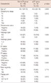
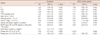
At diagnosis, most patients were presented with tumors of stages T2 to T4 and exhibited clinically detectable axillary lymph node involvement. The overall pCR rate in response to neoadjuvant chemotherapy was 11% (19 of 175 patients). Patients who achieved pCR were significantly more likely to be HER2-positive than were patients who did not attain pCR (p=0.005) (Table 1). By neoadjuvant chemotherapy regimen, 7% (3 of 42 patients) receiving anthracycline-based regimens, 8% (8 of 103) prescribed anthracycline and taxane based-regimens, and 27% (8 of 30) receiving herceptin-based regimens achieved pCR (p=0.012). In HER2+ group, patients treated with herceptin-based regimens had a somewhat higher pCR rate (27%, 8 of 30) than did those not treated with herceptin (14%, 5 of 36); yet, the difference was not statistically significant (p=0.227).
Association between tumor-associated lymphocytes and pathologic response
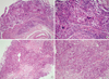
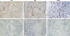
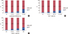
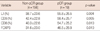
LI, the areal percentages infiltrated by CD3+ and CD8+ T-cells, and FOXP3 counts were significantly intercorrelated (p<0.001). Infiltration of large numbers of lymphocytes was observed in biopsy samples of patients who achieved pCR, whereas few lymphocytes were present in patients who did not attain pCR (Figure 1). Patients who achieved pCR showed significantly higher LI, CD3, and CD8 percentages as well as FOXP3 counts than did patients who did not achieve pCR (Table 2, Figure 2). Moreover, significant correlations were evident between the pCR rate and both LI (p=0.012) and CD8 counts (p=0.040, chi-square test) (Figure 3).
Univariate and multivariate analyses of factors predicting pCR in response to neoadjuvant chemotherapy
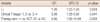
We performed univariate and multivariate analyses in order to assess the associations between pCR, tumor-associated lymphocytes, and other established clinicopathological parameters. Univariate analysis showed that high LI, the area infiltrated by CD3+ and CD8+ T-cells, high FOXP3+ lymphocyte counts, low clinical T stage, HER2 positivity, and treatment with herceptin-based regimens were significantly associated with pCR in all patients (Table 3). In the subgroup analysis regarding therapeutic regimen, LI was predictive of the response to anthracycline-based (odd ratio [OR], 1.91; 95% confidence interval [CI], 1.01-3.61; p=0.047) and anthracycline/taxane-based (OR, 1.51; 95% CI, 1.08-2.10; p=0.014) regimens. However, LI was not associated with pCR in patients receiving herceptin-based neoadjuvant chemotherapy (OR, 0.91; 95% CI, 0.65-1.30; p=0.634). A multivariate analysis revealed that LI (OR, 1.26; 95% CI, 1.03-1.55; p=0.024), clinical T stage (OR, 3.06; 95% CI, 1.08-8.92; p=0.041), and treatment with herceptin-based regimens (OR, 4.95; 95% CI, 1.65-14.84; p=0.004) were significant independent predictors of pCR (Table 4). However, in HER2-positive patients, only clinical T stage was independently associated with pCR to neoadjuvant chemotherapy.
Correlations between tumor-associated lymphocytes and clinicopathological data
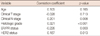
We also evaluated the association between tumor-associated lymphocytes and classic prognostic factors (Table 5). Higher LI values were observed in patients with nodal involvement, high-grade tumors, and ER/PR-negative but HER2-positive tumors.
DISCUSSION
Although relatively few patients achieve pCR in response to neoadjuvant chemotherapy, this remains the strongest indicator of excellent long-term prognosis in breast cancer patients [20]. Identification of factors that predict response to neoadjuvant chemotherapy is important in clinical practice. Although tumor-associated lymphocytes have been suggested to be an independent predictor of such a response in breast cancer patients, only limited data are available. We have shown herein that a good response to neoadjuvant chemotherapy correlates not only with the aspects of the tumor phenotype, such as small tumor size and HER2 overexpression, but also with the increased LI; these findings are in good agreement with those of previous studies [15,21]. The strong association between LI and the response to neoadjuvant chemotherapy suggests that parameters characterizing the host immune response may be as important in this respect as are phenotypic features of tumor cells.
The immune system becomes sensitized to tumor antigens before chemotherapy commences. In addition, chemotherapy-induced cell death releases tumor antigens that can be processed by antigen-presenting cells, leading to a direct lysis of tumor cells by activated CD8+ cytotoxic T-cells [22]. In this manner, chemotherapy serves as a form of immunotherapy [21]. Thus, the pretreatment immune response may enhance the ability of chemotherapy to eliminate cancer cells [23]. The manipulation of the immune system to recognize and eradicate breast cancer cells is a very attractive alternative approach to the current treatment regimens. Several types of anticancer vaccines based on dendritic cells, viruses, peptides, or whole cells are currently under development [24]. Our finding of a strong association between LI and the response to neoadjuvant chemotherapy may serve to establish therapeutic approaches that combine chemotherapy with immune therapy.
Tregs inhibit the development of cytotoxic T lymphocytes and play an immunosuppressive role in cancer tissues [25]. The FOXP3 transcription factor acts as a master regulator of Treg expression [26]. A drastic decrease in FOXP3+ T-cell numbers in the resected specimens examined after neoadjuvant chemotherapy has been associated with pCR in breast cancer patients [17]. We hypothesized that the infiltration of large numbers of FOXP3+ T-cells into pretreatment biopsy specimens might be associated with a reduced response to neoadjuvant chemotherapy. However, we found that the FOXP3+ T-cell count was significantly correlated with LI and was higher in patients with than without pCR. Recently, Oda et al. [27] also reported that the presence of both FOXP3+ and CD8+ T-cells was significantly associated with a high-pCR rate. It has been noticed that Tregs are sensitive to chemotherapy [28]. Therefore, they speculated that the inhibition of FOXP3+ T-cell by chemotherapy might reduce the FOXP3-dependent suppression of antitumor immunity, thus resulting in an elevated antitumor immunity leading to a high-pCR rate [27].
Although LI was predictive of the response to anthracycline-based (OR, 1.91; 95% CI, 1.01-3.61; p=0.047) and anthracycline/taxane-based (OR, 1.51; 95% CI, 1.08-2.10; p=0.014) regimens, this was not associated with pCR in patients receiving herceptin-based neoadjuvant chemotherapy (OR, 0.91; 95% CI, 0.65-1.30; p=0.634). In patients with HER2+ tumors, only clinical T stage was a significant predictor of pCR. Conversely, high-level expression of lymphocyte-associated genes and increased lymphocyte infiltration into node-negative HER2+ breast tumors have been shown to correlate with more favorable outcomes [29]. Larger prospective studies are needed to investigate the relationship between the amount of tumor-associated lymphocytes and the responsiveness to neoadjuvant chemotherapy in patients with HER2+ tumors.
LI, the percentages of tumor area infiltrated by CD3+ and CD8+ T-cells, and FOXP3+ cell counts were all significantly elevated in tumors of higher grade as well as in tumors of patients that were positive in terms of nodal status but were ER-negative, suggesting that tumor-associated lymphocytes are correlated with classical factors predictive of poor outcomes. An earlier study also showed that poor prognostic factors, including negative hormonal status, high tumor grade, and nodal involvement, were associated with significantly higher numbers of CD3+, CD8+, and FOXP3+ infiltrates prior to the commencement of neoadjuvant chemotherapy [17]. Increased infiltration of FOXP3+ T-cells was associated with shorter survival, rendering it possible to identify patients at risk of relapse after 5 years [18]. Poorly differentiated tumors and those of higher nuclear grade may be more sensitive to neoadjuvant chemotherapy than are tumors that are well-differentiated and of lower nuclear grade [30].
In patients with ovarian cancer, the presence of T-lymphocytes in the tumor cell islets, but not in the stroma, was predictive of a favorable prognosis [8]. In addition, high intratumoral T-lymphocyte counts have been associated with pCR in breast cancer patients. However, during therapy, stromal T-lymphocyte counts increased, perhaps reflecting the clearance of tumor cell islets infiltrated by T-lymphocytes [15]. In contrast, data from two large clinical trials revealed significant correlations between the percentage of tumor cell nests infiltrated by lymphocytes and the proportion of the stromal area that contained lymphocytes (Pearson correlation coefficients, 0.61 and 0.80; p≤0.005) [14]. Because we also found a significant correlation between these two parameters (data not shown), only the peritumoral lymphocyte numbers were assessed.
The limitations of this study include the retrospective nature of the study design and the relatively small sample size of the test participants. In addition, the fundamental problems of the immunohistochemical study, including the subjective scoring system, were not completely resolved. Further works standardizing the pathological methods to quantify lymphocytes and exploring the potential impact of tumor heterogeneity on lymphocytic infiltrate evaluation are necessary.
In conclusion, we found that tumor-associated lymphocytes are significantly linked to pCR in breast cancer patients on anthracycline- and anthracycline/taxane-based neoadjuvant chemotherapy regimens, and constitute an important pathologic factor predicting response to treatment. If validated prospectively, our findings may provide a basis for new therapeutic approaches consisting of combinations of chemotherapy and immunotherapy.
 XML Download