

 PDF
PDF ePub
ePub Citation
 Print
Print


INTRODUCTION
Using oncoplastic techniques can help maintain good cosmesis after large excisions exceeding 20% of the breast volume [1,2]. However, while many oncoplastic techniques are suitable for large breasts, many Japanese females have small and dense glandular breasts. We have therefore developed two oncoplastic techniques [3-5] for the breast-conserving reconstruction of small breasts.
If the excision will affect 20% to 30% of their breast volume, we propose that good cosmetic results can be obtained by using an extended glandular flap [5] or an adipofascial flap [3,4]. However, it is difficult to compensate with an extended glandular flap alone in cases where the defect includes about 40% of the breast volume. If the part of the breast that is excised is the lower portion, it may be possible to compensate for the defect with an inframammary adipofascial flap, even if the excision volume is about 40% of the breast volume. However, if the defect is in the outer portion of the breast, it is difficult to bring a large amount of tissue from the inframammary area.
Some volume replacement methods, such as the latissimus dorsi myocutaneous flap [6,7] and perforator flap [8,9], have been reported as oncoplastic techniques to repair defects in the outer portion of the breast affecting about 40% of the breast volume. These flaps are usually the best choice for defects that are too large to be corrected using local tissue. However, these techniques must be performed by plastic surgeons, and involve the loss of donor site muscle or lead to the formation of an additional donor-site scar. This often precludes the use of these techniques.
Therefore, we developed a method combining an adipofascial flap with an extended glandular flap for the reconstruction of a large breast partial resection comprising about 40% of the outer portion of the breast. The aim of this report is to describe the efficacy of the method combining these two oncoplastic techniques after a large excision of the breast volume for the breast-conserving reconstruction of small dense breasts.
CASE REPORT
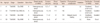
Four females with breast cancer in the outer portion of the breast underwent remodeling using the method combining two oncoplastic techniques (an extended glandular flap [5] and an inframammary adipofascial flap [3,4]) after an excision of about 40% of their breast volume. The excision volume compared to the total breast volume was estimated by using a preoperative photograph of the markings made for the partial resection area by seven independent observers (breast surgeons). Summary of the patient characteristics is shown in Table 1. In all patients, the size of the bra was an A cup, and the breast density on mammography was heterogeneously dense in all four cases. All patients had undergone a sentinel lymph node biopsy. Three patients had no metastasis in the sentinel lymph nodes, allowing them to avoid an axillary lymph node dissection. One patient with metastasis in the sentinel lymph nodes underwent an axillary lymph node dissection. The operations were performed by breast surgeons without the help of plastic surgeons.
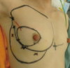
After marking the partial resection area with the patient in the supine position, the upper edge of the breast at the subclavicular area is drawn on the skin with the patient in the standing position (Figure 1). This marking is for the extended glandular flap. The extended glandular flap is perfused through the 2nd and/or 3rd internal mammary artery perforators. So a preoperative Doppler examination was performed to confirm the location of perforators from the internal mammary artery.
The inframammary groove and the area of the adipofascial flap are also drawn on the skin. At that moment, it is important to confirm the location of the perforators by Doppler sonography (Figure 1, x mark).
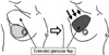
After a partial resection of the breast is performed, the extended glandular flap is made by freeing the breast from both the skin and the pectoralis fascia up to the subclavicular area that was marked before surgery. While making the flap, it is important to undermine the skin of the subclavicular area so that the subcutaneous fat might remain thick and taper gradually toward the head side. It is important to keep the perforators of the internal mammary artery intact as well. This flap is moved to the breast area where the tumor has been removed (Figure 2). After the flap is inserted, it is secured to the surrounding breast tissue with absorbable sutures.
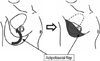
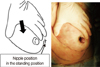
The skin of the inframammary area is undermined by subcutaneous fat. The subcutaneous fat, together with the fascia of the external oblique muscle and anterior sheath of the rectus abdominis muscle, is dissected semicircularly, resulting in the formation of an adipofascial flap. The flap is reflected back to the breast area remodeled using the extended glandular flap, and then is secured to the surrounding breast tissue with absorbable sutures (Figure 3). While making the breast mound, it is important to keep the perforators intact. In order to make a new inframammary line, the subcutaneous tissue of the skin is fixed to the muscle with absorbable sutures. While making the breast mound and re-shaping the inframammary line, the shape of the breast is checked when putting pressure from the upper side until the nipple is set at the position that had been marked before surgery when the patient was in the standing position (Figure 4). Finally, a suction tube was placed, and the skin was sutured.
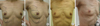
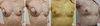
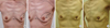
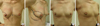
The cosmetic assessment of four patients was performed based on photographs taken more than 1 year after the operation. The photographs for case 1 was taken 6.6 years after the operation (Figure 5), the photographs for case 2 were taken 3.1 years after the operation (Figure 6), those for case 3 were taken 2.4 years after the operation (Figure 7), and the photographs for case 4 was taken 1.3 years after the operation (Figure 8). Photographs of the patients' breasts were then taken using frontal, left oblique and right oblique views without any identifying features. The cosmetic results were evaluated by seven independent observers (breast surgeons) as "excellent", "good", "fair", or "poor" using the Harvard Scale established by Harris et al. [10]. An excellent result was reported when the treated breast was almost identical to the untreated one, a good result was reported when the treated breast was slightly different from the untreated breast, a fair result when there was an obvious difference between the two sides without major distortion, and a poor result when the treated breast was seriously distorted. The observers were 'blinded' to the identity of the patients.
The blood loss and the length of the operation ranged from 31 to 186 g (average, 93.8 g) and from 116 to 168 minutes (average, 138.3 minutes), respectively. There was one case of invasive ductal carcinoma and three cases of noninvasive ductal carcinoma. None of the patients in this study complained of upper abdominal asymmetry. One patient had delayed wound healing, but eventually healed within 2 months. One patient refused radiation therapy. The other three patients received radiation therapy to the breast after wound healing, as usual. There was no fat necrosis of the flap and no dystrophic calcification on mammography in any of the patients. Depression on the donor area of the extended glandular flap was not noticeable, and furthermore, clothing that shows the décolletage is not popular in Japan, so there were no patient complaints. All of the patients are still alive without recurrence at time of the writing of this manuscript.
DISCUSSION
In European countries, oncoplastic techniques for the breast-conserving reconstruction have been popular for more than 20 years, and many oncoplastic techniques have been reported [2,11,12]. However, most of the reported oncoplastic techniques are suitable only for large breasts. Many Japanese patients have small breasts, therefore, it is necessary to develop new oncoplastic methods suitable for small breasts.
We have previously reported two oncoplastic techniques [3-5] for the breast-conserving reconstruction of small breasts. One technique employs an extended glandular flap [5], and the other uses an inframammary adipofascial flap [3,4]. The extended glandular flap comprises the mammary gland, including the fat in the subclavicular area, which is used for volume displacement for breast-conserving reconstruction in the upper portion of a dense breast. A dense glandular breast can be easily mobilized by advancing the breast tissue into the excision cavity, without a risk of fat necrosis. Thus, this procedure is suitable for small dense breasts. The inframammary adipofascial flap is used by inverting a tongue-shaped adipofascial flap in the upper abdominal area, which is used for volume replacement for breast-conserving reconstruction in the lower portion of the breast. These two techniques can be used for defects where it is often difficult to reshape the breast following large excisions exceeding 20% of the breast volume. However, it is difficult to fill up a defect comprising about 40% of the breast volume using an extended glandular flap alone. Since the inframammary adipofascial flap is a commonly used volume replacement technique, it could thus be used to replace a defect of about 40% of the breast volume if the tumor location is in the lower portion of the breast. However, if the tumor is in the outer portion of the breast, it is difficult to bring a sufficiently large amount of tissue from the inframammary area. For the tumors in the outer portion of the breast, other procedures for breast reshaping using a tissue flap, such as the latissimus dorsi myocutaneous flap [6,7] and perforator flap [8,9] have been reported. These flaps can be filled up for a large defect. However, these techniques must be performed by plastic surgeons and involve the loss of donor site muscle or the formation of an additional donor-site scar.
Using our new technique, we were able to fill up a defect of about 40% of the breast volume by using a method combining an adipofascial flap with an extended glandular flap. The advantages of this method are as follows: 1) It is not necessary to make an additional scar, since both an extended glandular flap and adipofascial flap can be created through the same skin incision; 2) If mastectomy is required due to the presence of a positive margin, it is possible to use a latissimus dorsi flap for reconstructive surgery; 3) Since not much tissue is taken from the inframammary area, deformation of the donor site is unlikely to occur; 4) This method can be performed by breast surgeons, without the help of a plastic surgeon. However, this method has limited indications for patients with a tumor located in the outer portion of a small dense breast. This is the greatest disadvantage.
We treated four patients with breast cancer in the outer portion of a small dense breast who underwent remodeling using a method combining the extended glandular flap and the inframammary adipofascial flap after an excision of about 40% of their breast volume. Even though more than 6 years had passed since the surgery in one case, there was no fat necrosis. In addition, no dystrophic calcification was observed by the mammography. This oncoplastic technique combining an adipofascial flap with an extended glandular flap has limited indications. However, this method is useful after a large excision of the breast volume for performing the breast-conserving reconstruction of small dense breasts.
 XML Download