

 PDF
PDF ePub
ePub Citation
 Print
Print


INTRODUCTION
Metastasis from extramammary malignancies, especially gastric carcinoma, is rare [1]. It is extremely rare for breast metastasis to present with pleomorphic microcalcification as this feature has been regarded as virtually pathognomonic of primary breast carcinoma. We describe a patient who presented with a breast mass containing microcalcification which was confirmed to be metastases from gastric carcinoma.
CASE REPORT
A 54-year-old pre-menopausal woman with unremarkable past health issues, presented with a 1-month history of abdominal distension and a mass in the right breast. On physical examination, a 5-cm mass with indistinct border was found in the upper outer quadrant of the right breast without evidence of axillary lymphadenopathy. A pelvic mass of 20-week size was also detected on physical examination. The CA125 was slightly elevated (41.7 IU/mL).
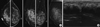
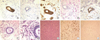
A mammogram revealed a large area with pleomorphic microcalcifications of architectural distortion in the upper outer quadrant of the right breast, suggestive of a malignant lesion (Figure 1A). The ultrasound demonstrated a 3.5-cm ill-defined hypoechoic lesion in the upper outer quadrant of the right breast, 1 to 4 cm from the nipple (Figure 1B). The provisional diagnosis given was primary breast carcinoma. A biopsy of the right breast mass was performed which showed scattered foci of microcalcification. An immunohistochemical stain showed that the cells were negative for estrogen receptor and c-erbB-2 staining (Figure 2A and B). The malignant cells showed positivity in cytokeratin AE1/AE3, PAS-D, and E-cadherin staining (Figure 2C-E). The malignant cells also showed positivity for cytokeratin 7 (CK7). The histological examination that followed confirmed the diagnosis of poorly cohesive carcinoma with signet ring cell features, compatible with metastasis from the stomach (Figure 2F and G).
Abdominal ultrasonography showed bilateral heterogeneous adnexal masses and ascites. An abdominal and pelvis computed tomography (CT) confirmed a large pelvic mass that likely had originated in the left ovary and a smaller right adnexal lesion which were both worrisome of malignancy. The clinical provisional diagnosis was ovarian carcinoma.
A total abdominal hysterectomy and bilateral salpingo-oophorectomy was performed. Intra-operatively, a large left ovarian lesion was found. A pathological examination revealed a poorly metastatisized differentiated adenocarcinoma with signet ring features (Figure 2H).
Immunohistochemical examination showed that the malignant cells were diffusely positive for CK7 and focally positive for cytokeratin 20 (CK20) (Figure 2I and J). Features were compatible with metastasis from the stomach or pancreaticobiliary system (Krukenberg's tumor).
An oesophago-gastric-duodenoscopy (OGD) was performed post-operative on day 4 which revealed an ulcerative tumor with elevated borders extending from the esophago-gastric junction to the higher lesser curvature. A biopsy of the tumor was performed which confirmed poorly differentiated adenocarcinoma.
The patient was treated with chemotherapy but her gastric cancer progressed with bone metastases and peritoneal dissemination. She died 11 months after the initial diagnosis.
DISCUSSION
Extramammary malignancy metastasis is rare, occurring in 0.5% to 2.0% of all breast malignancies [1]. The most common primary malignancy of a hematogeneous breast metastasis is melanoma [2]. Breast metastasis from gastric cancer occurs more often in Orientals [2]. The average age at presentation is 47 years [3]. Most cases are associated with advanced disease and a poor prognosis [4]. The first case was reported in 1999 and a limited number of similar cases have since been reported [5,6]. It has been suggested that an increased blood supply to the breast could be related to breast metastasis in premenopausal woman [5,6].
Only about 25% of metastasis from extramammary malignancies is the presenting feature of the occult extrammammary malignancy [7]. Metastatic breast cancer is usually non-tender, well-defined and mobile on physical examination [6]. Skin or nipple retraction and architectural distortion are rare clinical features [1]. Breast metastases are most often found in the upper outer quadrant of the left breast [5]. Multiple and bilateral metastases and axillary lymphadenopathy are rare [3]. A few cases of breast metastases have clinical features similar to inflammatory carcinoma of the breast [7]. In an ultrasonogram, breast metastases often have low-level internal echoes and sometimes posterior acoustic enhancement [1].
Since the first description of microcalcification in 1951, pleomorphic microcalcifications associated with a mammographically detected mass have been regarded as virtually pathognomonic of primary breast cancer [8-10]. It has been reported that breast metastases have mammographic features similar to benign breast lesions, such as fibroadenoma, being well-circumscribed without microcalcification [4,6,11]. A few cases of breast metastasis with microcalcifications from ovarian carcinoma have been reported. The presence of microcalcifications in mammography due to metastatic gastric cancer is extremely rare. Only a few cases of breast metastases from gastric cancer with microcalcifications have been reported in the literature, only 4 cases have been reported [12]. Our case is unusual as the breast metastasis shows an ill-defined border with microcalcifications and architectural distortion, all of which are features that mimic primary breast carcinoma.
A histopathological examination is useful for differentiating breast metastasis from primary breast cancer [6]. The absence of intraductal carcinoma or lobular carcinoma in situ is suggestive of metastasis [2,3]. Immunostaining for breast metastasis from gastric cancer is usually negative for c-erbB-2, estrogen and progesterone receptors and positive for epithelial markers like CEA, CK7, and CK20 [4,13]. Thus in our case, the combination of CK7 positive staining, negative estrogen receptor, and c-erbB-2 staining strongly supports the diagnosis of gastrointestinal primary adenocarcinoma rather than primary breast carcinoma. Also, the malignant cells in the breast biopsy in our case demonstrated a positivity in cytokeratin AE1/AE3 and PAS-D stain, further supporting the diagnosis of metastasis from gastric cancer. The E-cadherin positivity staining in our case was compatible with the diagnosis of metastasis from gastric cancer and rendered the possibility of invasive lobular carcinoma unlikely. The diagnosis of breast metastasis from gastric carcinoma is further supported by the fact that OGD found an ulcerative adenocarcinoma at the lesser curvature.
It has been shown that resection of the primary tumor may improve the patients survival with breast metastasis from gastric cancer [5]. Similar to our case, patients with breast metastasis from gastric cancer have been associated with a poor prognosis and the majority of the reported cases died within 1 year of diagnosis [5]. Differentiating a primary breast cancer and metastatic breast cancer would be important for the avoidance of unnecessary radical surgery in the case of primary breast cancer [13]. Also the hormonal treatment and chemotherapy of primary breast cancer and breast metastasis from gastric cancer are different [6].
A clinical history and pathological examination are helpful in differentiating primary breast cancer and metastasis from gastric cancer. Our case demonstrates the unusual mammographic features of an ill-defined mass lesion with pleomorphic microcalcification. Immunohistopathological examination is important in making a correct diagnosis. Accurate diagnosis of breast metastasis from gastric cancer is crucial for proper treatment.
 XML Download