

 PDF
PDF Citation
 Print
Print


Abstract
Purpose
The purpose of this study was to evaluate silver in situ hybridization (SISH) as an effective test to identify HER2 gene amplification in patients with breast cancer.
Methods
A systematic literature review was used to evaluate the effectiveness of SISH. The literature review covered from October 27, 2009 to December 1, 2009, and eight domestic databases including KoreaMed and foreign databases including Ovid-MEDLINE, EMBASE, and Cochrane Library were used. Keywords, such as 'silver in situ hybridization' and 'SISH', were used to search 63 documents. Ten studies regarding the evaluation of diagnostics were included in the final evaluation. The Scottish Intercollegiate Guidelines Network (SIGN) tool was used by two evaluators to independently evaluate the quality of the ten studies.
Results
A total of ten studies (nine diagnostic evaluation studies and one correlation study) were identified to evaluate SISH. The effectiveness of this test was evaluated based on diagnostic accuracy, concordance rate, and correlation with fluorescence in situ hybridization (FISH) results. The sensitivity of SISH was 0.81-1.00, and the specificity was 0.82-1.00. The positive predictive value was 0.95-1.00, negative predictive value was 0.81-1.00, and the test accuracy was 0.90-1.00. The concordance rate of SISH was 87.0-100% and two studies reported a correlation with FISH results. The body of evidence as a whole suggests a Grade D for SISH.
Figures and Tables
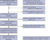 | Figure 1Documents selected for evaluation of silver in situ hybridization according to the literature search strategy.
|
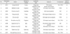
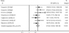 | Figure 2Diagnostic meaning of silver in situ hybridization (fixed-effect model).
OR=odds ratio; CI=confidence interval.
|
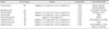
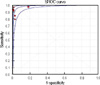 | Figure 3Receiver operating characteristics (ROC) curve of silver in situ hybridization (area under the curve [AUC]=0.9872).
|
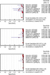
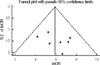
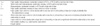
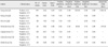
Table 3
Levels of evidence (From Scottish Intercollegiate Guidelines Network. SIGN 50: a guideline developer's handbook) (4)
![]()
Table 4
Grades of recommendations (From Scottish Intercollegiate Guidelines Network. SIGN 50: a guideline developer's handbook) (4)
![]()
References
1. Sińczak-Kuta A, Tomaszewska R, Rudnicka-Sosin L, Okoń K, Stachura J. Evaluation of HER2/neu gene amplification in patients with invasive breast carcinoma. Comparison of in situ hybridization methods. Pol J Pathol. 2007. 58:41–50.
2. Sung WJ, Park SJ, Gu MJ, Bae YK. Automated silver-enhanced in situ hybridization for evaluation of HER2 gene status in breast carcinoma: comparison with fluorescence in situ hybridization and immunohistochemistry. Korean J Pathol. 2010. 44:28–34.

3. Lee JW, Noh WC, Kim MS, Kim HA, Chang YH, Hong YJ, et al. Availability of fine needle aspirates for the assessment of HER2 gene amplification in invasive breast cancer patients. Korean J Lab Med. 2008. 28:392–399.
4. SIGN 50: a guideline developer's handbook. Scottish Intercollegiate Guidelines Network. Accessed January 22nd, 2011. http://www.sign.ac.uk/pdf/sign50.pdf.
5. Health Insurance Review Agency. A Study on the Construction and Management of Health Technology Assessment System. 2005. Seoul: Health Insurance Review Agency;227–228.
6. Bartlett JM, Campbell FM, Ibrahim M, Wencyk P, Ellis I, Kay E, et al. Chromogenic in situ hybridization: a multicenter study comparing silver in situ hybridization with FISH. Am J Clin Pathol. 2009. 132:514–520.
7. Francis GD, Jones MA, Beadle GF, Stein SR. Bright-field in situ hybridization for HER2 gene amplification in breast cancer using tissue microarrays: correlation between chromogenic (CISH) and automated silver-enhanced (SISH) methods with patient outcome. Diagn Mol Pathol. 2009. 18:88–95.
8. Kang J, Kwon GY, Lee YH, Gong G. Comparison of silver-enhanced in situ hybridization and fluorescence in situ hybridization for HER2 gene status in breast carcinomas. J Breast Cancer. 2009. 12:235–240.
9. Kim TJ, Kim TE, Jung ES, Yim HW, Song BJ, Jung SS, et al. The comparison of automated silver in situ hybridization and fluorescence in situ hybridization for evaluating HER2 gene amplification in breast carcinoma. J Breast Cancer. 2009. 12:295–301.
10. Shousha S, Peston D, Amo-Takyi B, Morgan M, Jasani B. Evaluation of automated silver-enhanced in situ hybridization (SISH) for detection of HER2 gene amplification in breast carcinoma excision and core biopsy specimens. Histopathology. 2009. 54:248–253.
11. Capizzi E, Gruppioni E, Grigioni AD, Gabusi E, Grassigli A, Grigioni WF, et al. Real time RT-PCR approach for the evaluation of ERBB2 overexpression in breast cancer archival samples: a comparative study with FISH, SISH, and immunohistochemistry. Diagn Mol Pathol. 2008. 17:220–226.
12. Carbone A, Botti G, Gloghini A, Simone G, Truini M, Curcio MP, et al. Delineation of HER2 gene status in breast carcinoma by silver in situ hybridization is reproducible among laboratories and pathologists. J Mol Diagn. 2008. 10:527–536.
13. Dietel M, Ellis IO, Höfler H, Kreipe H, Moch H, Dankof A, et al. Comparison of automated silver enhanced in situ hybridisation (SISH) and fluorescence ISH (FISH) for the validation of HER2 gene status in breast carcinoma according to the guidelines of the American Society of Clinical Oncology and the College of American Pathologists. Virchows Arch. 2007. 451:19–25.
 XML Download