

 PDF
PDF ePub
ePub Citation
 Print
Print


INTRODUCTION
Bilateral reduction mammoplasty (BRM) has long been used for cosmetic reasons. In recent years, BRM has also been used for the treatment of breast cancer. When used with radiotheraphy in breast cancer patients, local recurrence of this procedure has not been higher than that of modified radical mastectomy [1,2]. If there is discordance between tumor size and breast volume, the cosmetic results of traditional breast-conserving surgical techniques such as quadranectomy or lumpectomy have not been encouraging. The location of the primary tumor affects the cosmetic results. The amount of breast tissue removed with these techniques is much less than that in breast reduction, and symetrisation with the contralateral breast is therefore not a point at issue. If the breast volume is not reduced sufficiently with surgery, radiotherapy planning is quite difficult in breast cancer patients with macromastia. Given the proven safety of the oncological results of this procedure, it has been used with increasing frequency, particularly for macromastic women with breast cancer. Good cosmetic results have been obtained with symetrisation, as well as with macromasty-related symptoms like back, neck, shoulder and arm pain, which were also relieved, and the radiotherapy planning was also eased. Therefore, this procedure has been called "symetrisation mammoplasty" and using this procedure has created similar results as different oncoplastic techniques used in different tumor localisations. Consequently, the tumor localisation does not alter the amount of tissue removed or the cosmetic results.
The contralateral breast of a woman with breast carcinoma is at high risk for a new tumor. An important part of the contralateral breast can be removed with surgery, and quite a large specimen can be obtained for pathological examination. This study evaluated the importance of a routine pathological examination of contralateral breast specimens in breast cancer patients using reduction mammoplasty.
METHODS
Seventy-one patients operated on with BRM due to breast cancer between 2008 and 2010 were enrolled in the study. Upper or lower pediculated flap techniques were preferred according to the location of the primary tumor in the breast. The same technique has also been used for symetrisation and reduction purposes on the contralateral breast. The age, height, weight, and body mass index (BMI) of the patients, tumor features and stages, family history of breast cancer, the weight of breast tissues resected from the contralateral breast, and the number of tissue blocks prepared for pathological evaluation were recorded. Breast lesions found in the contralateral breast and accompanying lesions with tumors were also examined.
Breast reduction specimens were examined macroscopically and, if no lesion was identified, eight to nine random blocks of breast tissue were prepared from the contralateral breast. If a macroscopic lesion was present, sampling was concentrated on that area, with selection of an appropriate number of blocks as considered necessary by the pathologist. Biopsy samples were fixed in 10% formalin, processed routinely, and embedded in paraffin. Sections (5 µm thick) were taken from the paraffin blocks and stained with H&E.
Ductal and lobular in situ carcinomas, atypical epithelial, and ductal and lobular hyperplasias were evaluated in proliferative lesions from the high-risk group. Severe (florid) and moderate hyperplasias without atypia, sclerosing adenosis, and intraductal papillomas were evaluated in the low-risk lesions group. Fibrocystic changes, mild hyperplasias, and fibroadenomas were evaluated in lesions of patients not in any risk group.
Mammography and breast ultrasonography were used for preoperative contralateral breast evaluation. Patients with either clinical or radiological suspicious lesions in the contralateral breast were excluded. Therefore, patients requring breast MRI were also excluded.
A Student's t-test was used in the comparison of patients with high risk lesions and other patients using SPSS version 10.00 for Windows (SPSS Inc., Chicago, USA). p-values less than 0.05 were accepted as significant.
RESULTS
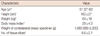
The median age of the patients was 51 years (range, 27-62 years), the mean BMI 29±4.8, and median weight of the tissue removed from contralateral breast was 1,080 g (range, 680-2,800 g). The general characteristics of the patients have been outlined in Table 1. Lower pediculated flaps in 53 patients (75%) and upper pediculated flaps in 18 patients (25%) were used. All patients were in T1 and T2 stage and sentinel lymph node biopsy was applied for axillary evaluation of tumor containing breasts. Fourteen patients with positive sentinel lymph node biopsy (SLNB) were operated on using axillary dissection, and all these patients were in stage N1. Incidentally discovered occult invasive carcinoma was not detected in the contralateral breast. Proliferative lesions with a high risk for breast carcinoma were reported in eight (11.2%) patients, three of whom exhibited ductal carcinoma in situ (DCIS) and the others, atypical ductal hyperplasia. Low-risk lesions were detected in 18 patients (25%) with the following distribution: severe epithelial hyperplasia in 10 patients, moderate epithelial hyperplasia in five patients, and sclerosing adenozis in three patients. Sixteen patients were reported to have lesions without any risk of malignancy, and their distribution was as follows: 11 fibrocystic changes, one mild epithelial hyperplasis, and one fibroadenoma. No lesions were reported in 29 patients (40.8%).
All three patients with atypical ductal hyperplasia were demonstrated to have a family history of breast cancer in first degree relatives; however, there were no lesions in another three patients with family histories. There were no patients with a family history of bilateral breast cancer in the study population.
The mean age of the patients with high-risk lesions was 45.6, while the mean age of the rest was 52.8 (p=0.036). The mean weight of the specimens of lower and upper pediculated flaps removed from contralateral breast were 1,086 g and 1,060 g, respectively.
The lower pediculated flap technique was used in seven patients with high-risk lesions. The mean number of tissue blocks prepared from the contralateral breast was 9.8±2.1. The mean tissue block number for patients with high-risk lesions was 11.2±2.4 (p=0.091). There was no difference between the patients with high-risk lesions and the others with respect to tumor size (p=0.076) and specimen size excised (p=0.082). The single parameter demonstrated to increase the incidence of high-risk lesions in contralateral breast was young age (Table 2). As the SLNB was negative at the site of the tumor-containing breast in eight patients with high-risk lesions in the contralateral breast, axillary dissection was not applied. Three DCIS cases showed low grade lesions, one of whom had simple mastectomy applied due to resection margin positivity. Adjuvant radiotheraphy and hormonotheraphy was added to the treatment of the other two patients with negative resection margins. Patients with atypical epithelial hyperplasia were followed-up.
Accompanying lesions in the tumor-containing breast of the patients included three ductal in situ carcinomas, two atypical ductal hyperplasias, one lobular carcinoma in situ (LCIS), and one invasive carcinoma. There were no high-risk lesions in the contralateral breast of these patients.
DISCUSSION
There have been no well-defined guidelines for the pathological examination of specimens retrieved from contralateral breast specimens produced by reduction mammoplasty. In this study, although there were no suspicious lesions in a macroscopic evaluation of the specimen, a microscopic evaluation was carried out with randomised 8-9 tissue blocks. When suspicious areas were defined, the specimens were evaluated in 3-mm-thick slices. The number of tissue blocks taken was not determined according to the size of the specimen because a huge specimen made up of mostly fatty tissues was not sampled like the fibrous rich ones. The series examining the lesions found after these surgeries have been mainly based on the series performed for benign reasons. The incidence of the occult invasive breast carcinoma detection rate after BRM surgeries for benign reasons has ranged between 0.06% to 0.4% [3,4]. These low rates may be explained by the use of these surgeries in younger patients and radiological surveillance used before surgery. Bondeson et al. [5] examined the lesions of 200 patients operated on with BRM for benign reasons and reported that while there was no pathological change in patients under the age of 30, LCIS was reported in 8% of the cases of patients over 40 years of age. Ambaye et al. [6] reported high-risk lesions (in situ carcinoma and atypical hyperplasias) in 6.2% of patients over 40 years of age and 7.9% of patients over 50 years of age. In another study, high-risk lesions were reported to be 6.4% in patients over 40 years of age [3]. In these studies, the authors suggested a more careful examination of benign BRM specimens in patients over 40 years of age. As the incidence of breast carcinoma increases with age, the increased frequency of high-risk lesions with increasing age is not surprising in patients with no diagnosis of breast cancer. The incidence of contralateral occult carcinoma in breast carcinoma patients operated on with BRM has increased up to 4% [6,7]. In a study comparing contralateral breast lesions in patients operated on for benign and malignant tumors, occult cancer and atypical proliferative lesion incidence in malignant cases were 2% and 7%, respectively. These rates for benign cases were 0.6% and 1%, respectively [8].
The annual absolute contralateral cancer risk for a patient with breast carcinoma is 0.7% [9]. The risk factors for bilateral breast cancer have been investigated extensively. Bilateral breast carcinoma risk has been demonstrated to be higher in patients with a diagnosis of breast carcinoma at an early age [10] of whom those with tumors greater than 2 cm are at higher risk [11] as those with familial bilateral breast cancer history, BRCA1 and BRCA2 gene mutations, and LCIS [12,13].
In the past, blind biopsies in the upper outer quadrant or in areas mirror imaging sites of primary tumors have been used for early diagnosis of contralateral breast lesions [14].
It can be said that upper outer quadrant lesions can be determined with the lower pediculated flap technique, while lower quadrant lesions can be determined with the upper pediculated flap technique. In a series by Petit et al. [7] the upper pediculated flap technique was used in 80% of the cases and most of the occult lesions were detected in the lower quadrants and central areas. In our series, there was no difference in the amount of tissues resected using the two techniques. We have observed that the lower pediculated flap technique was used for seven of eight high-risk lesions which can be explained by the increased frequency of breast carcinomas in the upper outer quadrant and our low rate of upper pediculated flaps.
Can BRM decrease metachronous tumor incidence? Ricci et al. [15] followed 114 breast cancer patients treated with BRM and 134 not treated with BRM of similar age and stages for 51 months. In the end, contralateral metachronous breast cancer incidence was 1.8% in the BRM group and 6.4% in the other group. Other studies have confirmed that this procedure decreases the risk of metachronous breast cancer [16]. In our series, there was no metachronous contralateral invasive and/or in situ carcinoma in our relatively short follow-up of 14 months.
The vast majority of cases of high-grade DCIS with comedonecrosis are recognised as microcalcifications in mammographic examinations. Nevertheless, low-grade DCIS cases are not detected in mammographies in 50-70% of cases [17]. We found that three DCIS cases detected were low-grade lesions. Further, in situ carcinomas that could not be recognised radiologically but were diagnosed within reduction specimens are expected to be low-grade lesions.
We did not encounter any contralateral occult carcinomas in our series however, the incidence of high-risk proliferative lesions was 11.2% even in patients without any radiological abnormalities, and these patients were considerably younger.
It has been found that the coincidence of high risk lesions in breast specimens of patients who underwent reduction mammoplasty for benign reasons such as macromastia increases with age [3,4]. However, the young age of the patients with breast cancer has been identified as a risk factor for high risk lesions in the contralateral breast [5]. It is well-known that genetic factors rather than enviromental factors are important in the etiology of breast cancer in young patients [12]. The tendency of these tumors for bilaterality and multicentricity might explain this occurence. Bilateral reduction mammoplasty is an effective measure to delineate the lesions in the contralateral breast. At least 8-9 tissue blocks should be taken to examine specimens that do not contain any macroscopic lesions. Further, young patients with a diagnosis of breast cancer and those with family histories of breast cancer are especially prone to high risk lesions in the contralateral breast. Therefore, contralateral specimens from these patients should be examined more carefully with a sufficient number of tissue blocks.

 XML Download