Journal List > J Breast Cancer > v.13(1) > 1036239

Lee, Gong, Park, Park, Jung, Lee, and The Korean Study Group for Breast Pathology: A Multi-institutional Study of Interlaboratory Variance in the Estrogen and Progesterone Receptor Assays

Abstract

Purpose

The expression of hormone receptors is the most reliable factor for predicting the responsiveness to hormonal therapy. At present, immunohistochemistry (IHC) is considered as a practically reliable method. This study was designed to examine the interlaboratory variance in immunohistochemical assays for estrogen receptor (ER) and progesterone receptor (PR) in Korea.

Methods

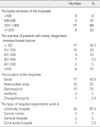
The Korean Study Group for Breast Pathology (KSGBP) made a questionnaire to know the current situation in HR assay in Korea. The questionnaire was sent to the members of KSGBP by e-mail, which were included eight questions relating to tissue handling, ER/PR IHC procedure and interpretation method. Forty laboratories replied with the completed questionnaire.

Results

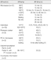
All 40 laboratories were using formalin as a fixative. Pretreatment was performed using six different methods including autoclave (25%), microwave (30%) and full autostainer (15%). Primary antibodies for ER were SP1 in 40%, 6F11 in 27.5% and 1D5 in 32.5%. Primary antibodies for PR were more variable (seven clones) than those for ER. Interpretation method used was Allred system in 20%, modified Allred system in 15%, report the % of positive tumor cells in 45%, positive/ negative in 15% and others in 5%. The expression rate of ER was ranged from 45.6% to 93% (mean 63.5%) and the expression rate of PR was ranged from 27% to 90% (mean 59.1%). The differences according to the numbers of breast cancer in each institute, primary antibodies, detection systems and interpretation methods did not influence to the expression rate of ER/PR, statistically (p>0.05).

Conclusion

In Korea, the interlaboratory variance in ER/PR IHC procedure was too huge to make a standardized method. We suggest the proper quality control program such as ER/PR staining with positive internal and external controls and negative control might be better to aim at getting similar results among the different laboratories rather than trying to standardize the procedure.

Figures and Tables

Figure 1
The differences according to the numbers of breast cancer cases at each institute (A), the antigen retrieval methods (B), the ER primary antibodies (C), the detection systems (D) and the interpretation methods (E) did not statistically influence the expression rate of ER and the differences according to the PR primary antibodies (F) did not statistically influence the expression rate of PR.
ER=estrogen receptor; PC=pressure cooker; polymer=polymer detection system; ABC/LSAB=avidin-biotin-enzyme complex/labeled streptavidin-biotin; %=% of positively stained tumor cells; Allred=Allred or modified Allred system; Dichotomy=posive vs negative.
jbc-13-46-g001
Table 1
Characteristics of responders
jbc-13-46-i001
Table 2
Applied antigen retrieval system, temperature and time for ER/PR IHC*
jbc-13-46-i002

ER=estrogen receptor; PR=progesterone receptor; IHC=immunohistochemistry; AR=antigen retrieval; Temp=temperature; n=number of hospital; PC=pressure cooker; A=autoimmunostainer.

*One hospital applied different methods for ER and PR and method for ER was described here; Includes Ventana autostainer and Leica autostainer; LabVision autostainer with PT module.

Table 3
The used antigen retrieval buffers for ER/PR IHC*
jbc-13-46-i003

ER=strogen receptor; PR=progesterone receptor; IHC=immunohistochemistry.

*One hospital applied different methods for ER and PR and method for ER was described here; DAKO product.

Table 4
Characteristics of primary antibody for ER/PR
jbc-13-46-i004

ER=estrogen receptor; PR=progesterone receptor.

Table 5
Detection systems for ER/PR IHC
jbc-13-46-i005

ER=estrogen receptor; PR=progesterone receptor; IHC=immunohistochemistry; ABC=Avidin-biotin-enzyme complex; LSAB=Labeled streptavidin-biotin-peroxidase.

Table 6
Interpretation methods for ER/PR IHC
jbc-13-46-i006

ER=estrogen receptor; PR=progesterone receptor; IHC=immunohistochemistry.

*Negative, positive (11-25%), positive (26-50%), positive (51-75%), positive (76-100%); 1 (<10%), 2 (11-1/3%), 3 (1/3-2/3%), 4 (>2/3%); 0, 1+ (<10%), 2+ (11-50%), 3+ (>50%); Included if information about fraction and intensity of stained tumor cells contained; Using TMA, negative, 1+ (>10% and mild intensity), 2+ (>10% and moderate intensity), 3+ (>10% and severe intensity); negative (>5%), 1+ (5-20% and weak intensity), 2+ (20-50% and inermediate intensity), 3+ (>50% and strong intensity).

References

1. Harvey JM, Clark GM, Osborne CK, Allred DC. Estrogen receptor status by immunohistochemistry is superior to the ligand-binding assay for predicting response to adjuvant endocrine assay for predicting response to adjuvant endocrine therapy in breast cancer. J Clin Oncol. 1999. 17:1474–1481.
2. Greene GL, Nolan C, Engler JP, Pjensen EV. Monoclonal antibodies to human estrogen receptor. Proc Natl Acad Sci U S A. 1980. 77:5115–5119.
crossref
3. Greene GL, Jensen EV. Monoclonal antibodies as probes for estrogen receptor detection and characterization. J Steroid Biochem. 1982. 16:353–359.
crossref
4. Alberts SR, Ingle JN, Roche PR, Cha SS, Wold LE, Farr GH Jr, et al. Comparison of estrogen receptor determinations by a biochemical ligand-binding assay and immunohistochemical staining with monoclonal antibody ER1D5 in females with lymph node positive breast carcinoma entered on two prospective clinical trials. Cancer. 1996. 78:764–772.
crossref
5. Barnes DM, Harris WH, Smith P, Millis RR, Rubens RD. Immunohistochemical determination of oestrogen receptor: comparison of different methods of assessment of staining and correlation with clinical outcome of breast cancer patients. Br J Cancer. 1996. 74:1445–1451.
crossref
6. Barnes DM, Millis RR, Beex LV, Thorpe SM, Leake RE. Increased use of immunohistochemistry for oestrogen receptor measurement in mammary carcinoma: the need for quality assurance. Eur J Cancer. 1998. 34:1677–1682.
crossref
7. Rhodes A, Jasani B, Barnes DM, Bobrow LG, Miller KD. Reliability of immunohistochemical demonstration of oestrogen receptors in routine practice: interlaboratory variance in the sensitivity of detection and evaluation of scoring systems. J Clin Pathol. 2000. 53:125–130.
crossref
8. Rhodes A, Jasani B, Balaton AJ, Barnes DM, Anderson E, Bobrow LG, et al. Study of interlaboratory reliability and reproducibility of estrogen and progesterone receptor assays in Europe. Am J Clin Pathol. 2001. 115:44–58.
crossref
9. Mann GB, Fahey VD, Feleppa F, Buchanan MR. Reliance on hormone receptor assays of surgical specimens may compromise outcome in patients with breast cancer. J Clin Oncol. 2005. 23:5148–5154.
crossref
10. Rudiger T, Hofler H, Kreipe HH, Nizze H, Pfeifer U, Stein H, et al. Quality assurance in immunohistochemistry: results of an interlaboratory trial involving 172 pathologists. Am J Surg Pathol. 2002. 26:873–882.
11. Joo HJ. Committee for Quality Control of the Korean Society of Pathologists. Quality control for immunohistochemistry. Manual for Quality Control. 2006. Seoul: Designmecca;139–156.
12. Taylor CR. FDA issues final rule for classification and reclassification of immunohistochemistry reagents and kits. Am J Clin Pathol. 1999. 111:443–444.
crossref
13. Rhodes A, Jasani B, Balaton AJ, Miller KD. Immunohitochemical demonstration of oestrogen and progesterone receptors: correlation of standards achieved on in house tumours with that achieved on external quality assessment material in over 150 laboratories from 26 countries. J Clin Pathol. 2000. 53:292–301.
crossref
14. Fitzgibbons PL, Page DL, Weaver D, Thor AD, Allred DC, Clark GM, et al. Prognostic factors in breast cancer. College of American Pathologists Consensus Statement 1999. Arch Pathol Lab Med. 2000. 124:966–978.
15. Goldhirsch A, Glick JH, Gelber RD, Coates AS, Thurlimann Bsenn HJ. Meeting highlights: international expert consensus on the primary therapy of early breast cancer 2005. Ann Oncol. 2005. 16:1569–1583.
crossref
16. Umemura S, Kurosumi M, Moriya T, Oyama T, Arihiro K, Yamashita H, et al. Immunohistochemical evaluation for hormone receptors in breast cancer: a practically useful evaluation system and handling protocol. Breast Cancer. 2006. 13:232–235.
crossref
17. Yun YH, Park SM, Noh DY, Nam SJ, Ahn SH, Park BW, et al. Trends in breast cancer treatment in Korea and impact of compliance with consensus recommendations on survival. Breast Cancer Res Treat. 2007. 106:245–253.
crossref
TOOLS
Similar articles