

 PDF
PDF ePub
ePub Citation
 Print
Print


Abstract
Purpose
Diagnosing benign or malignant papillary lesions of the breast through core needle biopsy (CNB) is often difficult. The purpose of this study was to identify the predictive factors of malignancy.
Methods
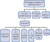
We retrospectively reviewed the pathology database and found 80 consecutive patients who had been diagnosed with breast papillary lesions prior to surgery at two medical centers from May 2004 through May 2009. Those patients who had undergone CNB and had been diagnosed with either intraductal papilloma or malignant lesions following surgical excision were included.
Results
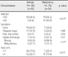
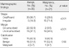
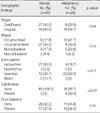
Forty-five cases were diagnosed as intraductal papilloma and 24 cases as malignant lesions. Malignancy was found to be related to being older than 60 years (p<0.01), having bloody nipple discharge (p=0.05), and a tumor size of more than 1 cm (p<0.01). Further, irregular shape (p<0.01) and uncircumscribed margin (p<0.01) on mammogram and irregular shape (p=0.04), calcification (p<0.01), and isoechoic pattern (p<0.01) on ultrasonogram were significantly related to malignancy.
Figures and Tables
References
1. Warren WC. The surgeon and the pathologist. JAMA. 1905. 45:149–165. Cited from Ibarra JA. Papillary lesions of the breast. Breast J 2006;12:237-51.
2. Kraus FT, Neubecker RD. The differential diagnosis of papillary tumors of the breast. Cancer. 1962. 15:444–455.

3. Arora N, Hill C, Hoda SA, Rosenblatt R, Pigalarga R, Tousimis EA. Clinicopathologic features of papillary lesions on core needle biopsy of the breast predictive of malignancy. Am J Surg. 2007. 194:444–449.
4. Rosen PP. Rosen's Breast Pathology. 2001. 2nd ed. Philadelphia: Lippincott Williams & Wilkins;78–85.
5. Ashkenazi I, Ferrer K, Sekosan M, Marcus E, Bork J, Aiti T, et al. Papillary lesions of the breast discovered on percutaneous large core and vacuum-assisted biopsies: reliability of clinical and pathological parameters in identifying benign lesions. Am J Surg. 2007. 194:183–188.
6. Lam WW, Tang AP, Tse G, Chu WC. Radiology-Pathology conference: papillary carcinoma of the breast. Clin Imaging. 2005. 29:396–400.
7. Rosen PP, Hoda SA. Breast Pathology: Diagnosis by Needle Core Biopsy. 2010. 3rd ed. Philadelphia: Lippincott Williams & Wilkins;171–178.
8. Bloodgood JC. Benign lesions of the female breast for which operation is not indicated. JAMA. 1922. 78:859–863.
9. Liberman L, Bracero N, Vuolo MA, Dershaw DD, Morris EA, Abramson AF, et al. Percutaneous large-core biopsy of papillary breast lesions. AJR Am J Roentgenol. 1999. 172:331–337.
10. Puglisi F, Zuiani C, Bazzocchi M, Valent F, Aprile G, Pertoldi B, et al. Role of mammography, ultrasound and large core biopsy in the diagnostic evaluation of papillary breast lesions. Oncology. 2003. 65:311–315.
11. Rosen EL, Bentley RC, Baker JA, Soo MS. Imaging-guided core needle biopsy of papillary lesions of the breast. AJR Am J Roentgenol. 2002. 179:1185–1192.
12. Mercado CL, Hamele-Bena D, Oken SM, Singer CI, Cangiarella J. Papillary lesions of the breast at percutaneous core-needle biopsy. Radiology. 2006. 238:801–808.
13. Ueng SH, Mezzetti T, Tavassoli FA. Papillary neoplasms of the breast: a review. Arch Pathol Lab Med. 2009. 133:893–907.
14. Agoff SN, Lawton TJ. Papillary lesions of the breast with and without atypical ductal hyperplasia: can we accurately predict benign behavior from core needle biopsy? Am J Clin Pathol. 2004. 122:440–443.
15. Sydnor MK, Wilson JD, Hijaz TA, Massey HD, Shaw de Paredes ES. Underestimation of the presence of breast carcinoma in papillary lesions initially diagnosed at core-needle biopsy. Radiology. 2007. 242:58–62.
16. Ganesan S, Karthik G, Joshi M, Damodaran V. Ultrasound spectrum in intraductal papillary neoplasms of breast. Br J Radiol. 2006. 79:843–849.
17. Kim TH, Kang DK, Kim SY, Lee EJ, Jung YS, Yim H. Sonographic differentiation of benign and malignant papillary lesions of the breast. J Ultrasound Med. 2008. 27:75–82.
18. Lee CS, Kook SH, Shin HJ, Moon WK, En EJ, Lee YU, et al. Papillary tumors of the breast: US findings of the benign and malignant lesions. J Korean Radiol Soc. 2000. 42:871–876.
19. Masood S, Loya A, Khalbuss W. Is core needle biopsy superior to fine-needle aspiration biopsy in the diagnosis of papillary breast lesions? Diagn Cytopathol. 2003. 28:329–334.
20. Park HL, Chang SY, Huh JY, Kim JY. Is further diagnostic surgery necessary for the benign papillary lesions that are diagnosed by large volume vacuum assisted breast biopsy? J Breast Cancer. 2010. 13:206–211.
 XML Download