

 PDF
PDF ePub
ePub Citation
 Print
Print


INTRODUCTION
The number of patients newly diagnosed with breast cancer in Korea exceeded 10,000 per annum in 2006 after a threefold increase in the decade from 1996. Westernized lifestyle, growing public concern about healthcare and prevalence of governmentally-led breast cancer screening programs are contributing to this rapid increase in newly diagnosed breast cancer.(1) The Korean Breast Cancer Society reported that the percentage of early stage breast cancer (Stage 0 & I) increased from 23.1% in 1996 to 47.1% in 2006. This increased the frequency of lumpectomy to 48.8% in 2006.(1) Metastatic lymph nodes are found in only 20% to 30% of early stage breast cancer patients. In other words, axillary lymphadenectomy is of no benefit to approximately 70% to 80% of patients, and associated adverse effects such as lymphedema decrease patients' quality of life (QOL).
The sentinel lymph node (SLN) concept was introduced in breast cancer care in the 1990s to overcome concern about lymphadenectomy(2,3) and it has become a routine procedure for the surgical staging of breast cancer patients.(4) Generally, the SLN evaluation is done intraoperatively using frozen section specimens, followed by the postoperative examination of permanent sections for a confirmatory diagnosis. However, it is commonly accepted that intraoperative examination is less sensitive for detection of metastasis than postoperative examination. The sensitivity of intraoperative SLNs examination has been reported to vary widely with a range of 44% to 95%, with most series reporting sensitivities between 60% and 75%.(5) While an intraoperative extensive SLN examination can reduce the chance of overlooking metastasis(6,7) such examination is difficult in practice because of time restrictions and the amount of work involved.
It is recommended that lymph nodes be examined by cutting at 2 mm intervals to find evidence of macrometastasis, which is reported to negatively impact the prognosis of patients.(8) Breast cancer patients with micrometastasis in lymph nodes are diagnosed as metastasis positive. While the clinical significance of micrometastasis is still controversial, patients who have only micrometastasis in lymph nodes (pN1mi) have until recently been classified in the same risk group as those with macrometastasis. A recent SEER database analysis denied the validity of classifying pN1mi patients as stage II, showing that, compared to patients with metastasis negative lymph nodes, 5- and 10-yr survival rates of patients with lymph node metastases less than 2 mm decreased by only 1%. Based on this fact, the 7th edition of the American Joint Committee on Cancer (AJCC) guidelines (published in 2009) have reclassified patients with lymph node micrometastases (pN1mi) as Stage Ib, with a view of indicating the association with better prognosis and promoting further investigation.(8) Additionally, research on the feasibility of predicting the metastatic status of axillary lymph nodes of patients with micrometastasis in SLNs is ongoing, and a method for accurate detection of micrometastasis in SLNs is required.
While, histopathology is currently regarded as the gold standard for the examination of metastasis in lymph nodes, it is inherently limited by its examination of limited portions of lymph nodes and by the subjectivity of its results, with findings often varying with examiner. Substantial time and effort are needed to obtain highly accurate examination results. Recently, a new molecular biological method named one-step nucleic acid amplification (OSNA) was developed to resolve the inherent limitations of histopathological examination.(9) The OSNA assay targets CK19 mRNA and detects it in a sample after amplification by the reverse-transcription loop-mediated isothermal amplification (RT-LAMP) method. Basic study has demonstrated high specificity of the OSNA assay(9) and some clinical studies have demonstrated its clinical significance and utility, showing high concordance with the results of histopathological examination of permanent lymph node slices sectioned at 1mm to 2 mm intervals. OSNA assay has also been deemed intraoperatively applicable on account of its ease-of-use and rapidity.(10-12) Molecular biological methods like the OSNA assay have the advantages of the feasibility of extensive examination (through lymph node homogenization), the objectivity of results (through reduction of human intervention by automation of the analysis process), and the ability to accurately estimate the volume of metastasis.(9)
The objective of this study was to assess the clinical utility and applicability of OSNA assay in breast cancer treatment in Korea by comparing it with histopathological examination. Lymph nodes used in the evaluation were cut into 4 pieces, at 2 mm intervals or less, and alternative blocks were provided to the two methods. Four levels (1 level for intraoperative and 3 levels for postoperative examination) of specimens were prepared from each lymph node block for histopathological examination. Next, we investigated the feasibility for implementation of OSNA assay by comparing its results with those of histopathological examination and also by assessing the turnaround time from lymph node homogenization to receiving measurement results.
METHODS
Patients and source of lymph nodes
Two-hundred-and-eighty-six fresh lymph nodes were obtained from 200 breast cancer patients undergoing SLN dissection from July 2009 to November 2009. One patient was excluded because she was finally diagnosed as not having breast cancer but large B cell lymphoma. The clinicopathologic characteristics of 199 patients are shown in Table 1. Patients were staged according to the TNM classification. Prior to the conduct of the present study, the institutional review board examined the contents of study-related documents including study protocol, informed consent document and sample case report form from ethical and scientific standpoints and approved the implementation of the study. For the detection of sentinel node, we used both radioisotope and blue dye in 159 patients, and radioisotope only in 40 patients. One to six hr prior to surgery, subareolar intradermal injection of Tc99m-antimony sulfate colloid (0.4 mCi) was performed in the quadrant where the tumor was located. After approximately 40-50 min, numbers and locations of SLN were checked with a gamma camera. Subareolar intradermal injection of 0.8% indigiocarmine (0.8 cc) in four parts of the periareola was performed immediately prior to surgery. SLN was defined as any blue-stained nodes or any nodes with radioactive counts of 10% or great. The included patients were suspected as negative for lymph node metastasis from initial clinical assessment, and scheduled for SLN biopsies. The patients receiving neoadjuvant therapy before undergoing SLN biopsy, and those who had already undergone SLN biopsy were excluded from the study. Lymph nodes excised from enrolled patients for the purpose of treatment or diagnosis and identified and dissected as SLNs were included. Lymph nodes which were not identified by radioisotope or dye method, but identified as SLNs by intraoperative findings and dissected were also included in this study.
Study design
Resected lymph nodes were equally sectioned into blocks along their long axis at 2 mm intervals with a special cutting device (Figure 1A).(9) Blocks a and c were subjected to OSNA assay, and blocks b and d to intra- and postoperative histopathological examination. If lymph nodes were less than 4 mm in the short axis, they were cut in half. One half was subjected to OSNA assay, and the other half to histopathological examination. Each lymph node was subjected to OSNA assay and histopathological examination, and presence/absence of metastases for each patient was judged from those results. By comparing the judgment results from both methods on a patient basis, the sensitivity, specificity, positive predictive value (PPV), negative predictive value (NPV) and concordance rate of OSNA assay compared to histopathological examination were determined. Also the rapidity of OSNA assay was investigated by measuring the turnaround time, i.e., the time between starting homogenization and obtaining the results of OSNA assay. In discordant cases, clinical information, status of non-SLNs, and expression of CK19 protein in lymph node metastasis foci were evaluated on a patient basis.
OSNA
Each lymph node was homogenized in glycine buffer, Lynorhag, pH 3.5 (Sysmex, Kobe, Japan). The solutions (10-time diluted and 100-time diluted solution) and the gene amplification reagent Lynoamp BC (Sysmex, Kobe, Japan) were set in dedicated device (RD-100i; Sysmex) and the following steps were automatically done. The solutions were mixed with six different CK19 primers, four deoxynucleoside triphosphates, reverse transcriptase, DNA synthetase and magnesium sulfate. The resulting solution reacted at a constant temperature of 65℃.(13) cDNA was synthesized from CK19 mRNA in the lymph node homogenized solution using reverse transcriptase. The gene amplification was preceded by DNA synthetase based on this synthesized cDNA. The degree of DNA amplified product was detected by measuring the time until the turbidity caused by magnesium pyrophosphate (white precipitate), a by-product of the reaction, exceeded a given threshold (rise time). There is known to be a linear relationship between CK19 mRNA concentrations and the rise time. Therefore, it is possible to calculate CK19 mRNA concentrations in the analysis samples (10-time diluted solution and 100-time diluted solution) by using a calibration curve made by measuring calibrators (three levels) with known CK19 mRNA concentrations. Whether each lymph node was metastasis positive or negative was judged from based on the calculated CK19 mRNA concentration and the threshold value which was set on the basis of previous data obtained from a series of lymph nodes dissected from breast cancer patients.(9) Negative was the case when both CK19 mRNA concentrations of 10-time diluted solution and that of 100-time diluted solution were <250 copies/µL. OSNA assay can classify the positive result into 3 categories: (++), (+), and (+I; positive with reaction inhibited). Positive (++) was the case when CK19 mRNA concentration in the 10-time diluted solution was ≥5,000 copies/µL. Positive (+) was the case when CK19 mRNA concentration in the 10-time diluted solution <5,000 and ≥250 copies/µL. Positive (+I) was the case when CK19 mRNA concentration in the 10-time diluted solution was <250 copies/µL and CK19 mRNA concentration in the 100-time diluted solution was ≥250 copies/µL. Patients were regarded as positive if more than 1 SLN was judged as positive by OSNA assay. Positive patients were further classified as follows: patients were regarded as (++) if more than 1 SLN was judged as (++) by OSNA assay; patients were regarded as (+) if more than 1 SLN was judged as (+) and no SLN was judged as (+I) or (++) by OSNA assay; patients were regarded as (+I) if more than 1 SLN was judged as (+I), and no SLN was judged as (++) by OSNA assay. Patients were regarded as negative if all SLNs were judged as negative by OSNA assay.
Histopathological examination
The short axis of each sentinel node was measured and each SLN cut along its longitudinal axis into sections of 1.5-2.0 mm thickness. For the intraoperative histopathological examination, the frozen sections for H&E staining were prepared from one level of two cutting surfaces of blocks b. (no.1 [red] and 3 [red] in Figure 1B), and the pathologists performed careful gross examination of the sections to detect focal lesions. After intraoperative examination, the lymph node sections were fixed in formalin for paraffin section histology. For the postoperative histopathological examination, three level sections [green] from blocks b and d were prepared at 200 µm intervals (Figure 1A). And three sections were obtained at each level for H&E staining, anti-cytokeratin antibody (AE1/ AE3) immunohistochemical (IHC) staining and unstaining (Figure 1B). Presence/absence of metastases was judged by observing H&E staining and AE1/3 staining slides.
In accordance with the TNM classification of AJCC 7th edition,(14) metastatic deposits were recorded as isolated tumor cells (ITC) if their largest diameter was smaller than 0.2 mm, as micrometastases if they were larger than 0.2 mm but not larger than 2 mm, and as macrometastases if they were larger than 2 mm. In concordance with the TNM designation of ITC as pN0 (i+), lymph node samples were only regarded as positive if at least one micrometastasis or macrometastasis was found. Consequently, lymph nodes with ITC were considered as negative in this study. Macrometastasis or micrometastasis was confirmed by both or either of intraoperative histopathological examination of frozen section specimens and postoperative histopathological examination with permanent tissue specimens. Patients were regarded as positive if more than one SLN was judged as positive by histopathological examination. Positive patients were further classified as follows: patients were regarded as having macrometastasis if more than one SLN contained macrometastasis; patients were regarded as having micrometastasis if more than one SLN contained micrometastasis and no SLN contained macrometastasis. Patients were regarded as negative if all the SLNs were judged as 'negative' by histopathological examination. Negative patients were further classified as follows: patients were regarded as 'patients with ITC' if no SLN was judged as having macro- or micrometastasis and more than 1 SLN was judged as ITC; patients were regarded as 'negative patient' when no SLN was judged as macro- or micrometastasis or ITC.
RESULTS
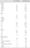
The age distribution of the 199 enrolled patients roughly matched the distribution in breast cancer patients nationwide (Table 1). Most patients were classified as lower than stage II. The histological type was classified by WHO classification and approximately 80% were invasive ductal carcinoma, and 5% were ductal carcinoma in situ patients.
OSNA performance
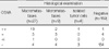
OSNA performance was evaluated by considering the results of both intraoperative and postoperative histology as the gold standard (Table 2). The sensitivity, specificity, PPV, and NPV of OSNA assay compared to histopathological examination were, respectively: 77.8% (28/34; 95% confidence interval [CI], 0.61-0.90), 96.3% (157/163; 95% CI, 0.92-0.99), 82.4% (28/34; 95% CI, 0.65-0.93), and 95.2% (157/165; 95% CI, 0.91-0.98). The concordance rate of OSNA assay and histopathological examination in terms of detecting breast cancer metastasis to lymph nodes was 93.0% (185/199; 95% CI, 0.88-0.96). The kappa statistic analysis indicated substantial agreement of both methods with a value of 0.76 (95% CI, 0.64-0.88). In 19 of 23 patients with (++) on OSNA assay, histologically detectable macrometastases was confirmed. That is, macrometastasis predictive value of (++) on OSNA assay was 82.6% (19/23; 95% CI, 0.61-0.95). The mean turnaround time of 199 patients was 39.0 min, breaking down to 35.2 min for patients with 1 lymph node, 44.8 min with 2 lymph nodes, 50.4 min with 3 lymph nodes and 50.0 min with 4 lymph nodes. There was no statistical difference in turnaround time for analyzing the same number of lymph nodes among the three sites (39.5, 40.2, and 35.9 min, respectively).
Analysis of causes of discordance
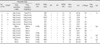
To investigate, on a patient basis, discordant results between OSNA assay and histological examination, clinical information, status of non-SLNs and expression of CK19 protein in metastasis foci of lymph nodes were evaluated (Table 3). For case 1, OSNA assay provided (++) judgment with 14,000 copies/µL, indicating high probability of macrometastasis. Considering the histopathological result of ITC detection as well as the existence of metastasis in other SLNs (which were not used for this study), the cause of the discordance was attributed to the localization of metastasis foci in the lymph node. For cases 2, 3, and 5, OSNA assay provided (+) judgment with 950, 460, and 250 copies/µL, respectively, indicating the existence of weak positivity in the sample. For case 4, even though OSNA assay provided (++) judgment with 14,000 copies/µL indicating high probability of macrometastasis, we were unable to find any other findings in this evaluation to support this judgment. We supposed the cause of this discordance to be localization of metastasis foci in the lymph node as the probability of OSNA false positives is considered to be very low based on the results of previous studies. For case 6, OSNA assay provided positive results for each of 2 lymph nodes from this patient. Considering the existence of metastasis in other SLNs (which were not used for this study), we supposed the cause of this discordance to be localization of metastasis foci in a lymph node. OSNA negative and histopathology positive cases were investigated immunohistochemically using CK19 antibody to confirm the protein level of CK19 in tumors. Cases 7, 10, 11, and 13 showed expression of CK19 protein in less than 10% of the tumor. The cause of these discordances may have been the inability of OSNA assay to detect metastasis of breast cancers with low CK19 expression. For cases 8 and 9, metastases were histopathologically found by the postoperative examination of permanent specimens, while intraoperative histopathological examinations using the frozen specimens of the cutting surface adjacent to the lymph node block used for OSNA assay were negative. CK19 IHC of case 8 and 9 has not been done due to insufficient tissue. So, we cannot say the reason of discordant for case 8 and 9 was due to CK19 low expression. For case 12, one SLN used for this study was judged as 'positive' by postoperative permanent-section even though it was judged 'negative' by both intraoperative frozen-section and OSNA. And other 2 SLNs which were not used for this study were also judged as 'positive.' In addition, 2 non-SLNs were judged as 'positive.' As a result, this patient had total 5 positive nodes (3 SLNs and 2 non-SLNs), and one of them were infraclavicular lymph node, so this patient was categorized into pN3 according to the AJCC criteria.
Consequently the causes of these discordant cases (case 8, 9, and 12) attributed to the localization of metastasis foci in lymph nodes. For case 14, the OSNA result was negative with a flag indicating weak expression of CK19 mRNA (under cutoff level), and the histopathological examination result was micrometastasis. The cause of this discordance was thus supposed to be localization of metastasis foci in the lymph node.
DISCUSSION
In Korea, the increase of early stage breast cancer patients has directly influenced the incidence-rise and importance of intraoperative SLN evaluation. The sensitivity of intraoperative histological examination is relatively low with a 19-42% false-negative rate.(15-18) Rapid immunohistochemistry and extensive serial sectioning increase the sensitivity compared to frozen section alone, (19,20) but overburdened pathology resources make this an impractical option. Several studies have shown the clinical feasibility of molecular detection of lymph node metastasis using reverse transcription-PCR with a variety of combinations of several markers.(21-23) Of particular note, a system using CK19 and mammaglobin is now being used clinically with high reliability.(24) The OSNA assay is an automated system for rapid and quantitative detection of CK19 mRNA with the RT-LAMP method, and has been shown to have high specificity and accuracy in Japan, the Netherlands, and Germany.(10-12)
The results of this study indicate that the OSNA assay has equivalent accuracy to histopathology in detecting breast cancer metastasis to lymph nodes. Specificity of OSNA assay against the histopathological examination result was 96.3%, indicating a low probability of OSNA assay being falsely positive. Meanwhile, the sensitivity of OSNA assay against histopathological examination was 77.8% and this unsatisfactory result is assumed to be due to the protocol of this study. In this study, lymph nodes were divided equally between OSNA assay and histopathology in order to evaluate the performance of the OSNA assay. It is commonly known that this evaluation design causes discordance in the results of the two methods due to the localization of metastasis foci in lymph nodes. However, the OSNA assay, which in theory can analyze a major portion of a lymph node by homogenizing it, is anticipated to have higher sensitivity for metastasis compared to histopathology which examines a limited portion of a lymph node only. On the comparison between OSNA and frozen histology, we were unable to demonstrate OSNA superiority (data not shown). This could be due to the study protocol which required frozen histology to be performed on almost the same portion of the same lymph node block used for permanent histopathology even though different lymph node blocks were used for OSNA. Turnaround time of OSNA assay with one to four lymph nodes was 35.2 min to 50.4 min in the three sites used in this evaluation. OSNA assay had less than 40 min turnaround time for approximately 70% of subjects enrolled in this evaluation, indicating the feasibility of it being applied to intraoperative settings. Longer turnaround time was associated with higher numbers of lymph nodes and shorter turnaround time was associated with accumulation of operator experience. Further, it was demonstrated that ease-of-use of the OSNA assay enables operators with little training to provide an equivalent level of metastasis detection to operators with more experience.
It is anticipated that OSNA assay will contribute to improved treatment outcomes and QOL of patients by providing higher accuracy in metastasis detection during surgery than that of the combination of intra- and postoperative histopathology. In addition, the automated nature of the OSNA assay will help standardize the diagnosis of breast cancer metastasis in lymph nodes. The result of the present evaluation demonstrated that (++) judgments of OSNA assay suggest existence of macrometastasis with high reliability. Conventional histopathology judges the size of metastasis foci from an examination of a limited portion of lymph nodes on a slide specimen, and there is a possibility of underestimation of macrometastasis as micrometastasis. The OSNA assay, on the other hand, can in theory examine a larger proportion of lymph nodes, and has the potential to be the first method for assessing the volume of metastasis in lymph nodes. In this study, 14 cases of discordance were detected and on analysis most discordance was attributed to the localization of metastasis foci in a lymph node. Since alternate blocks of the lymph node were used for OSNA and histology in this evaluation, it is self-evident that in some instances tumor deposits might have been confined to the slices analyzed by OSNA or to the blocks used for histology. The discordance of three of eight OSNA negative/histology positive discordant cases might be due to inability of OSNA assay to detect metastasis of breast cancer with low CK19 expression. Furthermore, it is known that some breast cancers do not express CK19.(9,25) An inherent limitation of the OSNA assay is therefore that CK19 mRNA is the only target marker for metastasis detection. Some clinical institutions have started their own attempts to overcome this issue by simultaneous intraoperative imprint cytology or preoperative confirmation of CK19 expression by core-biopsy specimen, and the establishment of an alternative assay is under discussion.
CONCLUSION
In summary, the OSNA assay proved to be a reliable and easy-to use tool for the intraoperative detection of SLN metastases in breast cancer patients at multiple institutes. Since OSNA was shown to have equivalent accuracy to in-depth, heavily used histological analyses routinely performed in Korea, its clinical usage could lead to improved diagnosis and easing of resource constraints. We anticipate that the OSNA assay will be of great clinical significance to breast cancer treatment in Korea.

 XML Download