

 PDF
PDF ePub
ePub Citation
 Print
Print


INTRODUCTION
Intraductal papilloma of the breast (IDP) is the most common cause of bloody nipple discharge, and IDP accounts for about 10% of all benign breast tumors.(1-3) It usually developes in a central or peripheral site of the lactiferous duct, and the tumors can be solitary or multiple.(1,2) IDP is known to be associated with breast cancer, though the previous reports have varied according to their findings.(4-10) Based on Korean studies, the rate of co-existence of intraductal papillomas and cancer stands at 22.7%, while according to one study, the rate was 28% for women over 40 yr old (the Seoul National University Hospital, 105 cases, 1962).(4) Yet further reports on the co-existence of intraductal papillomas and cancer have been rare. The lack of sufficient information on IDP as well as the potentially significant clinical implications led us to examine the clinicopathologic results of papillary lesions and the pathologic impression of the surrounding lesions that have been identified in the operated specimen along with the papillary lesions.
METHODS
One hundred sixty-one IDP cases were observed from February 2003 to November 2008. A retrospective chart review was performed on the patients who underwent operation for IDP at the Breast Clinic, Konyang University Hospital. We collected the information on the presenting symptoms, the image findings (mammographic, ductographic and sonographic) and the pathology.
Mammography and ultrasonography were routinely perfomed. Ductography was performed on all 83 patients with abnormal nipple discharge. We performed microdochectomy on intraductal papillomas of the breast in all patients confirmed by needle biopsy. A focused excision of affected ducts can also be accomplished following radiographic visualization of the depth of the lesion or duct can be cannulated with a fine probe. The suspicious duct was dissected along its entire length and removed with a small cone of the breast tissue surrounding the duct. Whole specimen as well as mass and around the periphery of the involved spaces was evaluated.
Chi-square test was used to examine any differences by SPSS version 13.0 for Windows (SPSS Inc., Chicago, USA). A p-value of <0.05 defined a statistically significant result.
RESULTS
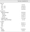
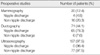
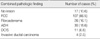
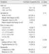
All 161 patients were female and their ages raged from 14 to 81 (mean±SD, 43.2±10.1). No difference was found in age between the group with solitary IDP and the group with papillomatosis. Sixty-six cases (40.7%) showed the presence of lesions in their right breast, 83 (51.2%) showed the presence of lesions in their left breast and 12 (7.4%) showed the presence of lesions in both of their breasts. The presenting symptom was nipple discharge and this was identified in 83 patients (51.6%), and the majority were further categorized into (a) a group with bloody nipple discharge (37 cases, 44.6%) and (b) a group having nipple discharge with more diversity, e.g., yellowish or serous (46 cases, 55.4%) (Table 1). Mammography and ultrasonography were routinely perfomed. And ductography was performed on all 83 patients with abnormal nipple discharge. The number of cases of diagnostically significant findings for each of the performed tests was 20 cases on mammography, 71 on ductography and 157 on ultrasonography (Table 2). Mammographic abnormalities were seen in 20 and ultrasonographic masses in 157 of the 161 patients. When ductography were obtained, abnormalities were seen in 71 of the 83 patients. The abnormalities included filling defects, dilated ductes and ductal cutoff. The mean size of the lesions that were confirmed via ultrasonography was 0.94 cm (SD, ±0.5). The biopsy showed that 148 cases (91.9%) had solitary IDP and 13 cases (8.1%) had papillomatosis. One hundred and forty-four cases (89.4%) had pathologic lesions found in the surrounding tissue of the operated specimens (Figure 1), with 107 (66.5%) fibrocystic changes (FCC), 26 (16.1%) fibroadenomas, 30 (18.6%) atypical ductal hyperplasias (ADH), 11 (6.8%) carcinomas in situs and 4 (2.5%) invasive ductal carcinomas (Table 3). Among the 161 patients who underwent surgery for IDP, 41 (25.5%) patients had precancerous lesions (ADH & carcinoma in situ) and 4 (2.5%) patients had invasive ductal carcinomas. No differences were found in the co-presence of lesions that varied according to the symptoms, solitary IDP or papillomatosis and size of masses (p=0.140, 0.539, and 0.482). However, a difference was found in the rate of the presence of precancerous lesions according to age: 11.8% (6/51) for those under 40 yr of age and 31.8% (35/110) for those 40 yr old or older (p=0.001). Invasive ductal carcinoma was found only in the patients who were 40 yr old or older. Bilateral IDP (7/12) had more precancerous lesions and carcinomas (p=0.037) (Table 4).
We recommended follow-up ultrasonography after 6 month to all patients, and then annual follow-up was recommended to the patients without any lesions. During six to forty-four months of follow-up, 7 patients (4.3%) developed recurrent IDP (median, 36 months). At 6 to 38 months of the follow up, 3 patients developed ADH and 5 patients developed carcinoma in situ (4 ductal carcinomas in situ [DCIS] and 1 lobular carcinoma in situ [LCIS]).
DISCUSSION
IDP accounts for about 10% of all benign breast tumors, and IDP is most commonly observed among pre- and post-menopausal women between the ages of 35 to 55.(1-3) In 2 previous studies, the most common symptom was nipple discharge in 64% to 88% of the IDP patients. Breast masses were also observed, though rarely.(1,2) In this present study, the patients with nipple discharge (83 cases or 51.6%) were the major group, and among them there were 37 cases (44.6%) with bloody discharge and 46 cases (55.4%) showed a greater variety, e.g., clear or yellowish, and their feature was almost always mucoid. Twenty-four cases (14.9%) also had breast masses, while 2 cases showed both nipple discharge and breast masses.
Mammography usually reveals breast masses around the nipples and in rare cases, calcifications as well. Breast ultrasonography typically shows well-defined, smoothwalled, solid and hypoechoic masses within the dilated ducts.(1,2,6,7) Along with nipple discharge, ductography can be done to confirm the intraluminal smooth or irregular filling defects that are associated with obstructed or dilated ducts. Ductography may be useful in identifying and localizing the lesion prior to operation.(6) Though MRI has turned out to be a highly sensitive diagnostic tool for diagnosing IDP among younger women with dense breasts, the technique is not a perfect tool in differentiating IDP from breast cancer.(9) Ductoscopy is also an excellent tool for visualizing within the dilated duct and it is a useful way to diagnose IDP.(7,9) Fine-needle aspiration cytology and needle biopsy are commonly used to examine lesions in breasts. The needle biopsy, with its higher sensitivity and specificity, is becoming a more accepted form of examination and it is being reported to be a better tool for differentiating papillary lesions.(3) Mammography and ultrasonography were routinely performed. And ductography was performed on all 83 patients with abnormal nipple discharge. Significant diagnostic results were obtained from each of the tests: (a) 20 cases with mammography; (b) 71 cases with ductography and (c) 157 cases with breast ultrasonography. The size of the lesions identified by ultrasound examination was 0.94 cm on average. For the cases with preoperative nipple discharge, ductography (78.3%) and ultrasonography (96.4%) were found to be effective diagnostic tools. For those cases without nipple discharge, ultrasonography-guided needle biopsy (97.5%) was the most helpful tool for making the diagnosis.
IDP can be found in the central or peripheral site of the mammary duct, and it can be solitary or multiple. The reported frequency of solitary IDP has varied from 33% to 75%, whereas that of papillomatosis is from 31% to 67%.(7) We observed solitary papilloma in 148 cases (91.9%), while the papillomatosis was found in 13 cases (8.1%).
Appropriate management of IDP is controversy. Many authors have reported that surgical excision may not always be necessary for papillary lesion of the breast that is diagnosed on core-needle biopsy and surgical excision is considered in patients with papillomatosis or papillary lesions with atypism.(3) But it is well recognized that distinguishing benign from malignant papillary lesions on needle biopsy may have difficult diagnostic problem and IDP have a increasing the risk of malignancy. Page et al.(10) reported that individuals with IDP had twice the risk of developing breast cancer as compared to the non-IDP individuals. Other lesions in the surrounding tissue such as ADH and DCIS/LCIS, accompany papillary lesions and their presence has been reported to increase the risk of breast cancer 4 to 7.5 times.(10-12) These conditions have also been reported to be associated with tumor recurrence.(10) We recommended surgical excision of intraductal papillomas of the breast in all patients confirmed by needle biopsy.
Whang(4) found out that the rate of the co-presence of IDP and cancer was 22.7% and it was up to 28% among women over 40 yr old. We discovered 144 cases (89.4%) of pathologic lesions in the surrounding tissue from the operated specimen. Among these cases, 107 cases (66.5%) were FCC, 26 (16.1%) were fibroadenoma, 30 (18.6%) were ADH, 11 (6.8%) were carcinoma in situ and 4 (2.5%) were invasive ductal carcinoma. Among the 161 patient who underwent operation for IDP, 25.5% of these patient had precancerous lesion (ADH and carcinoma in situ) and invasive ductal carcinoma was found in 2.5% of them. Of the 25.5% who had precancerous lesions, only 6 patients (6/51, 11.8%) were under 40 yr old, and 35 patients (35/110, 31.8%) were 40 yr old or older. Invasive ductal carcinoma was found only in the patients who were 40 yr old or older. Papillomatosis is commonly associated with a higher rate of cancer,(7,9,13) though some reports have found no significant difference in the rates between the two groups.(14) In our data, we observed precancerous lesions in 30.8% (4/13) of the women with papillomatosis, and this percentage was higher than that for women with the solitary papilloma (28.4%). But the difference was not significant (p=0.539). No differences were found in the co-presence of lesions that varied according to the symptoms and size of masses (p=0.140 and 0.482).
We recommended follow-up ultrasonography after 6 month to all the patients. Annual follow-up was then recommended to the patients who were without any lesions. At six to forty-four months of the follow-up, 7 patients (4.3%) developed recurrent IDP (median, 36 months). At 6 to 38 months of the follow up, 3 patients developed ADH and 5 patients developed carcinoma in situ (4 DCIS and 1 LCIS).
These findings suggest that a more thorough examination of the surrounding tissue is necessary for those patients with IDP. A careful and more detailed follow-up survey is also recommended for the women who are aged 40 or older or for those women with intraductal papillomatosis.
CONCLUSION
IDP is often found in patients without nipple discharge; in these cases, ultrasonography-guided needle biopsy plays a critical role in among the diagnosis. IDP should be closely followed up due to its malignant potential and the surrounding breast tissue with IDP should be carefully evaluated due to the high rate of developing other precancerous lesions.
 XML Download