

 PDF
PDF ePub
ePub Citation
 Print
Print


INTRODUCTION
Gynecomastia is a benign clinical condition which is unilateral or bilateral enlargement of the male breast due to hormonal imbalance at the breast tissue.(1) The incidence of this common condition varies 30% to 50% of normal male,(2) but sometimes it causes significant psychological and physical stress in young male and it brings phobia of malignancy in elderly male. The causes are so complex and various including idiopathic, obesity, consumption of estrogens, anabolic steroids, cancer, drugs, and supplements.(3) The treatment depends on the causes and may vary from observation, withdrawal of drugs, to correction of underlying conditions with surgical excision. The surgical removal is the best option for longstanding gynecomastia or glandular hypertrophied male breast that is failed to non-surgical therapy.(4) The surgical procedures for gynecomastia include conventional surgical excision (subcutaneous mastectomy), liposuction through a periareolar incision, and vacuum-assisted biopsy device excision as a new trial. As one of appropriate cosmetic surgical approaches for gynecomastia, I chose vacuum-assisted biopsy device excision technique for simultaneous removal of glandular and fatty tissues through a single 2-3 mm sized incision. I evaluated the usefulness and safety of the vacuum-assisted biopsy device excision for treating gynecomastia.
METHODS
Twenty-two cases of gynecomastia in 18 male who did not have severe underlying causes had been treated by vacuum-assisted biopsy device excision from November 2005 to June 2007 at Chungmu General Hospital, Cheonan, Korea. Their ages was between 14 and 72 (mean age, 34 yr). Patients under psychological and physical distress with gynecomastia were chose for treating. During this period, vacuum-assisted biopsy device excision for breast mass was performed in about 700 times at the hospital. To more readily categorize the degree of deformity and possibility of vacuum-assisted biopsy device excision in each patient, Simon's classification was used. I tried vacuum-assisted biopsy device excision in patient with Simon's grade 1 and 2A.(5) The choice of probe scale (11G or 8G) was depended on size of breast and experience of operator.(6) At the initial trial, both 11G and 8G probe were used, but later period 8G probe was used only. The procedure was performed by a fully accredited surgeon under local anesthesia. The postoperative follow-up period was performed after 3 months. Surgical procedures were described below: First, in supine position with raising both hands on head, the whole resection range was marked with ultrasound guide. Second, one small incision (2-3 mm) at the lateral side of nipple after local anesthesia on skin was made for removing patient's hypertrophied glandular and fatty tissue. Then, the marked resection range was removed with guidance of ultrasound using vacuum-assisted biopsy device. After completion of vacuum-assisted biopsy device excision, the incision site was sutured or taped by surgical strip. The patients were instructed to wear a vest compression garment for 2 days. Oral antibiotics and pain medications were given for 3 days after procedure. Patient satisfaction score was checked after procedure in all patient, the definition of satisfaction score was not satisfied (less 5), satisfied (5-8), very satisfied (more than 8).(7)
RESULTS
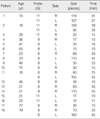
The average lesion size checked by preoperative ultrasound was 3.5 × 2.0 cm in vacuum-assisted biopsy device, while 7.0 × 5.6 cm in subcutaneous mastectomy. The duration of vacuum-assisted biopsy device procedure was from 10 min to 55 min (average, 25). The numbers of removed tissue pieces were 12 to 160 (average, 69) (Table 1). There were no significant early and late (3 months follow-up) complications such as open operation for bleeding control, re-operation for remaining of glandular tissue of breast, and sensation change of nipple. All patients were satisfied (scores of 8) or very satisfied (scores of 9 or 10). In comparing the subcutaneous mastectomy results underwent during same period, the vacuum-assisted biopsy device procedure showed acceptable result as one of surgical methods for gynecomastia.
DISCUSSION
Gynecomastia is the development of enlarged male breast. These may be quiet small (only a few cubic centimeters in volume) but located right at the nipple-areolar complex, causing a pointed protrusion, although they can be massive subcutaneous fat and glandular tissues.(8) Regardless of size, gynecomastia with accompanying pain may sometimes causes significant physical and psychological problem. In young men, physical feminization is needed to correct cosmetically. Correction of this condition is changing a physical appearance normally found in women from men. In old men, pain and phobia for cancer need to confirm tissue diagnosis.(9) It is not only fixing a physical problem but also making a profound impact on the patient's mind.(1) Traditionally, surgery such as subcutaneous mastectomy has been the treatment of choice for gynecomastia but cosmetically unsatisfactory results are reported to occur in as much as 50% of all patients,(10) so recently there has been an increase in use of minimally invasive techniques such as vacuum-assisted biopsy device, laser therapy, radio-frequency ablation, cryotherapy, focused ultrasound waves and liposuction.(11,12)
Vacuum-assisted biopsy device excision is one of convenient and effective method to small gynecomastia patients for whom might be stressed with post operative anterior chest wall scar after conventional subcutaneous mastectomy (Figure 1). The vacuum-assisted biopsy device excision method is more easy and convenient than liposuction and surgery because of no need for general anesthesia and admission. It is performed under ultrasound guide with local anesthesia, and it is possible to check the remained tissue during procedure.(13) The extend of procedure depends on both the amount of breast tissue to be removed and the degree of skin redundancy. With recent advancement of vacuum-assisted biopsy device technology, newly developed probe (EX type) can make possible to remove larger gynecomastia with shortening of duration time.

 XML Download