

 PDF
PDF ePub
ePub Citation
 Print
Print


INTRODUCTION
Axillary lymph node metastasis (ALNM) is the single most important prognostic factor for patients with breast cancer.(1) The incidence of ALNM in breast cancer usually increases according to the tumor size, but the phenomena is inconsistent due to the fact that ALNM in breast cancer patients is the result of a complex of factors composed of genetic, epigenetic, ethnic, environmental and biological characteristics.
Determining the factors affecting ALNM in patients with early breast cancer is essential for personalized therapy with a precise risk assessment of each patient. González-Vela et al.(2) reported that the predictors of ALNM are a tumor size over 2 cm, an infiltrative margin and a high Ki-67 index. Some investigators have suggested that ALNM is related with a young age at presentation. One study in Southeast Asia has reported that the tumor size, the invasive ductal and lobular cell type, lymphovascular invasion (LVI) and the progesterone receptor status are independent predictors of ALNM in Asian patients with breast cancer.(3) LVI has also been proved to be a strong predictive factor for ALNM in many studies, but it has still not been fully investigated.(4-6)
After sentinel lymph node biopsy was introduced in breast cancer surgery, it has been replacing the standard level I/II axillary lymph node dissection (ALND) for the staging work-up and regional therapy in the clinically negative axilla. The American Society of Clinical Oncology (ASCO) guidelines recommend ALND as the primary treatment for sentinel node micrometastases (SNMM) in breast cancer patients.(7) However, there is great variation in the management of SNMM in breast cancer patients and we need more accurate clues to skip ALND in the presence of sentinel node metastasis of clinically node negative breast cancers.(8-10)
Despite many productive studies and confirmative results, we still do not have valid information about the predictive factors for ALNM in patients with breast cancer sized smaller than 2 cm. We designed the current study to analyze the predictive factors of ALNM in patients with T1 breast cancer.
METHODS
We reviewed the medical records and pathologic reports of all the breast cancer patients who had undergone a surgical procedure between 2001 and 2006. We retrieved 206 cases of T1 (tumor size less than 2 cm) breast cancer that were diagnosed as invasive ductal carcinoma or mixed invasive ductal and lobular carcinoma, and we divided them into two groups depending on the absence or presence of ALNM. Initially, 48 cases of invasive lobular, tubular, papillary, medullary, mucinous, apocrine, cribriform, mucinous signet ring cell like and mixed type of lobular and tubular carcinoma were excluded. The node negative group was composed of T1N0 breast cancers and the node positive group was composed of T1N1, T1N2, or T1N3 breast cancers.
Clinicopathologic parameters
We analyzed the prognostic factors such as age at diagnosis, the operative methods, tumor size (T1a, T1b, and T1c), multiplicity, the histologic grade (HG), the nuclear grade (NG), the presence of lymphovascular invasion (LVI), the estrogen receptor (ER) and progesterone receptor (PgR) status, the HER2/neu expression, the Ki-67 labelling index and the bcl-2 expression in each group.
The operative methods were breast conserving surgery (BCS) or mastectomy with sentinel node biopsy in all cases. Standard ALND was performed when the sentinel LN biopsy was positive in the frozen biopsy. We measured the primary tumors to the nearest 0.1 cm increment and subdivided them into T1a, T1b, and T1c according to the definition of tumor size. T1a was defined as tumor larger than 0.1 cm but not larger than 0.5 cm, T1b was defined as larger than 0.5 cm but not larger than 1 cm, and T1c was defined as larger than 1 cm but not larger than 2 cm.
We divided the patients by the age of 40 yr and we analyzed the trend of age distribution in each group. The pathologic prognostic determinants such as tumor size, multiplicity, HG, NG, ER, PgR, HER2/neu, Ki-67 and bcl-2 were evaluated by two pathologists. The HG and NG were determined according to the modified Bloom and Richardson-Scarff histologic grading system and Black's nuclear grading system. We evaluated the ER, PgR, Ki-67 and bcl-2 expressions by immunohistochemistry and the HER2/neu amplification was assessed by fluorescence in situ hybrization (FISH).
Immunohistochemistry (IHC) for ER, PgR, bcl-2 and Ki-67
Liquid mouse monoclonal antibody ER NCL-1-ER-6F11 (Leica Microsystems Inc., Newcastle Upon Tyne, UK) and PR NCL-L-PGR-312 (Leica Microsystems Inc.) diluted 1:80 with normal goat serum (NGS diluted 1 in 5 TBS) was used as the primary antibody for the ER and PgR assay. The secondary antibody was goat anti-mouse peroxidase conjugated immunoglobulines and we used 3, 3'-diaminobenzidine tetrahydrochloride (DAB) as the chromogen. ER and PR were scored as 0, 1+, 2+ and 3+ according to the intensity with the description of the percentage according to the proportion of stained nuclei in ten high power fields. The ER and PR positivities were defined as any positive scores or percentage over zero.
Immunohistochemical staining for bcl-2 and Ki-67 was performed by the avidin-biotin peroxidase complex method with aminoethylcarbazole as a chromogen and using the Vectastain ABC Elite kit (Vector Laboratories, Burlingame, USA). The sections were counterstained with Mayer's hematoxylin. For bcl-2 measurement, the sections were incubated in monoclonal mouse anti-human bcl-2 oncoprotein (1:100; Dako, Glostrup, Denmark), and the brown nuclear immunostaining was examined. For the Ki-67 measurement, the sections were incubated with monoclonal mouse anti-human Ki-67 antigen (1:100; Dako), and the brown nuclear immunostaining was examined.(11) Ki-67 of >10% and bcl-2 of >33% were as considered positive expressions
Fluorescence in situ hybridization (FISH) for HER2/neu
Two-color FISH was done on 3.5 µm consecutive sections of the TMA paraffin blocks using 20 µL of LSI HER2/CEP17 probes (Vysis Inc., Downers Grove, USA). At least a 2-fold increase of the HER2 signals over the CEP 17 signals with using the LSI HER2 probe in the tumor cell was considered the criterion for gene amplification.
Statistical methods
We used Pearson's chi-square test and Fisher's exact test to determine the statistical significance of the clinicopathologic characteristics affecting the absence or presence of axillary lymph node metastasis within the T1 breast cancer. Multivariate analysis was performed with logic regression analysis to identify the independent predictors of ALNM.
RESULTS
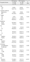
Thirty-eight patients (18.4%) were younger than 40 yr while 168 patients (81.6%) were older than 40 yr of their age. BCS was performed for 190 patients (92.2%) and mastectomy was performed for 16 patients (7.8%). Thirty seven patients (18%) had HG 1 tumors, while 169 patients (82%) had HG 2 or 3 tumors. ER positive tumors were 151 (73.3%) and HER2/neu was amplified in 71 (34.5%). Ki-67 labeling index was increased in 132 (64.1%), bcl-2 expression was increased in 103 (50.0%), and LVI was confirmed in 83 patients (39.7%).
One hundred thirty nine cases were node negative (T1N0) and 67 cases were node positive (T1N1-3). In younger group (age <40 yr), nodal metastasis was more frequently observed (p=0.011) and the proportion of HG 2 or 3 increased in node positive group (p=0.019). The presence of LVI (p<0.001) and amplification of HER2/neu (p=0.005) were more frequently observed in node positive than negative group. Ki-67 labeling index (p=0.012) significantly increased but bcl-2 expression (p=0.026) decreased in the node positive group (Table 1). There were no statistically significant correlations between ALNM and the tumor size (T1a, T1b, and T1c) (p=0.462), multiplicity (p=0.473), NG (p=0.157), ER (p=0.383), PgR (p=0.145) and ALNM.
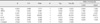
On the univariate analysis, age at diagnosis, HG, LVI, HER2/neu expression, the Ki-67 labeling index and the bcl-2 expression were statistically significant prognostic factors that were related to node metastasis in the T1 breast cancer patients. On the multivariate analysis, LVI (p<0.001) and HER2/neu expression (p=0.009) were significantly associated with node metastasis in T1 breast cancer patients (Table 2).
DISCUSSION
The ALNM is still the single most important prognostic factor of breast cancer albeit numerous biologic and genetic prognostic factors have been investigated. The incidence of ALNM is known to be increased as tumor size increases since the tumor size is the chronological indicator for the presence of the tumor. A recent study reported that SLNM is correlated with tumor size in T1 breast cancer.(12) When we subdivided the T1 tumor into T1a, T1b and T1c there was no statistically significant correlation between tumor size and the node positivity within T1 tumor (T1N0 vs. T1N1-3). Other investigators reported that tumor size is related with ALNM in T1 breast cancer.(13) Association between ALNM and tumor size in T1 breast cancer cannot be determined at this stage, since most studies were performed on relatively small sample size.
The finding that ALNM is not uncommon even in small breast cancers indicates that ALNM is a result of different biological property than chronological result of individual tumors. HER2/neu amplification and LVI were the significant indicators of ALNM in the current study. Other biological factors such as HG, Ki-67 labeling index and bcl-2 overexpression lost their significance in multivariate analysis, although they were associated with ALNM in univariate analysis. The results suggest that the other biological factors were the indirect index of invasive nature of small breast cancers. LVI has been extensively investigated regard to their effect on ALNM in breast cancer.(5,14-16) The prognostic implication of LVI has been confirmed in node-negative as well as node-positive breast cancers.(17) Other investigators also suggested that the presence of LVI can predict a worse outcome of invasive breast cancer and it can be used as an indicator of aggressive behavior and the metastatic ability of the primary malignancy.(18) Based on these investigations, LVI has been included in the risk assessment for the early breast cancer at St. Gallen Consensus Conference.(19) Result of the current study was consistent with aforementioned results.
Detailed serial sectioning of the mastectomy specimens identifies additional separate tumor deposits in approximately 30% of the women with breast cancer. Many studies have demonstrated the associations of tumor multiplicity with LN metastasis,(20,21) while there have been some studies showing that multifocality or multicentricity of breast cancer do not affect the ALNM in early breast cancer patients.(22,23) When we assessed the association between tumor multiplicity and ALNM for each group of tumors, tumor multiplicity didn't show a relation with the presence of ALNM. Regarding the operative methods, BCS was performed for 92.2% of the patients and mastectomy was performed for 7.8% of the patients and both types of operations showed a similar percentage of multiplicity (11.1% for BCS and 12.5% for mastectomy, p=0.860). But because the BCS group has the possibility of incidental multiple tumors that were not detected on preoperative evaluations, this can be a limitation of our study.
The importance of the HG as a prognostic factor in breast cancer has been clearly proved in numerous clinical studies. The higher grade tumors (poorly differentiated) in breast cancer patients show the higher rate of distant metastasis and poorer survival than do the lower grade tumors.(24,25) Our study also demonstrated that the HG 2 and 3 tumors had more increased in the node positive than the node negative T1 group and this was statistically significant on univariate analysis, but not on multivatiate analysis. This is also considered as a biased result that was possibly caused by the small sample size of this study.
Our study showed that the Ki-67 expression was increased in the node positive group and it was statistically significant on univariate analysis, but not on multivariate analysis. When we used two cutoff values of 10% or 15% to analyze the relation between Ki-67 and ALNM, the 10% cutoff value was more statistically significant than the 15% cutoff value. There are some controversies about the proper cutoff values of Ki-67. In one meta-analysis, some studies used 10% as the cut-off (arbitrary value), whereas others chose the mean, the median, the optimal cut-off value or arbitrary values, and these differences might be responsible for the difficulty to determine a standard threshold in daily practice. However, some authors have described that the choice of the cut-off point for IHC may depend on the clinical objective.(26)
HER2/neu amplification was useful to predict ALNM in the T1 breast cancer patients in our study. Breast cancer with HER2/neu amplification has aggressive biologic behavior and is associated with high tumor grade and absence of hormone receptor.(27) Numerous biologic factors other than LVI and HER2/neu lost their predictive power on multivariate analysis in the current study. High HG and Ki-67 labeling index are the relevant markers for rapidly progressing tumors and are the biological characteristics of HER2/neu-amplified breast cancer. This might be the reason why biological factors other than LVI and HER2/neu amplification lost their predictive power for ALNM on multivariate analysis in the current study.
An interesting finding of our study was an association between ALNM and bcl-2 overexpression. The finding was not expected one. A high bcl-2 expression in breast cancer appears to be associated with favorable prognostic factors in many studies,(28,29) whereas bcl-2 is known as an anti-apoptotic factor in cancer cells, thus potentially allowing malignant cells to proliferate. Berardo et al.(30) had reported that for lymph node positive breast cancer patients, a high bcl-2 expression is associated with a number of good prognostic factors and it is independently associated with a better clinical outcome. Charpin et al.(31) reported that bcl-2 may have some limited practical clinical relevance for the management of patients with breast carcinomas. They used 15% as the cutoff value and this was significantly correlated with longer disease-free survival and longer recurrence-free survival in the entire cohort of patients. There were other investigators who used 33% as the cutoff value for bcl-2 to assess the prognostic value of bcl-2 in breast cancer patients who were treated with neo-adjuvant anthracycline chemotherapy.(32) We scored bcl-2 as the proportion of stained tumor cells and we analyzed it using the following cutoff values <1%, 10%, 33%, 80%. When we used 33% as the cutoff values of bcl-2, it was statistically significant on univariate analysis (p=0.026), but not on multivatiate analysis. Our results are not in agreement with the previous studies that the bcl-2 expression is increased in the node negative group of T1 breast cancer, which has a more favorable prognosis than does node positive T1 breast cancer. The biological role of bcl-2 in breast cancer cannot be determined at this stage since the different cut-off values for the bcl-2 expression among studies has hampered direct comparison between studies.
Metastasis to regional lymph node or distant organ depends on invasiveness of primary breast cancer. We cannot evaluate the invasiveness of the tumor accurately by a few biological markers, since tumor invasiveness is determined by both biological property of individual tumor and complex interactions between primary tumor and peritumoral environment. Among the biological markers studied in the current study, LVI and HER/neu amplification were the significantly associated with ALNM in T1 breast cancer. The LVI is a pathologic result from tumor invasion whereas HER2/neu amplification is a clonal characteristic of individual breast cancer. Metastatic potential of breast cancer could be assessed more accurately by genetic and phenotypic classification of individual tumor.
 XML Download