

 PDF
PDF ePub
ePub Citation
 Print
Print


Abstract
Purpose
This comparative study analyzed the relationship between the preoperative diagnostic tumor size and the postoperative pathologic tumor size for breast cancer patients and benign breast tumor patients.
Methods
We analyzed the clinicopathological information of 191 breast cancer patients and 187 benign breast tumor patients by conducting a retrospective chart review. The preoperative diagnostic tumor sizes were measured using physical examination, mammography and sonography in the benign breast tumor patients and they were additionally measured by computerized tomography and magnetic resonance imaging in the breast cancer patients. Body mass index (BMI) was defined as the ratio of the body weight in kilograms to the square of height in meters.
Results
The tumor sizes measured by mammography (r=0.66) and physical examination (r=0.87) were highly correlated to the pathologic tumor size in the breast cancer patients and benign the breast tumor patients, respectively. Physical examination and magnetic resonance imaging had a tendency to overestimate the tumor size and sonography underestimated the pathologic tumor size in the breast cancer patients. The correlation coefficient for the physical examination was increased when the patient age was less than 50 years and the BMI was less than 25. Multiple regression analysis revealed that assessing the tumor size according to physical examination, mammography and sonography were effective for determining estimation of pathologic tumor size in the benign breast tumor patients, but assessing the tumor size by physical examination and sonography was not effective for determining the tumor size in breast cancer patients.
Conclusion
Mammography and physical examination can be useful to estimate the pathologic tumor size in breast cancer patients and benign breast tumor patients, respectively. Physical examination can be useful to estimate the size when a breast tumor is palpable, the age of a patient is less than 50, and the BMI is less than 25.
Figures and Tables
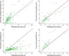 | Figure 1A linear regression scatter plot between the pathologic tumor size and tumor sizes measured using physical examination, mammography, sonography and breast magnetic resonance imaging in breast cancer patients. The Pearson correlation coefficients were 0.55, 0.66, 0.43 and 0.58 for the size based on physical examination, mammography, sonography and breast magnetic resonance imaging respectively compared to pathologic tumor size in breast cancer patients.
......=line of linear regression; —=line of equation.
|
 | Figure 2A linear regression scatter plot between the pathologic tumor size and tumor sizes measured using physical examination, mammography and sonography in benign breast tumor patients. The Pearson correlation coefficients were 0.87, 0.72 and 0.79 for the size based on physical examination, mammography and sonography, respectively compared to the pathologic tumor size in benign breast tumor patients.
......=line of linear regression; —=line of equation.
|
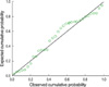 | Figure 3A Normal probability plot of regression standardized residual by linear regression analysis in benign breast tumor patients. The pathologic tumor size can be calculated using the following equation; Pathologic tumor size=0.014+0.049×[tumor size on physical examination]-0.490×[tumor size by mammography]+1.361×[tumor size by sonography].
|
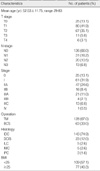
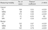
Table 2
Results of correlation analysis between pathologic tumor size and other tumor sizes measured using physical examination, mammography, ultrasonography, computerized tomography, and magnetic resonance imaging in breast cancer and benign breast tumor patients
![]()
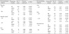
Table 3
Resutls of correlation analysis between pathologic tumor size and other tumor sizes measured using physical examination, mammography, sonography, computerized tomography, and magnetic resonance imaging in breast cancer patients (n=191) according to various clinicopathological features
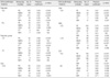
PE=physical examination; MMG=mammography; USG=ultrasonography; CT=computerized tomography; MRI=magnetic resonance imaging; NS= not significant; NA=not applicable; BMI=body mass index; IDC=invasive ductal carcinoma.
*Pearson's coefficient and p-value cannot be calculated because the number of cases is 2.
![]()
References
1. Greene T, Cocilovo C, Estabrook A, Chinitz L, Giuliano C, Rosenbaum Smith S, et al. A single institution review of new breast malignancies identified solely by sonography. J Am Coll Surg. 2006. 203:894–898.

2. Buchberger W, DeKoekkoek-Doll P, Springer P, Obrist P, Dünser M. Incidental findings on sonography of the breast: clinical significance and diagnostic workup. AJR Am J Roentgenol. 1999. 173:921–927.
3. Gordon PB, Goldenberg SL. Malignant breast masses detected only by ultrasound. A retrospective review. Cancer. 1995. 76:626–630.
4. Tohno E, Ueno E, Watanabe H. Ultrasound screening of breast cancer. Breast Cancer. 2009. 16:18–22.
5. Uchida K, Yamashita A, Kawase K, Kamiya K. Screening ultrasonography revealed 15% of mammographically occult breast cancers. Breast Cancer. 2008. 15:165–168.
6. Honjo S, Ando J, Tsukioka T, Morikubo H, Ichimura M, Sunagawa M, et al. Relative and combined performance of mammography and ultrasonography for breast cancer screening in the general population: a pilot study in Tochigi Prefecture, Japan. Jpn J Clin Oncol. 2007. 37:715–720.
7. Uematsu T, Yuen S, Kasami M, Uchida Y. Comparison of magnetic resonance imaging, multidetector row computed tomography, ultrasonography, and mammography for tumor extension of breast cancer. Breast Cancer Res Treat. 2008. 112:461–474.
8. Price J, Chen SW. Screening for breast cancer with MRI: recent experience from the Australian Capital Territory. J Med Imaging Radiat Oncol. 2009. 53:69–80.
9. Almubarak M, Osman S, Marano G, Abraham J. Role of positron-emission tomography scan in the diagnosis and management of breast cancer. Oncology (Williston Park). 2009. 23:255–261.
10. Lee CS, Bong JG, Park JH, Lee YS, Paik SM, Shin MJ, et al. The accuracy of the physical examination, mammography, and ultrasonography in the assessment of tumor size and axillary lymph node metastasis in breast cancer patient. J Korean Breast Cancer Soc. 2003. 6:87–94.
11. Herrada J, Iyer RB, Atkinson EN, Sneige N, Buzdar AU, Hortobagyi GN. Relative value of physical examination, mammography, and breast sonography in evaluating the size of the primary tumor and regional lymph node metastases in women receiving neoadjuvant chemotherapy for locally advanced breast carcinoma. Clin Cancer Res. 1997. 3:1565–1569.
12. Fornage BD, Toubas O, Morel M. Clinical, mammographic, and sonographic determination of preoperative breast cancer size. Cancer. 1987. 60:765–771.
13. Bosch AM, Kessels AG, Beets GL, Rupa JD, Koster D, van Engelshoven JM, et al. Preoperative estimation of the pathological breast tumour size by physical examination, mammography and ultrasound: a prospective study on 105 invasive tumours. Eur J Radiol. 2003. 48:285–292.
14. Forouhi P, Walsh JS, Anderson TJ, Chetty U. Ultrasonography as a method of measuring breast tumour size and monitoring response to primary systemic treatment. Br J Surg. 1994. 81:223–225.
15. Jiang YX, Liu H, Liu JB, Zhu QL, Sun Q, Chang XY. Breast tumor size assessment: comparison of conventional ultrasound and contrast-enhanced ultrasound. Ultrasound Med Biol. 2007. 33:1873–1881.
16. Cheung YC, Wan YL, Lo YF, Leung WM, Chen SC, Hsueh S. Preoperative magnetic resonance imaging evaluation for breast cancers after sonographically guided core-needle biopsy: a comparison study. Ann Surg Oncol. 2004. 11:756–761.
17. Weatherall PT, Evans GF, Metzger GJ, Saborrian MH, Leitch AM. MRI vs. histologic measurement of breast cancer following chemotherapy: comparison with x-ray mammography and palpation. J Magn Reson Imaging. 2001. 13:868–875.
18. Davis PL, Staiger MJ, Harris KB, Ganott MA, Klementaviciene J, McCarty KS Jr, et al. Breast cancer measurements with magnetic resonance imaging, ultrasonography, and mammography. Breast Cancer Res Treat. 1996. 37:1–9.
19. Choi GH, Bae JW, Lee JB, Koo BH. Clinical, mammographic, and ultrasonographic assessment of breast cancer size. J Korean Surg Soc. 2000. 58:331–336.
20. Meden H, Neues KP, Röben-Kämpken S, Kuhn W. A clinical, mammographic , sonographic and histologic evaluation of breast cancer. Int J Gynaecol Obstet. 1995. 48:193–199.
21. Pain JA, Ebbs SR, Hern RP, Lowe S, Bradbeer JW. Assessment of breast cancer size: a comparison of methods. Eur J Surg Oncol. 1992. 18:44–48.
22. Devolli-Disha E, Manxhuka-Kërliu S, Ymeri H, Kutllovci A. Comparative accuracy of mammography and ultrasound in women with breast symptoms according to age and breast density. Bosn J Basic Med Sci. 2009. 9:131–136.
23. Fasching PA, Heusinger K, Loehberg CR, Wenkel E, Lux MP, Schrauder M, et al. Influence of mammographic density on the diagnostic accuracy of tumor size assessment and association with breast cancer tumor characteristics. Eur J Radiol. 2006. 60:398–404.
24. Saarenmaa I, Salminen T, Geiger U, Heikkinen P, Hyvärinen S, Isola J, et al. The effect of age and density of the breast on the sensitivity of breast cancer diagnostic by mammography and ultrasonography. Breast Cancer Res Treat. 2001. 67:117–123.
25. Watermann DO, Tempfer C, Hefler LA, Parat C, Stickeler E. Ultrasound morphology of invasive lobular breast cancer is different compared with other types of breast cancer. Ultrasound Med Biol. 2005. 31:167–174.
26. Evans N, Lyons K. The use of ultrasound in the diagnosis of invasive lobular carcinoma of the breast less than 10 mm in size. Clin Radiol. 2000. 55:261–263.
27. Kim DY, Moon WK, Cho N, Ko ES, Yang SK, Park JS, et al. MRI of the breast for the detection and assessment of the size of ductal carcinoma in situ. Korean J Radiol. 2007. 8:32–39.
 XML Download