Journal List > J Breast Cancer > v.13(2) > 1036197

Lee, Kim, Suh, and Shim: Predictors of 4 or More Positive Axillary Nodes in Patients with Nodepositive T1-2 Breast Carcinoma: The Indications for Adjuvant Irradiation of the Level III Axilla and Supraclavicular Fossa

Abstract

Purpose

We evaluate the predictors of 4 or more involved axillary nodes in patients with node-positive T1-2 breast carcinoma to select a group of patients who are indicated for adjuvant irradiation of the level III axilla and supraclavicular fossa (SCF).

Methods

We analyzed 286 patients with positive axillary nodes and who were without distant metastases and who underwent breast conserving surgery and axillary lymph node dissection or modified radical mastectomy. We investigated the relationship between the patients and the tumor factors and 4 or more positive axillary nodes.

Results

On the multivariate logistic-regression analysis, an increased tumor size (p=0.002), the presence of lymphovascular space invasion (LVSI) (p<0.001) and a palpable mass p<0.001) were positively associated with involvement of 4 or more axillary lymph nodes. In our study, 86.1% of the patients with all the unfavorable factors had involvement of 4 or more nodal metastases.

Conclusion

Our data suggest that for patients with node-positive T1-2 breast cancer, the presence of 4 or more involved nodes is frequently observed for the patients with an increased tumor size, the presence of LVSI and a palpable mass at the time of diagnosis, and we recommend that they undergo irradiation of the high axilla and SCF for adjuvant care, if they do not undergo complete axillary dissection.

REFERENCES

1. Moran MS, Haffty BG. Local-regional breast cancer recurrence: prognostic groups based on patterns of failure. Breast J. 2002; 8:81–87.
crossref
2. Recht A, Gray R, Davidson NE, Fowble BL, Solin LJ, Cummings FJ, et al. Locoregional failure ten years after mastectomy and adjuvant chemotherapy with or without tamoxifen without radiation: experience of the Eastern Cooperative Oncology Group. J Clin Oncol. 1999; 17:1689–1700.
3. Shim SJ, Kim YB, Keum KC, Lee IJ, Lee HD, Suh CO. Validation of radiation volume by analysis of recurrence pattern in breast-conserving treatment for early breast cancer. J Breast Cancer. 2009; 4:257–264.
crossref
4. Galper S, Recht A, Silver B, Manola J, Gelman R, Schnitt SJ, et al. Factors associated with regional nodal failure in patients with early stage breast cancer with 0-3 positive axillary nodes following tangential irradiation alone. Int J Radiat Oncol Biol Phys. 1999; 45:1157–1166.
crossref
5. Halverson KJ, Taylor ME, Perez CA, Garcia DM, Myerson R, Philpott G, et al. Regional nodal management patterns of failure following conservative surgery and radiation therapy for stage I and II breast cancer. Int J Radiat Oncol Biol Phys. 1993; 26:593–599.
6. Grills IS, Kestin LL, Goldstein N, Mitchell C, Martinez A, Ingold J, et al. Risk factors for regional nodal failure after breast conserving therapy: regional nodal irradiation reduces rate of axillary failure in patients with four or more positive lymph nodes. Int J Radiat Oncol Biol Phys. 2003; 56:658–670.
7. Schlembach PJ, Buchholz TA, Ross MI, Kirsner SM, Salas GJ, Strom EA, et al. Relationship of sentinel and axillary level I-II lymph nodes to tangential fields used in breast irradiation. Int J Radiat Oncol Biol Phys. 2001; 51:671–678.
crossref
8. Coen JJ, Taghian AG, Kachnic LA, Assaad SI, Powell SN. Risk of lymphedema after regional nodal irradiation with breast conservation therapy. Int J Radiat Oncol Biol Phys. 2003; 55:1209–1215.
9. Kim HJ, Jang WI, Kim TJ, Kim JH, Kim SW, Moon SH, et al. Radiation-induced pulmonary toxicity and related risk factors in breast cancer. J Breast Cancer. 2009; 2:67–72.
crossref
10. Johansson S, Svensson H, Denekamp J. Dose response and latency for radiation-induced fibrosis, edema, and neuropathy in breast cancer patients. Int J Radiat Oncol Biol Phys. 2002; 52:1207–1219.
crossref
11. Frederick LG, David LP, Irvin DF, April F, Charles MB, Daniel GH, et al. In: AJCC Cancer Staging Manual/American Joint Committee on Cancer. 6th ed. Philadelphia: Lippincott Williams & Wilkins; 2002. pp. 223-240.
12. Kambouris AA. Axillary node metastases in relation to size and location of breast cancers: analysis of 147 patients. Am Surg. 1996; 7:519–524.
13. Fein DA, Fowble BL, Hanlon AL, Hooks MA, Hoffman JP, Sigurdson ER, et al. Identification of women with T1-T2 breast cancer at low risk of positive axillary nodes. J Surg Oncol. 1997; 1:34–39.
crossref
14. Cetintaş SK, Kurt M, Ozkan L, Engin K, Gökgöz S, Taşdelen I. Factors influencing axillary node metastasis in breast cancer. Tumori. 2006; 92:416–422.
crossref
15. Jonjic N, Mustac E, Dekanic A, Marijic B, Gaspar B, Kolic I, et al. Predicting sentinel lymph node metastases in infiltrating breast carcinoma with vascular invasion. Int J Surg Pathol. 2006; 14:306–311.
crossref
16. Silverstein MJ, Skinner KA, Lomis TJ. Predicting axillary nodal positivity in 2282 patients with breast carcinoma. World J Surg. 2001; 25:767–772.
crossref
17. Shahar KH, Hunt KK, Thames HD, Ross MI, Perkins GH, Kuerer HM, et al. Factors predictive of having four or more positive axillary lymph nodes in patients with positive sentinel lymph nodes: implications for selection of radiation fields. Int J Radiat Oncol Biol Phys. 2004; 59:1074–1079.
crossref
18. Katz A, Niemierko A, Gage I, Evans S, Shaffer M, Smith FP, et al. Factors associated with involvement of four or more axillary nodes for sentinel lymph node-positive patients. Int J Radiat Oncol Biol Phys. 2006; 65:40–44.
crossref
19. Katz A, Smith BL, Golshan M, Niemierko A, Kobayashi W, Raad RA, et al. Nomogram for the prediction of having four or more involved nodes for sentinel lymph node-positive breast cancer. J Clin Oncol. 2008; 26:2093–2098.
crossref

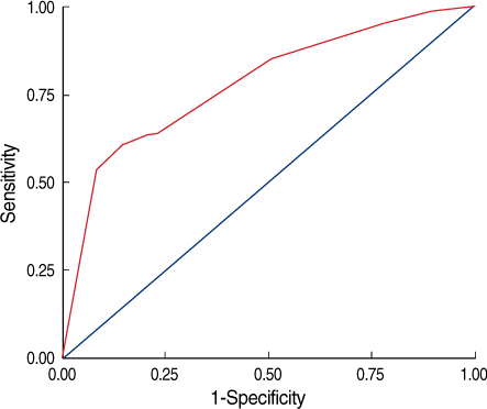
Fig 1.
It shows the receiver operating curve (ROC) that corresponds to the multiple logistic model we applied to our data set of 286 patients. The area under the ROC is 0.80 (p<0.001; 95% CI, 0.742-0.858), which indicates the potentially promising predictive power of the multivariate logistic-regression model.
jbc-13-167-g001.tif
Table 1.
Patient and tumor characteristics (n=286)
Characteristic No. %
Age groups (yr)
 0-50 158 55.2
 >50 128 44.8
Tumor size (mm)
 T1a (0-5.0) 14 4.9
 T1b (5.1-10) 19 6.6
 T1c (10.1-20.0) 73 25.6
 T2 (20.1-50.0) 180 62.9
Histologic grade
 1 20 7.0
 2 67 23.4
 3 83 29.0
 Unknown 116 40.6
Lymphovascular invasion
 Negative 138 48.2
 Positive 106 37.1
 Unknown 42 14.7
ER status
 Negative 112 39.2
 Positive 169 59.1
 Unknown 5 1.7
PR status
 Negative 111 38.8
 Positive 170 59.5
 Unknown 5 1.7
HER2 status
 Negative 156 54.6
 Positive 125 43.7
 Unknown 5 1.7
EGFR status
 Negative 140 49.0
 Positive 23 8.0
 Unknown 123 43.0
Tumor calcification
 Negative 86 30.1
 Positive 109 38.1
 Unknown 91 31.8
Palpable mass
 Negative 60 21.0
 Positive 226 79.0

ER=estrogen receptor; PR=progesterone receptor; HER2=human epidermal growth factor receptor 2; EGFR=epidermal growth factor receptor 1.

Table 2.
Involvement of 4 or more axillary nodes with regard to the patient and tumor characteristics
Characteristic Four or more involved nodes % p-value
Age groups (yr) 0.140
 0-50 78/158 49.4
 >50 52/128 40.6
Tumor size (cm) <0.001
 T1 (≤2) 22/106 20.8
 T2 (>2, ≤5) 108/180 60.0
Histologic grade 0.404
 1 8/20 40.0
 2 38/67 56.7
 3 42/83 50.6
Lymphovascular invasion <0.001
 Negative 44/138 31.9
 Positive 78/106 73.6
ER status 0.663
 Negative 52/112 46.4
 Positive 74/169 43.8
PR status 0.044
 Negative 58/111 52.3
 Positive 68/170 40.0
HER2 status 0.144
 Negative 76/156 47.7
 Positive 50/125 40.0
EGFR status 0.752
 Negative 78/140 55.7
 Positive 12/23 52.2
Tumor calcification 0.102
 Negative 48/86 55.8
 Positive 48/109 44.0
Palpable mass <0.001
 Negative 12/60 20.0
 Positive 118/226 52.2

ER=estrogen receptor; PR=progesterone receptor; HER2=human epidermal growth factor receptor 2; EGFR=epidermal growth factor receptor 1.

Table 3.
Univariate logistic-regression analysis for factors associated with involvement of 4 or more axillary nodes
Variable Unadjusted odds ratio (95% of confidence interval) p-value
Tumor size (cm)
 T1 (≤2) 1.00 (referent)
 T2 (>2, ≤5) 5.72 (3.28-9.98) <0.001
Lymphovascular invasion
 Negative 1.00 (referent)
 Positive 5.95 (3.39-10.42) <0.001
PR status
 Negative 1.00 (referent)
 Positive 0.60 (0.37-0.98) 0.037
Palpable mass
 Negative 1.00 (referent)
 Positive 4.37 (2.20-8.66) <0.001

ER=estrogen receptor; PR=progesterone receptor.

Table 4.
Multivariate logistic-regression analysis for factors associated with involvement of 4 or more axillary nodes
Variable Adjuated odds ratio (95% of confidence interval) p-value
Tumor size (cm)
 T1 (≤2) 1.00 (referent)
 T2 (>2, ≤5) 2.91 (1.52-5.57) 0.001
LVSI
 Negative 1.00 (referent)
 Positive 4.70 (2.58-8.57) <0.001
Palpable mass
 Negative 1.00 (referent)
 Positive 5.01 (2.19-11.45) <0.001

LVSI=lymphovascular space invasion.

Table 5.
Involvement of 4 or more axillary nodes with regard to the combinations of the significant factors identified on the multivariate analysis
Characteristic Four or more involved nodes %
No LVSI, nonpalpable, T1 mass 2/20 10.0
No LVSI, nonpalpable, T2 mass 4/23 21.1
No LVSI, palpable, T1 mass 13/49 17.4
No LVSI, palpable, T2 mass 14/33 42.4
LVSI, nonpalpable, T1 mass 0/3 0
LVSI, nonpalpable, T2 mass 5/19 26.3
LVSI, palpable, T1 mass 9/18 50.0
LVSI, palpable, T2 mass 68/79 86.1

LVSI=lymphovascular space invasion.

TOOLS
Similar articles