

 PDF
PDF ePub
ePub Citation
 Print
Print


INTRODUCTION
Breast cancer is the most common malignant tumor found in women throughout the world.(1) Korea has one of the lowest incidence rates of breast cancer (40.5 per 100,000 women-years) of any country, yet the incidence rates in Korea are rapidly increasing because of increased cancer detection activities, and notably screening mammography or breast ultrasonography.(2) According to the cancer registration report issued by the Ministry of Health and Welfare, the breast is the most common site of primary cancer for women in Korea since 2001.(3)
Veronesi et al.(4) and Fisher et al.(5) have reported the results of large studies on breast-conserving surgery (BCS) in the 1980s. Because of these studies, BCS has become the standard approach to surgery for treating early breast cancer. This surgery was originally designed to improve the cosmetic outcomes of patients who undergo early breast cancer treatment, and this has resulted in improved patient satisfaction with the surgical outcome.(6) Even though BCS can maintain the natural shape of the breast, a long noticeable surgical scar on the skin of the breast can result in a poor cosmetic outcome. One of the main advantages of endoscopic surgery is that it can be performed with making only small incisions that become inconspicuous after surgery.(7,8) Endoscopy-assisted breast surgery (EABS) has been recognized as a useful approach for performing aesthetic procedures such as augmentation mammoplasty.(6) Many studies have reported on its application to breast disease, and even for malignant diseases, without worsening the therapeutic outcome.(9) Therefore, we adopted performing EABS for those patients with early breast cancer with the objective of improving the cosmetic results. We report herein on the surgical method of endoscopy-assisted BCS (EA-BCS) that we used the aesthetic outcomes and patient outcomes after EA-BCS.
METHODS
Patients
Twenty two patients that were underwent EA-BCS for breast cancer at the Department of Breast Care Center, Myongji Hospital, Kwandong University College of Medicine from June 2006 to February 2008. These patients were selected for EA-BCS according to the following criteria: 1) early breast cancer without any severe co-morbid conditions such as heart disease, liver dysfunction, renal failure, or a poor performance status, 2) no tumor extension to the nipple or direct invasion to the skin and pectoralis muscles, 3) no axillary lymph node metastasis, 4) no multifocal lesions and 5) no microcalcified lesions which could not localize by preoperative ultrasonography. The patients were evaluated by preoperative studies such as mammography, ultrasonography, chest computed tomography (CT), and magnetic resonance imaging (MRI). Informed consent was obtained from all the patients prior to performing EA-BCS.
Instruments for surgery
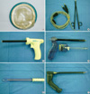
In addition to the basic endoscopic monitoring system (Stryker®; Stryker Endoscopy, San Jose, USA), Figure 1 shows the special tools we used for this surgery: 1) an Alexis® Wound Retractor (2-4 cm, XS; Applied Medical, Rancho Santa Margarita, USA) (Figure 1A) was used for axillary incision wound protection 2) Bipolar scissors BiSect (ERBE, Tuebingen, Germany) (Figure 1B) and a Harmonic Scalpel™ (Ethicon Endo-Surgery, Cincinnati, USA) were used for accurate resection and hemostasis 3) a Visiport™ Plus (5-11 mm; Tyco Healthcare, Norwalk, USA) (Figure 1C) was used for sentinel lymph node biopsy (SLNB) and axillary lymph node dissection (ALND) 4) an ENDOPATH® (Ethicon Endo-Surgery) (Figure 1D) was used for penetration of subcutaneous tissue, 5) an Endosector LE (Curexo, Anyang, Korea) (Figure 1E) and an Ultra Retractor (Johnson & Johnson KK, Tokyo, Japan) (Figure 1F) were used for dissection of the retromammary space. The endoscope is a rigid, straight instrument that is 5-mm in diameter, it is oblique at 30° and it is 10-mm in diameter and flat at 0°.
Surgical procedure
Under general anesthesia, the patient was placed in the supine position with the ipsilateral arm abducted to 90° without disturbing the operative field. The breast, chest wall, axilla, and ipsilateral arm were prepared and draped in the usual sterile fashion. The location of the tumor was confirmed by means of palpation and performing intraoperative ultrasonography, even if the tumor was non-palpable lesion. The tumor location and the quadrant to be excised were marked on the skin. A SLNB was performed with the dye-staining method or radioisotope-guided method, or a combined method at the beginning of the operation before gland resection on a case by case basis. For the cases using the dye staining method, we subcutaneously injected 5 mL of 0.8% indigocarmine in the periareolar region, and we performed breast massage for 10 min. A 3.5-cm skin incision was made along the line of the axillary skin crease. A Visiport™ Plus and 10-mm/0° scope was inserted from the axillary incision, and the stained lymph nodes were found with using endoscopy assistance. In the patients who were found to have a positive sentinel lymph node, an ALND was carried out at levels I and II through the same incision under endoscopy assistance. After completing the SLNB, the Alexis® Wound Retractor was inserted into the incision. Through the axillary incision, we cut the adipose tissue deep into the shallow lateral chest fascia, and we slide it to the lateral edge of the major pectoral muscle; we then bluntly dissected the fascia beyond the retromammary space of the quadrant where the tumor was to be excised by using an Endosector LE or an Ultra Retractor under 5-mm/30° scope assistance. The fiberoptic breast retractor and a 10-mm/0° scope were inserted into the subpectoral pocket, and further dissection was performed using the Bipolar scissors BiSect or Harmonic Scalpel™ under endoscopic guidance. After completing the dissection of the retromammary space, we went through the subcutaneous tissue. A skin flap was made by the tunnel method.(10) Further, we made a 3-cm periareolar skin incision in the direction of the tumor. The ENDOPATH® which has an opening that accommodates a 10-mm/0° scope was next inserted beneath the skin flap of the periareolar skin incision. The skin flap was made by blunt thrusting of the ENDOPATH®. We completed the subcutaneous flaps of the tumor quadrant with using the Bipolar scissors BiSect or the Harmonic Scalpel™. The Bipolar scissors BiSect or Harmonic Scalpel™ were also used to vertically dissect the mammary gland. We partially removed the mammary gland with free surgical margins at least 1 cm away from the tumor border. The resected gland was pulled out through the axillary Alexis® Wound Retractor port. We stained the superficial cut surface of the resected gland with India ink, with placing one or more sutures for directional orientation. The resected gland was brought to the pathology department and the frozen sections of its cut margin were immediately examined by a pathologist. If the cut margin was positive, then additional resection was required until a negative cut margin was confirmed. After the resection was completed, meticulous dissection of the residual tissue and hemostasis could be achieved with using the Harmonic Scalpel™ and an electrocoagulator under endoscopic guidance. After saline irrigation, the remaining mammary glands were mobilized and sutured together to preserve the original breast shape. A suction drainage was left in place before closing the surgical wound. We reconstructed the breast by immediate volume replacement as needed. We placed an absorbable implant into the dead space. The absorbable implant was made of Vicryl mesh® (polyglactin 910 Mesh; Ethicon, Johnson & Johnson, Somerville, USA) wrapped with Interceed® (Oxidized regenerated cellulose; Ethicon, Johnson & Johnson).(11) In cases of immediate volume replacement, however, we did not use a suction drainage to preserve breast contour by seroma formation. The surgical wound was closed with a subcutaneous reversed suture using 4-0 Vicryl. Compression was provided with a surgi-bra to prevent postoperative bleeding and hematoma formation.
Cosmetic evaluation and the patient satisfaction
All the patients were regularly examined and questioned about their satisfaction with the aesthetic results every six months follow-up. Many methods of cosmetic assessment have been reported.(12-14) We used the 5-item-4-step method (ABNSW) designed by Yamashita.(12) The method of ABNSW is the modified method of the MDACS (malposition, distortion, asymmetry, contour deformity and scar) grading system.(13) ABNSW contains 5 items: asymmetry (A), breast shape (B), nipple deformation (N), skin condition (S) and wound scar (W). All items can be scored by the patients themselves under the 4 steps of the grading system as follows: 0: poor, 1: fair, 2: good, 3: excellent. The scores for the 5 items were then combined to obtain a total score. On a scale of 15 points, the summed points were defined as excellent: 15 points, good: 11-14 points, fair: 6-10 point and poor: <5 points. The patients were also requested to evaluate the subjective satisfaction of their postoperative aesthetic results as poor, fair, good, or excellent using 4-step method of ABNSW at every six months after surgery.
RESULTS
Clinicopathologic features of patients
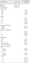
The clinical/pathological features of all the patients are presented in Table 1. The average age was 52.0 yr (range, 36-74 yr). The locations of the tumors are summarized in Table 1. The upper-outer quadrant was the most common site (50.0%). The mean tumor size was 2.2 cm (range, 0.7-5.5). TNM staging was done according to the American Joint Committee on Cancer staging system. All the patients were negative for lymph node metastasis on the preoperative evaluation, and this included ultrasonography or MRI. A SLNB was performed in all patients. Four patients who had positive sentinel lymph node frozen biopsy results underwent ALND, and only one out of four patients had positive axillary node biopsy results at level I and II. For the postoperative diagnosis, 81.8% (18 out of 22) of the patients were negative for metastatic lymph nodes; however, 18.2% (4 out of 22) of the patients were positive for lymph node metastasis; distant metastasis was not observed in any patient. The disease in all the patients with malignancies was classified as earlier than stage IIB. Estrogen receptor positive results were obtained in 86.4% (19 out of 22) of the patients and the progesterone receptor status was positive in 95.5% (21 out of 22) of the patients. We defined more than three scores at the sum of proportional and intensity score as hormone receptor positive. An over-expression of HER2/neu was observed in 40.9% (9 out of 22) of the patients. The histopathological diagnoses of treated diseases are summarized in Table 1.
Operative methods
EA-BCS was performed for 22 patients with breast cancer. A SLNB was performed in 22 patients. An ALND was performed in four patients who had positive sentinel lymph node frozen biopsy results. Immediate volume replacement treatment was carried out in 11 patients with an absorbable implant that was made of Vicryl mesh® wrapped with Interceed®. The mean operative time was 144 min (range, 105-190 min). The mean volume of the extracted mass was 219.2 cm3 (range, 56-1332 cm3).
Satisfaction level for surgery
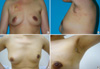
Ninety five percent (21 out of 22) of the patients were questioned about their satisfaction with the aesthetic results every six months follow-up. There was one patient who underwent a skin sparing mastectomy (SSM) with immediate breast reconstruction due to a positive resection margin of the nipple specimen on the permanent report was excluded in this assessment. The postoperative evaluations after the procedures are shown in Figure 2. The scar from the wound was inconspicuous. The shape of the breast appeared natural and nearly symmetrical. The patients were satisfied with the operation. The aesthetic results were evaluated using the original scoring system ABNSW.(12) The mean total ABNSW score was 12.2 (Table 2). By comparing with 4-step method, scores greater than 11 points were considered good or excellent; subsequently, 95% of the evaluated cases had good or excellent results. In addition, the degree of satisfaction was evaluated using 4-step method of ABNSW. 100% (21 out of 21) of the patients scored good or excellent results. Almost all the patients were satisfied with the aesthetic results of their surgery.
Table 3 summarizes the surgical complications. The postoperative complications were as follows: lymphedema developed in 9.1% (2 out of 22), skin retraction developed in 9.1% (2 out of 22) and wound infection developed in 13.6% (3 out of 22). Two out of these 3 patients had wound infection because of the prosthetic infection. All cases of wound infection were developed in diabetes patients. Skin retraction was caused by reoperation of infected prosthesis removal. Three patients underwent reoperation. There were 9.1% (2 out of 22) of the patients who had to have their prosthesis removed because of infection of the Vicryl mesh®. We did not additional procedure to preserve breast shape after the infected prosthesis removal. In addition, 4.5% (1 out of 22) of the patients had a positive resection margin of the nipple specimen on the permanent pathology report. Therefore, we performed a SSM with immediate breast reconstruction nine days after the initial operation. To detect local recurrence and distant metastasis, after surgery, ultrasonography was performed every six months and positron emission tomography-CT was performed one year after the operation. The patients underwent standard adjuvant therapy based on the recommendation of St. Gallen.(15) After a mean follow-up period of 24.0 months (range 12-32 months), neither local/regional recurrence nor distant metastases were detected.
DISCUSSION
Preservation of the natural shape of the breast is very important for women who suffer with breast disease that requires surgery. BCS was originally introduced to improve the cosmetic outcomes of breast cancer treatment by Veronesi et al.(4) and Fisher et al.,(5) and it has greatly improved the patient satisfaction with these procedures.(6) However, when a long marked operation scar occurs this reduces the patient satisfaction with the cosmetic outcome.
Endoscopic surgery has been established as a treatment modality for abdominal and thoracic disorders.(16, 17) One of the main advantages of endoscopic surgery is that it can be performed via small incisions that become inconspicuous after the surgery.(7,8) EABS was first developed for performing augmentation mammoplasty.(6) In 1992, Kompatscher(18) described the endoscopic capsulotomy technique for treating capsular contracture that occurs after breast augmentation. Following that numerous studies regarding endoscopy-assisted breast augmentation have been reported. EABS has now become the standard technique for breast augmentation. Although it is challenging as a surgical technique, it has the potential of becoming an important alternative approach for treating both benign and malignant tumors of the breast.(6)
Kitamura et al.(19) reported on endoscopic extirpation of benign breast tumors. In the past few years, endoscopic surgery has been successfully used for benign breast disease such as breast lump excision, breast augmentation, subcutaneous mastectomy for gynecomastia, and axillary dissection.(20) Friedlander et al.(21) first reported on EABS for breast cancer in 1995. They initially performed experimental surgery using an endoscope on porcine models and thereafter on cadavers. Yamagata and Iwai(22) reported on endoscopic partial mastectomy and axillary dissection for breast cancer in 1997. Subsequently, there have been numerous reports on EABS for treating breast cancer.(7) As Fukuma et al.(23) and Yamagata et al.(10) have suggested, almost all surgeries can be performed through a 3.5-cm axillary incision and a 3-cm periareolar skin incision in the direction of the tumor. However, the periareolar wound scar is more conspicuous than the axillary wound scar, and the former often distorts the nipple and the areola, and causes sensory disturbance around the areola. In the properly selected cases, we attempt to make only an axillary incision for those cases where the tumor is located in the outer-upper or inner-upper quadrant and the tumor is thought to be early breast cancer; all surgical procedures are currently performed with endoscopic assistance. Reconstruction of the conserved breast is simultaneously performed during the EABS at our center; an absorbable implant is placed into the dead space during the procedure. This likely contributes to the excellent cosmetic results after EABS (Figure 2A). We experienced two cases of prosthetic wound infection. All of these cases were developed in diabetes patients. After that, we did not use Vicryl mesh® replacement in diabetes patients.
The status of the surgical margins is very important for assessing the risk of disease recurrence. To obtain accurate surgical margins, we used ultrasonography or MRI preoperatively and we performed accurate dissection under intraoperative ultrasonography with an endoscopic monitor. The surgical edge of the tumor was examined during surgery by fast-frozen section analysis. The permanent pathology report revealed one case with a positive resection margin in the nipple. In this case, we performed SSM with immediate breast reconstruction nine days after the initial operation.
Endoscopy-assisted SLNB and ALND is another application of endoscopic surgery for treating breast cancer. These techniques have been mainly developed in Europe, and they appear to have significant advantages from a cosmetic point of view.(6) SLNB has been established as an alternative treatment option for patients with early stage breast cancer. The oncological safety of SLNB was recently confirmed by a randomized controlled trial.(24, 25) Some investigators have recently reported on endoscopic SLNB and they have confirmed its superior, higher rate of identifying lymph nodes with tumor.(6) The procedure was initially introduced in 1993 by Suzanne et al.(26) who reported on the simplicity and safety of endoscopic axillary lymphadenectomy with fat aspiration. In 1996, Salvat et al.(27) reported on the results of a randomized study that compared endoscopic sampling and open surgery for the axillary lymph nodes in patients with breast cancer; there were no statistical differences identified for the operation time between the two groups, as well as the duration of the hospital stay, the immediate post-operative complications, the number of lymph nodes removed, and the size of the lymph nodes. Most reports have concluded that endoscopic axillary lymphadenectomy produced a better cosmetic outcome with fewer sensory disturbances, and the number of removed lymph nodes did not differ from that of conventional surgery.(28,29) However, some investigators have doubts about the clinical benefits of endoscopic axillary lymphadenectomy.(6,28) We had 22 cases that underwent endoscopic-assisted SLNB. Four patients were found to have a positive sentinel lymph node on the frozen section, and ALND could be carried out at levels I and II through the same incision under endoscopic-assistance. Using an incision for the SLNB to insert the Alexis® Wound Retractor, we were further able to reduce the injury to the skin envelope of the breast, which apparently improved the cosmetic appearance after the surgery. Yet other than the cosmetic benefits, the advantages of endoscopy-assisted ALND remain unclear, and so further studies are needed to determine this.
Overall the cosmetic results and patient satisfaction are excellent. In the cosmetic evaluation by using ABNSW, more patient selected good score than excellent score in the items of the asymmetry (A), breast shape (B). Patients think that they will have the preoperative breast contour after operation by using the new operative techniques; such as EABS, Vicryl mesh® replacement. The postoperative results fall short of patients' expectation. We think that these higher expectation lead up to these results. In practice, the semicircular surgical wound around the nipple becomes conspicuous after the surgery. For the properly selected cases, we started to make only an axillary incision to improve the cosmetic result. We are now developing another method of breast reconstruction to further the outcomes.
There are several factors to consider before performing EA-BCS instead of conventional BCS, and these factors include a longer operation time, difficulty in handling instruments and the safety from point of view of tumor recurrence. We can ask: Is the extra time and cost of EABS worth the benefit of reducing the size of the scar on the breast? However, endoscopic surgery has been used for a variety of breast surgery procedures over the past 10 yr and it is expected to be used for almost all surgeries of the breast in the future.(9,12)
Our study had several limitations. First, evidence obtained from retrospective studies, like ours, is statistically weak. Second, the follow-up period was relatively short, and additional careful follow-up is needed to ascertain the long-term follow-up results of this procedure. Third, our wound infection rate was not lower than that reported in previous literature in cases where immediate reconstruction was performed. However, new specialized instruments must be developed for improving this technique and a large clinical trial with adequate follow up and then comparing the results with conventional BCS are needed to confirm the safety of EA-BCS.
CONCLUSION
EA-BCS was feasible and effective for treating breast cancer and it can be regarded as possibly the best surgical option for obtaining better aesthetic results as it can be performed via a small and remote wound that becomes inconspicuous after surgery. EA-BCS has promising potential though there are several important factors to consider when performing this procedure. Further study with more patients and long-term follow-up is needed.


 XML Download