

 PDF
PDF ePub
ePub Citation
 Print
Print


INTRODUCTION
An angiolipoma is an unusual, benign variant of a lipoma with a hemangiomatous component. The tumor can occur anywhere in the body, but an angiolipoma of the breast is rare.(1,2)
The mammographic findings of an angiolipoma are variable, and the lesion can sometimes mimic a carcinoma.(2) However, in most cases, sonography shows the tumor to be a homogeneously well-circumscribed hyperechoic lesion that is usually benign.(3,4)
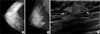
We report a rare case of an angiolipoma of the breast that presented with a non-tender palpable mass. Although no focal abnormal findings were seen on mammography, the lesion appeared as a single homogeneously irregular hyperechoic lesion with an extension to the subcutaneous layer on ultrasound.
CASE REPORT
A 40-yr-old woman presented with a complaint of a painless, palpable mass in the right breast for one-month duration. The family history was unremarkable. There was no nipple discharge or skin change.
Mammography showed heterogeneously dense breast without focal abnormality or microcalcification in the region of the palpable breast mass (Figure 1A, B). On ultrasound examination of the 9-o'clock position of the right breast, an approximate 2 cm sized partly indistinct, irregular shaped homogeneously hyperechoic intraparenchymal mass (arrow) without definite internal vascularity was detected, extending to the subcutaneous layer (Figure 1C).
An ultrasonography-guided 14-gauge core needle biopsy was performed. The pathological description was dilatation and proliferation of the capillaries, and the possibility of a malignant lesion, such as angiosarcoma, cannot be totally excluded, suggesting total removal of mass. Due to patient request, an ultrasonography-guided 8-gauge vacuum-assisted biopsy (mammotome) was ultimately performed.
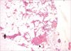
Pathologic description of the lesion was adult adipose tissue with proliferating capillaries in the fibrous stroma, combined with surrounding mammary glands. The final diagnosis of an angiolipoma of the breast was confirmed (Figure 2).
DISCUSSION
Angiolipomas account for 5-17% of benign fatty tumors, and were first established as a pathological entity by Howard and Helwig in 1960.(5) The lesion is a benign variant of a lipoma with a hemangiomatous component that is located in the subcutis. It typically occurs in the trunk, abdomen, or extremities. Common clinical symptoms associated with an angiolipoma are pain and tenderness.(2) Although the tumor can be found anywhere in the body, an angiolipoma of the breast is rare.(4)
The tumor may be seen as a solitary mass or may present with multiple breast masses. Reported ages for diagnosis range from one year to 82 yr. On physical examination, the lesion can be either palpable or nonpalpable. Angiolipomas at a non-breast site present with pain or tenderness, but lesions in the breast have typically been painless in most reported cases.(2-5) As with the present case, skin changes are unusual.(3,4)
On mammography, the tumor may appear as a dense nodule with poorly defined margins, and may be confused with malignant conditions based on both clinical and radiological evaluations.(2) However, there is no typical mammographic appearance of an angiolipoma. The lesion may appear normal or may appear as a nodule with calcification.(3,4) Our case showed only a heterogeneously dense breast with no definite focal abnormality on mammography.
Computed tomography (CT) findings of a breast angiolipoma have rarely been described. In 1980, Sibala et al.(2) reported CT findings of a breast angiolipoma, which was considered as a malignancy on physical examination and mammography. On CT mammography, the lesion showed intermediate enhancement whereas other malignant lesions typically show strong enhancement.(2)
Almost all angiolipomas of the breast appear as homogeneously hyperechoic masses on ultrasound. Fat-containing lesions in the body appear echogenic and, in contrast to breast tissue, fat-containing lesions are hypoechoic relative to fibroglandular components.(3) Hyperechoic breast masses are relatively rare, and most of the homogeneously hyperechoic masses are benign and show especially well circumscribed margins (100% negative predictive value for malignancy).(4,6) In our case, although the mass had an irregular margin, the lesion appeared with homogeneously hyperechoic features and we considered the mass to be a benign lesion such as a focal mastitis or fat necrosis rather than a malignancy. As depicted on ultrasound, a spindle cell lipoma and a hemangioma may appear as an homogeneously hyperechoic mass, as for an angiolipoma. However, hemangiomas may appear with posterior acoustic shadowing or, occasionally, as an hypoechoic mass.(7,8)
An angiolipoma is an encapsulated, intimate admixture of fat cells and blood vessels lacking atypia of endothelial cells. The lesion can be distinguished from an aggressive angiosarcoma.(9) The established pathological criteria include the following: 1) gross evidence of a tumor with or without a capsule; 2) microscopic evidence of mature lipomatocytes as the major population (at least 50%) of the tumor; 3) microscopic evidence of angiomatous proliferation inside the tumor.(10)
There are two types of angiolipomas. These are infiltrating and noninfiltrating types. Breast angiolipomas have been reported as the noninfiltrating type and treatment is simple excision. For the infiltrating type, wide excision may be required.(3) Noninfiltrating angiolipoma occurs after puberty and tends to involve subcutaneous tissue, whereas infiltrating angiolipoma usually involves the muscles.(4) In our case, although an irregular shape was seen on ultrasonogram, a relatively well defined margin was apparent under microscopy, so the diagnosis was noninfiltrating type.
The cause of angiolipoma is unknown, but repeated minor trauma is one of the possible causes.(5)
In conclusion, a breast angiolipoma may show various appearances on mammography, but may appear as an homogenously hyperechoic lesion on ultrasound. Thus, the lesion can be distinguished from a malignancy.
 XML Download