

 PDF
PDF ePub
ePub Citation
 Print
Print


Abstract
Purpose
This study was to investigate the clinical significance of diffusely increased F-18 FDG uptake in the thyroid gland as an incidental finding on F-18 FDG PET/CT imaging in patients with breast carcinoma.
Methods
One hundred four patients with breast carcinoma who had no prior history of thyroid disease were enrolled. All patients underwent F-18 FDG PET/CT, ultrasound and thyroid function test (TFT-TSH, FT4, and T3), anti-TPO antibody test within 2 weeks. Also we checked estrogen (ER) and progesterone receptors (PR). We classified all patients into subgroups according to the existence and degree of F-18 FDG uptake in the thyroid gland, and evaluated the difference between subgroups.
Results
Of the 104 patients, 42 (40.4%) subjects showed diffusely increased thyroid uptakes. There was no significant difference in rate of abnormality in TFT and thyroid US, and existence of anti-TPO antibody and ER/PR between two groups. Of 42 patient who showed diffuse uptake, 12 (28.5%), 13 (31.0%), and 17 (40.5%) subjects demonstrated hypointense, isointense, and hyperintense thyroid uptake compared with activity of mediastinal blood pool. Thirteen (76.4%) of 17 subjects in the hyperintense thyroid uptake group revealed abnormality in various tests (US, TFT, and anti-TPO antibody). The rate of abnormality in this group was significantly different with the other two groups (p=0.002).
Conclusion
Our data suggested that the rate of diffuse thyroid uptakes on F-18 FDG PET/CT imaging of patients with breast carcinoma was higher than healthy subjects. In case of someone who had no prior thyroid disease showed diffuse thyroid uptakes more than activity of mediastinal blood pool on F-18 FDG PET/CT imaging, it should be considered further evaluation about the thyroid gland.
Figures and Tables
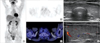 | Figure 1A case of diffusely increased F-18 FDG uptake, but less than activity of mediastinal blood pool, in the thyroid gland of 55-yr-old patient. On ultrasound examination, echogenecity of thyroid gland was homogeneous and blood flow was normal. Serum TSH was 2.61 mIU/L (reference range, 0.17-4.05 mIU/L), free T4 was 10.9 ng/dL (reference range, 9.4-25 ng/dL) and T3 was 2.03 ng/dL (reference range, 0.8-2.2 ng/dL), suggestive of normal state of thyroid function. Shown are 3D MIP image (A), transaxial PET image (B), transaxial fused PET/CT image (C), ultrasound image (D), and doppler ultrasound image (E).
|
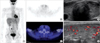 | Figure 2A case of diffusely increased F-18 FDG uptake, similar intensity with mediastinal blood pool, in the thyroid gland of 46-yr-old patient. On ultrasound examination, thyroid gland was homogeneous and blood flow was mildly increased. Serum TSH was 2.09 mIU/L, free T4 was 11.3 ng/dL and T3 was 1.22 ng/dL, suggestive of normal state of thyroid function.
|
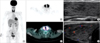 | Figure 3A case of intensely and diffusely increased F-18 FDG uptake, more than activity of mediastinal blood pool, in the thyroid gland of 58-yr-old patient. On ultrasound examination, thyroid gland was prominent and heterogeneous. Blood flow of thyroid gland was slightly increased. Serum TSH was 2.09 mIU/L, free T4 was 11.3 ng/dL, T3 was 1.22 ng/dL and TPO antibodies were elevated at 100 IU/mL (reference range, 0-0.3 IU/mL), suggestive of diagnosis of Hashimoto's thyroiditis.
|
Table 1
Number of patients with abnormal laboratory data, surgical data, and US finding in two groups, classified by diffuse thyroid uptake

![]()
Table 2
Number of patients with abnormal laboratory data, surgical data, and US finding in three groups, classified by degree of thyroid uptake

TFT=thyroid function test; anti-TPO Ab=anti-thyroid peroxidase antibody; US=ultrasound; ER/PR=estrogen receptor/progesterone receptor; Group 1=less than blood pool activity of mediastinum; Group 2=equal to blood pool activity of mediastinum; Group 3=higher than blood pool activity of mediastinum.
*Fisher's exact test.
![]()
Table 3
Laboratory, surgical data, and US findings in 17 patients who showed diffuse thyroid uptake more than blood pool activity of mediastinum
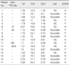
US=ultrasound; Anti-TPO Ab=anti-thyroid peroxidase antibody; T3=triiodothyronine; FT4=free thyroxine; TSH=thyroid-stimulating hormone; ER/PR=estrogen receptor/progesterone receptor; NS=not significant; P=positive; N=negative.
*Reference values=TSH, 0.17-4.05 mIU/L; T3, 0.8-2.2 ng/dL; FT4, 9.4-25 ng/dL; TPO antibody, 0-0.3 IU/mL.
![]()
References
1. KIM TS, Moon WK, Lee DS. FDG-PET for the detection of recurrent or metastatic breast cancer. J Korean Breast Cancer Soc. 2000. 3:25–33.

2. Noh DY, Yun IJ, Kang HS, Kim JS, Lee DS, Chung JK, et al. The diagnostic value of positron emission tomography in detecting the breast cancer. J Korean Breast Cancer Soc. 1998. 1:6–12.
3. Fletcher JW, Djulbegovic B, Soares HP, Siegel BA, Lowe VJ, Lyman GH, et al. Recommendations on the use of 18F-FDG PET in oncology. J Nucl Med. 2008. 49:480–508.
4. Field J. Astwood EB, Greep RO, editors. Intermediary metabolism of the thyroid. American Physiological Society Handbook of Physiology: Endocrinology-Section 7. 1974. Volume III, Thyroid. Washington, DC: American Physiological Society;147–159.
5. Hosaka Y, Tawata M, Kurihara A, Ohtaka M, Endo T, Onaya T. The regulation of two distinct glucose transporter (GLUT1 and GLUT4) gene expressions in cultured rat thyroid cells by thyrotropin. Endocrinology. 1992. 131:159–165.
6. Nakamoto Y, Tatsumi M, Hammoud D, Cohade C, Osman MM, Wahl RL. Normal FDG distribution patterns in the head and neck: PET/CT evaluation. Radiology. 2005. 234:879–885.
7. Karantanis D, Bogsrud TV, Wiseman GA, Mullan BP, Subramaniam RM, Nathan MA, et al. Clinical significance of diffusely increased F-18 FDG uptake in the thyroid gland. J Nucl Med. 2007. 48:896–901.
8. Kurata S, Ishibashi M, Hiromatsu Y, Kaida H, Miyake I, Uchida M, et al. Diffuse and diffuse-plus-focal uptake in the thyroid gland identified by using FDG-PET: prevalence of thyroid cancer and Hashimoto's thyroiditis. Ann Nucl Med. 2007. 21:325–330.
9. Salvatori M, Melis L, Castaldi P, Maussier ML, Rufini V, Perotti G, et al. Clinical significance of focal and diffuse thyroid diseases identified by 18F-fluorodeoxyglucose positron emission tomography. Biomed Pharmacother. 2007. 61:488–493.
10. Liu Y. Clinical significance of thyroid uptake on F-18 fluorodeoxy-glucose positron emission tomography. Ann Nucl Med. 2009. 23:17–23.
11. Yasuda S, Shohtsu A, Ide M, Takagi S, Takahashi W, Suzuki Y, et al. Chronic thyroiditis: diffuse uptake of FDG at PET. Radiology. 1998. 207:775–778.
12. Chen YK, Chen YL, Cheng RH, Yeh CL, Lee CC, Hsu CH. The significance of FDG uptake in bilateral thyroid glands. Nucl Med Commun. 2007. 28:117–122.
13. Giani C, Fierabracci P, Bonacci R, Gigliotti A, Campani D, De Negri F, et al. Relationship between breast cancer and thyroid disease: relevance of autoimmune thyroid disorders in breast malignancy. J Clin Endocrinol Metab. 1996. 81:990–994.
14. Giustarini E, Pinchera A, Fierabracci P, Roncella M, Fustaino L, Mammoli C, et al. Thyroid autoimmunity in patients with malignant and benign breast diseases before surgery. Eur J Endocrinol. 2006. 154:645–649.
15. Tateishi U, Gamez C, Dawood S, Yeung HW, Cristofanilli M, Inoue T, et al. Chronic thyroiditis in patients with advanced breast carcinoma: metabolic and morphologic changes on PET-CT. Eur J Nucl Med Mol Imaging. 2009. 36:894–902.
16. Kim TY, Kim WB, Ryu JS, Gong G, Hong SJ, Shong YK. 18F-fluorodeoxyglucose uptake in thyroid from positron emission tomogram (PET) for evaluation in cancer patients: high prevalence of malignancy in thyroid PET incidentaloma. Laryngoscope. 2005. 115:1074–1078.
17. Wolf G, Aigner RM, Schaffler G, Schwarz T, Krippl P. Pathology results in [18F]fluorodeoxyglucose positron emission tomography of the thyroid gland. Nucl Med Commun. 2003. 24:1225–1230.
 XML Download