Journal List > J Breast Cancer > v.12(4) > 1036164

Oh, Yang, Lee, Park, Park, and Lee: The Prognostic Significance of Survivin Expression in Breast Cancer

Abstract

Purpose

Survivin is a member of the inhibitors of apoptosis family. It has recently comes into the limelight as a promising tumor marker, but many previous reports have shown controversial results regarding the significance and prognostic value of a survivin expression. In this study we determined the correlation between the survivin expression and the conventional prognostic markers and we also investigated the outcomes according to the localization of the survivin expression.

Methods

Tissue microarray (TMA) blocks were made with formalin-fixed paraffin-embedded tissues from 185 breast cancer patients and the immunohistochemical staining was done using an anti-survivin antibody. Among these, 157 patients were available for a survivin expression. The conventional clinicopathologic features and overall survival were correlated with the localization of the survivin expression.

Results

Survivin was expressed in 101 breast cancers (64.3%). A higher cytoplasmic survivin expression were noted in the older group (p=0.003), in the node-negative cancers (p= 0.012), in the earlier tumor stages (p=0.012) and in the cancers that had not been treated with adjuvant chemotherapy (p=0.014). On the contrary, a higher nuclear survivin expression was inversely correlated with an estrogen expression (p=0.006) and a progesterone receptor (p=0.043) expression. In terms of survival, a cytoplasmic expression was associated with improved overall survival (p=0.01) but a nuclear survivin expression was correlated with unfavorable overall survival (p=0.002). A high cytoplasmic to nuclear ratio of survivin was associated with improved overall survival (p=0.001) conversely, increased nuclear to cytoplasmic survivin ratio was correlated with unfavorable overall survival (p<0.0001). Multivariate analysis revealed that nuclear survivin expression (p= 0.001) and high nuclear to cytoplasmic survivin ratio (p=0.012) were independent predictor of overall survival.

Conclusion

Survivin is frequently expressed in primary breast cancer. A cytoplasmic survivin expression is a good prognostic predictor for patients with axillary node negative early breast cancers and a nuclear survivin expression is a worse independent predictor of overall survival for patients with axillary node positive breast cancers.

REFERENCES

1. Jiang X, Wilford C, Duensing S, Munger K, Jones G, Jones D. Participation of survivin in mitotic and apoptotic activities of normal and tumor-derived cells. J Cell Biochem. 2001; 83:342–354.
crossref
2. Li F, Ambrosini G, Chu EY, Plescia J, Tognin S, Marchisio PC, et al. Control of apoptosis and mitotic spindle checkpoint by survivin. Nature. 1998; 396:580–584.
crossref
3. LaCasse EC, Baird S, Korneluk RG, MacKenzie AE. The inhibitors of apoptosis (IAPs) and their emerging role in cancer. Oncogene. 1998; 17:3247–3259.
crossref
4. Altieri DC. Validating survivin as a cancer therapeutic target. Nat Rev Cancer. 2003; 3:46–54.
crossref
5. Ambrosini G, Adida C, Altieri DC. A novel anti-apoptosis gene, survivin, expressed in cancer and lymphoma. Nat Med. 1997; 3:917–921.
crossref
6. Kawasaki H, Altieri D, Lu CD, Toyoda M, Tenjo T, Tanigawa N. Inhibition of apoptosis by survivin predict shorter survival rates in colorectal cancer. Cancer Res. 1998; 58:5071–5074.
7. Ikeguchi M, Kaibara N. Survivin messenger RNA expression is a good prognostic biomarker for oesophageal carcinoma. Br J Cancer. 2002; 87:883–887.
crossref
8. Wall NR, O\'Connor DS, Plescia J, Pommier Y, Altieri DC. Suppression of survivin phosphorylation on Thr34 by flavopiridol enhances tumor cell apoptosis. Cancer Res. 2003; 63:230–235.
9. Zaffaroni N, Pennati M, Colella G, Perego P, Supino R, Gatti L, et al. Expression of the anti-apoptotic gene survivin correlates with taxol resistance in human ovarian cancer. Cell Mol Life Sci. 2002; 59:1406–1412.
crossref
10. Zhang W, Chen X, Qiu F. An antisense plasmid targeting survivin expression induces apoptosis and sensitizes hepatocarcinoma cells to chemotherapy. J Huazhong Univ Sci Technolog Med Sci. 2003; 23:387–391.
11. Cao C, Mu Y, Hallahan DE, Bo Lu. XIAP and survivin as therapeutic targets for radiation sensitization in preclinical models of lung cancer. Oncogene. 2004; 23:7047–7052.
crossref
12. Kayaselcuk F, Nursal TZ, Polat A, Noyan T, Yildirim S, Tarim A, et al. Expression of survivin, bcl-2, P53 and bax in breast carcinoma and ductal intraepithelial neoplasia (DIN 1a). J Exp Clin Cancer Res. 2004; 23:105–112.
13. Kennedy SM, O\'Driscoll L, Purcell R, Fitz-Simons N, McDermott EW, Hill AD, et al. Prognostic importance of survivin in breast cancer. Br J Cancer. 2003; 88:1077–1083.
crossref
14. Ryan BM, Konecny GE, Kahlert S, Wang HJ, Untch M, Meng G, et al. Survivin expression in breast cancer predicts clinical outcome and is associated with HER2, VEGF, urokinase plasminogen activator and PAI-1. Ann Oncol. 2006; 17:597–604.
crossref
15. Altieri DC. Validating survivin as a cancer therapeutic target. Nat Rev Cancer. 2003; 3:46–54.
crossref
16. Bao R, Connolly DC, Murphy M, Green J, Weinstein JK, Pisarcik DA, et al. Activation of cancer-specific gene expression by the survivin promoter. J Natl Cancer Inst. 2002; 94:522–528.
crossref
17. Span PN, Sweep FC, Weigerinck ET, Tjan-Heijnen VC, Manders P, Beex LV, et al. Survivin is an independent prognostic marker for risk stratification of breast cancer patients. Clin Chem. 2004; 50:1986–1993.
crossref
18. Asanuma H, Torigoe T, Kamiguchi K, Hirohashi Y, Ohmura T, Hirata K, et al. Survivin expression is regulated by coexpression of human epidermal growth factor receptor 2 and epidermal growth factor receptor via phosphatidylinositol 3-kinase/AKT signalling pathway in breast cancer cells. Cancer Res. 2005; 65:11018–11025.
19. Tsuji N, Furuse K, Asanuma K, Furuya M, Kondoh K, Kamagata C, et al. Mutations of the p53 gene and loss of heterozygosity at chromosome 17p13. 1 are associated with increased surviving expression in breast cancer. Breast Cancer Res Treat. 2004; 87:23–31.
20. Tanaka K, Iwamoto S, Gon G, Nohara T, Iwamoto M, Tanigawa N. Expression of survivin and its relationship to loss of apoptosis in breast carcinomas. Clin Cancer Res. 2000; 6:127–134.
21. Chu JS, Shew JY, Huang CS. Immunohistochemical analysis of survivin expression in primary breast cancers. J Formos Med Assoc. 2004; 103:925–931.
22. O\'Driscoll L, Linehan R, M Kennedy S, Cronin D, Purcell R, Glynn S, et al. Lack of prognostic significance of survivin, survivin-deltaEx3, survivin-2B, galectin-3, bag-1, bax-alpha and MRP-1 mRNAs in breast cancer. Cancer Lett. 2003; 201:225–236.
23. Hinnis AR, Luckett JC, Walker RA. Survivin is an independent predictor of short-term survival in poor prognostic breast cancer patients. Br J Cancer. 2007; 96:639–645.
crossref
24. Okada E, Murai Y, Matsui K, Isizawa S, Cheng C, Masuda M, et al. Survivin expression in tumor cell nuclei is predictive of a favorable prognosis in gastric cancer patients. Cancer Lett. 2001; 163:109–116.
crossref
25. Grabowski P, Kühnel T, Mühr-Wilkenshoff F, Heine B, Stein H, Höpfner M, et al. Prognostic value of nuclear survivin expression in oesophageal squamous cell carcinoma. Br J Cancer. 2003; 88:115–119.
crossref
26. Lu B, Gonzalez A, Massion PP, Shyr Y, Shaktour B, Carbone DP, et al. Nuclear survivin as a biomarker for non-small-cell lung cancer. Br J Cancer. 2004; 91:537–540.
crossref
27. Li F, Yang J, Ramnath N, Javle MM, Tan D. Nuclear or cytoplasmic expression of survivin: what is the significance? Int J Cancer. 2005; 114:509–512.
crossref
28. Brennan DJ, Rexhepaj E, O\'Brien SL, McSherry E, O\'Connor DP, Fagan A, et al. Altered cytoplasmic-to-nuclear ratio of survivin is a prognostic indicator in breast cancer. Clin Cancer Res. 2008; 14:2681–2689.
crossref
29. Fortugno P, Wall NR, Giodini A, O\'Connor DS, Plescia J, Padgett KM, et al. Survivin exists in immunochemically distinct subcellular pools and is involved in spindle microtubule function. J Cell Sci. 2001; 115:575–585.
crossref
30. Mahotka C, Wenzel M, Springer E, Gabbert HE, Gerharz CD. Survivin-deltaEx3 and survivin-2B: two novel splice variants of the apoptosis inhibitor survivin with different antiapoptotic properties. Cancer Res. 1999; 59:6097–6102.
31. Badran A, Yoshida A, Ishikawa K, Goi T, Yamaguchi A, Ueda T, et al. Identification of a novel splice variant of the human anti-apoptosis gene survivin. Biochem Biophys Res Commun. 2004; 314:902–907.

Fig 1.
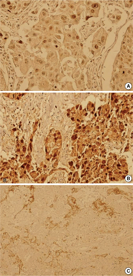
Immunohistochemical staining results for survivin (DAB with hematoxylin counterstaining). (A) Nuclear staining of survivin (×200), (B) Cytoplasmic and nuclear staining of survivin (×200), (C) Cytoplasmic staining of survivin (×200) in invasive ductal carcinoma.
jbc-12-285-g001.tif
Fig 2.
Kaplan-Meier survival estimates for positive and negative cases of breast cancer regarding (A) cytoplasmic survivin expression and (B) nuclear survivin expression.
jbc-12-285-g002.tif
Fig 3.
Kaplan-Meier survival estimates for using a ratio of 2 as a cutoff. (A) cytoplasmic to nuclear survivin expression ratio and (B) nuclear to cytoplasmic survivin expression ratio.
jbc-12-285-g003.tif
Fig 4.
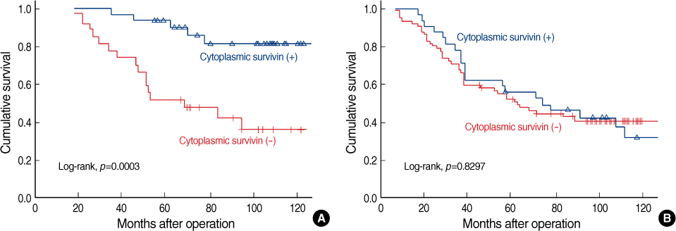
Kaplan-Meier survival curve according to lymph node negative group (A) and lymph node positive group (B) categorized by cytoplasmic survivin expression.
jbc-12-285-g004.tif
Fig 5.
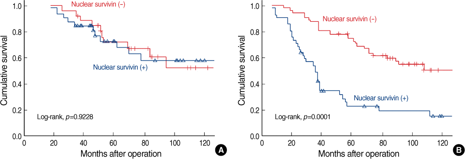
Kaplan-Meier survival curve according to lymph node negative group (A) and lymph node positive group (B) categorized by nuclear survivin expression.
jbc-12-285-g005.tif
Fig 6.
Kaplan-Meier survival curve according to TNM stage categorized by cytoplasmic survivin expression.
jbc-12-285-g006.tif
Fig 7.
Kaplan-Meier survival curve according to TNM stage categorized by nuclear survivin expression.
jbc-12-285-g007.tif
Table 1.
Patients characteristics
No. of patients %
Age (yr)
 <35 30 19.1
 ≥35 127 80.9
Size
 <2 cm 52 33.1
 2-5 cm 95 60.5
 >5 cm 10 6.4
Lymph node status
 Negative 59 37.6
 Positive 98 62.4
TNM stage
 1 23 14.6
 2 84 53.5
 3 50 31.8
Histologic grade
 I 38 30.6
 II+III 86 69.4
ER
 Negative 81 51.6
 Positive 76 48.4
PR
 Negative 100 63.7
 Positive 57 36.3
HER2
 Negative 46 52.3
 Positive 42 47.7
Hormonal therapy
 No 84 55.3
 Yes 68 44.7
Radiation therapy
 No 99 66.9
 Yes 49 33.1
Chemotherapy
 No 48 31.2
 Yes 106 68.8

ER=estrogen receptor; PR=progesterone receptor.

Table 2.
Localization of survivin expression by immunochemical methods
No. of patients %
Cytoplasm and/or nucleus
 Negative 56 35.7
 Positive 101 64.3
Cytoplasm
 Negative 93 59.2
 Positive 64 40.8
Nucleus
 Negative 83 52.9
 Positive 75 47.1
Both cytoplasm and nucleus
 Negative 35 22.3
 Positive 19 12.1
Cytoplasmic to nuclear ratio
 <2 105 66.9
 ≥2 52 33.1
Nuclear to cytoplasmic ratio
 <2 116 73.9
 ≥2 41 26.1
Table 3.
Association between survivin expression and tumor characteristics
C-expression
N-expression
Negative (93) Positive (64) p-value Negative (83) Positive (74) p-value
Age (yr) 0.003 0.688
 <35 25 (26.9) 5 (7.8) 17 (20.5) 13 (17.6)
 ≥35 68 (73.1) 59 (92.2) 66 (79.5) 61 (82.4)
Tumor size 0.385 0.274
 <2 cm 30 (32.3) 22 (34.4) 30 (36.2) 22 (29.7)
 2-5 cm 55 (59.1) 40 (62.5) 50 (60.2) 45 (60.8)
 >5 cm 8 (8.6) 2 (3.1) 3 (3.6) 7 (9.5)
LN status 0.012 0.189
 Negative 27 (29.0) 32 (50.0) 27 (32.5) 32 (43.2)
 Positive 66 (71.0) 32 (50.0) 56 (67.5) 42 (56.8)
TNM stage 0.012 0.547
 1 13 (14.0) 10 (15.6) 13 (15.7) 10 (13.5)
 2 42 (45.2) 42 (65.6) 41 (49.4) 43 (58.1)
 3 38 (40.8) 12 (18.8) 29 (34.9) 21 (28.4)
Histologic grade 0.297 0.051
 I 21 (27.3) 17 (36.2) 25 (39.1) 13 (21.7)
 II+III 56 (72.7) 30 (63.8) 39 (60.9) 47 (78.3)
HER2 status 0.194 0.099
 Negative 23 (48.9) 23 (56.1) 22 (55.0) 24 (50.0)
 Positive 24 (51.1) 18 (43.9) 18 (45.0) 24 (50.0)
ER 0.108 0.006
 Negative 53 (57.0) 28 (43.7) 34 (41.0) 47 (63.5)
 Positive 40 (43.0) 36 (56.3) 49 (59.0) 27 (36.5)
PR 0.129 0.030
 Negative 64 (68.8) 36 (56.3) 46 (55.4) 54 (73.0)
 Positive 29 (31.2) 28 (43.7) 37 (44.6) 20 (27.0)
HTx 0.124 0.441
 No 51 (58.0) 33 (51.6) 48 (60.0) 36 (50.0)
 Yes 37 (42.0) 31 (48.4) 32 (40.0) 36 (50.0)
RTx 0.339 0.738
 No 55 (64.0) 44 (71.0) 50 (64.1) 49 (70.0)
 Yes 31 (46.0) 18 (29.0) 28 (35.9) 21 (30.0)
CTx 0.014 0.602
 No 21 (23.3) 27 (42.2) 23 (28.8) 25 (33.8)
 Yes 69 (76.7) 37 (57.8) 57 (71.2) 49 (66.2)

C-expression=cytoplasmic expression; N-expression=nuclear expression; LN=lymph node; ER=estrogen receptor; PR=progesterone receptor; HTx=hormone therapy; RTx=radiation therapy; CTx=chemotherapy.

Table 4.
Prognostic significance of clinicopathologic variables
Variables Risk rate 95% CI p-value
Age (<35 vs ≥35) 0.532 0.320-0.887 0.013
Tumor size (<2 cm vs ≥2 cm) 0.926 0.584-1.468 0.743
LN metastasis (negative vs positive) 1.889 1.147-3.114 0.013
Histologic grade (I vs II, III) 1.786 1.040-3.067 0.035
Cytoplasmic survivin (negative vs positive) 0.540 0.336-0.868 0.011
Nuclear survivin (negative vs positive) 2.132 1.363-3.335 0.001
Cytoplasmic to nuclear ratio (<2 vs ≥2) 0.402 0.234-0.688 0.001
Nuclear to cytoplsmic ratio (<2 vs ≥2) 2.642 1.658-4.210 <0.001
ER status (negative vs positive) 0.654 0.419-1.022 0.059
PR status (negative vs positive) 0.514 0.313-0.843 0.008

LN=lymph node; ER=estrogen receptor; PR=progesterone receptor.

Table 5.
Multivariate Cox proportional hazard model for overall survival
Variables Risk rate 95% CI p-value
Age (<35 vs ≥35) 0.566 0.330-0.972 0.039
LN metastasis (negative vs positive) 2.046 1.141-3.668 0.016
Histologic grade (I vs II, III) 1.551 0.889-2.706 0.122
Cytoplasmic survivin (negative vs positive) 0.571 0.324-1.008 0.053
Nuclear survivin (negative vs positive) 2.258 1.370-3.721 0.001
Cytoplasmic to nuclear ratio* (<2 vs 2≥) 0.524 0.274-1.001 0.051
Nuclear to cytoplsmic ratio* (<2 vs 2≥) 2.010 1.116-3.466 0.012
PR status (negative vs positive) 0.678 0.392-1.170 0.163

CI=confidence interval; LN=lymph node; PR=progesterone receptor.

* Adjusted for age, lymph node metastasis, histologic grade and PR status.

TOOLS
Similar articles