

 PDF
PDF ePub
ePub Citation
 Print
Print


Abstract
Purpose
We wanted to identify the clinicopathologic factors that predict the presence of invasive cancer after core biopsy for ductal carcinoma in situ (DCIS).
Methods
The patients diagnosed with ductal carcinoma in situ on core biopsy (stereotactic or ultrasound) from February 2003 to May 2007 were identified by retrospectively reviewing the collected data. We analyzed the demographic data, the characteristics on the imaging studies and the histologic features on DCIS. We assessed the factors that included age, the physical examination, the radiologic findings, the biopsy method, and the histologic findings related to the presence of invasive cancer after core biopsy.
Results
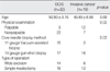
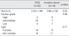
Fifty-one patients were diagnosed with DCIS after core biopsy. Of the 51 patients, 19 patients had invasive carcinoma diagnosed on final excision. The factors that correlated with invasion were the biopsy method, a palpable mass and a mammographic mass, regardless of calcification. A high nuclear grade, the comedo type, age, and the tumor size were not related to presence of invasive cancer.

References
1. Korea Central Cancer Registry. 2002 Annual report of the Korea Central Cancer Registry. 2003. Gwacheon: Ministry of Health and Welfare, Republic of Korea.
2. Emster VL, Ballard-Barbash R, Barlow WE, Zheng Y, Weaver DL, Cutter G, et al. Detection of ductal carcinoma in situ in women undergoing screening mammography. J Natl Cancer Inst. 2002. 94:1546–1554.

3. Fentiman IS. The dilemmaof in situ carcinoma of the breast. Int J Clin Pract. 2001. 55:680–683.
4. Wahedna Y, Evans AJ, Pinder SE, Ellis IO, Blamey RW, Geraghty JG. Mammographic size of ductal carcinoma in situ does not predict the presence of an invasive focus. Eur J Cancer. 2001. 37:459–462.
5. Dillon MF, McDermott EW, Quinn CM, O'Doherty A, O'Higgins N, Hill AD. Predictors of invasive disease in breast cancer when core biopsy demonstrates DCIS only. J Surg Oncol. 2006. 93:559–563.
6. Lee CH, Carter D, Philpotts LE, Couce ME, Horvath LJ, Lange RC, et al. Ductal carcinoma in situ diagnosed with stereotactic core needle biopsy: can invasion be predicted. Radiology. 2000. 217:466–470.
7. Rutstein LA, Johnson RR, Poller WR, Dabbs D, Groblewski J, Rakitt T, et al. Predictors of residual invasive disease after core needle biopsy diagnosis of ductal carcinoma in situ. Breast J. 2007. 13:251–257.
8. Verkooijen HM, Peeters PH, Buskens E, Koot VC, Borel Rinkes IH, Mali WP, et al. Diagnostic accuracy of large-core needle biopsy for nonpalpable breast disease: a meta-analysis. Br J Cancer. 2000. 82:1017–1021.
9. Leifland K, Lagerstedt U, Svane G. Comparison of stereotactic fine needle aspiration cytology and core needle biopsy in 522 non-palpable breast lesions. Acta Radiol. 2003. 44:387–391.
10. Crowe JP, Rim A, Patrick RJ, Rybicki LA, Grundfest-Broniatowski SF, Kim JA, et al. Does core needle breast biopsy accurately reflect breast pathology? Surgery. 2003. 134:523–528.
11. Jackman RJ, Burbank F, Parker SH, Evans WP 3rd, Lechner MC, Richardson TR, et al. Stereotactic breast biopsy of nonpalpable lesions: determinants of ductal carcinoma in situ underestimation rates. Radiology. 2001. 218:497–502.
12. Yen TW, Hunt KK, Ross MI, Mirza NQ, Babiera GV, Meric-Bernstam F, et al. Predictors of invasive breast cancer in patients with an initial diagnosis of ductal carcinoma in situ: a guide to selective use of sentinel lymph node biopsy in management of ductal carcinoma in situ. J Am Coll Surg. 2005. 200:516–526.
13. Houssami N, Ciatto S, Ellis I, Ambrogetti D. Underestimation of malignancy of breast core-needle biopsy: concepts and precise overall and category-specific estimates. Cancer. 2007. 109:487–495.
14. Liberman L, Smolkin JH, Dershaw DD, Morris EA, Abramson AF, Rosen PP. Calcification retrieval at stereotactic, 11-gauge, directional, vacuum-assisted breast biopsy. Radiology. 1998. 208:251–260.
15. Silverstein MJ, Poller DN, Waisman JR, Colburn WJ, Barth A, Gierson ED, et al. Prognostic classification of breast ductal carcinoma-in-situ. Lancet. 1995. 345:1154–1157.
16. Fisher ER, Costantino J, Fisher B, Palekar AS, Redmond C, Mamounas E. Pathologic findings from the National Surgical Adjuvant Breast Project (NSABP) Protocol B-17. Intraductal carcinoma (ductal carcinoma in situ). The National Surgical Adjuvant Breast and Bowel Project Collaborating Investigators. Cancer. 1995. 75:1310–1319.
17. Hoorntje LE, Schipper ME, Peeters PH, Bellot F, Storm RK, Borel Rinkes IH. The finding of invasive cancer after a preoperative diagnosis of ductal carcinoma-in-situ: causes of ductal carcinoma-in-situ underestimated with stereotactic 14-gauge needle biopsy. Ann Surg Oncol. 2003. 10:748–753.
18. Bagnall MJ, Evans AJ, Wilson AR, Pinder SE, Denley H, Geraghty JG, et al. Predicting invasion in mammographically detected microcalcification. Clin Radiol. 2001. 56:828–832.
19. Renshaw AA. Predicting invasion in the excision specimen from breast core needle biopsy specimens with only ductal carcinoma in situ. Arch Path Lab Med. 2002. 126:39–41.
20. Huo L, Sneige N, Hunt KK, Albarracin CT, Lopez A, Resetkova E. Predictors of invasion in patients with core-needle biopsy-diagnosed ductal carcinoma in situ and recommendations for a selective approach to sentinel lymph node biopsy in ductal carcinoma in situ. Cancer. 2006. 107:1760–1768.
21. Lagios MD, Margolin FR, Westdahl PR, Rose MR. Mammographically detected duct carcinoma in situ. Frequency of local recurrence following tylectomy and prognostic effect of nuclear grade on local recurrence. Cancer. 1989. 63:618–624.
22. King TA, Farr GH, Cederbom GJ, Smetherman DH, Bolton JS, Stolier AJ, et al. A mass on breast imaging predicts coexisting invasive carcinoma in patients with a core biopsy diagnosis of ductal carcinoma in situ. Am Surg. 2001. 67:907–912.
23. Hoda SA, Rosen PP. Practical considerations in the pathologic diagnosis of needle core biopsies of breast. Am J Clin Pathol. 2002. 118:101–108.
 XML Download