

 PDF
PDF ePub
ePub Citation
 Print
Print


Abstract
Purpose
We purpose to determine the correlation of HER-2/neu and paxillin expression in ductal carcinoma in situ (DCIS), invasive ductal carcinoma with ductal carcinoma in situ (IDC with DCIS) and mucinous carcinoma.
Methods
To evaluate the expression of HER-2/neu and paxillin, the immunohistochemical staining was performed for 13 cases of DCIS, 13 cases of IDC with DCIS and 6 cases of mucinous carcinoma.
Results
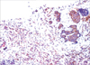
The DCIS and IDC were associated with infiltration of the inflammatory cells, especially in the comedo type and solid type of tumor. In cases with infiltration of the inflammatory cells, HER-2/neu and paxillin were strongly expressed. When comparing the expression level of HER-2/neu from adjacent normal tissue between DCIS and IDC with DCIS, expression of HER-2/neu was similar to that of normal tissue adjacent to DCIS. However, in the adjoining normal ductal epithelial cells, paxillin was highly expressed in cells of all of the tumor types, and especially for IDC with DCIS. HER-2/neu and paxillin were not expressed in mucinous carcinoma cells in all cases.
Conclusion
HER-2/neu in the DCIS and IDC with infiltration of inflammatory cells shows higher expression than non-inflammatory DCIS and IDC. If normal duct epithelial cells show a high level of HER-2/neu expression, the epithelial cells have a high probability of transformation into anaplastic cells. However, paxillin appears to have no value as a prognostic factor. The difference of expression of HER-2/neu between IDC with DCIS and DCIS suggests a different origin of tumor cells. The growth pattern of mucinous carcinoma cell is different from the that of DCIS or IDC cell, which grow slowly.
Figures and Tables
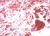 | Fig 2The Paxillin showed score 3 pattern in solid type DCIS portion and invasive portion in area in Fig 1 (×40).
|
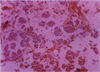
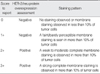
Table 4
Expression of HER-2/neu and paxillin in part of infiltrating ductal carcinoma and part of ductal carcinoma in situ

![]()


References
1. Turner CE. Paxiliin. Int J Biochem Cell Biol. 1998. 30:955–959.
2. Madan R, Smolkin MB, Cocker R, Fayyad R, Oktay MH. Focal adhesion proteins as markers of malignant transformation and prognostic indicators in breast carcinoma. Hum Pathol. 2006. 37:9–15.

3. Schaller MD. Paxillin: a focal adhesion-assocatied adaptor protein. Oncogene. 2001. 20:6459–6472.
4. Scibelli A, d'Angelo D, Pelagalli A, Tafuri S, Avallone L, Della Morte R, et al. Expression levels of focal adhesion-associated proteins paxillin and p130CAS in carine and feline mammary tumors. Vet Res. 2003. 34:193–202.
5. Yamaguchi R, Mazaki Y, Hirota K, Hashimoto S, Sabe H. Mitosis specific serine phosphorylation and downregulation of one of the focal adhesion proteins, paxillin. Oncogene. 1997. 15:1753–1761.
6. Salgia R, Li JL, Ewaniuk DS, Wang YB, Sattler M, Chen LB, et al. Expression of the focal adhesion protein paxillin in lung cancer and its relation to cell motility. Oncogene. 1999. 18:67–77.
7. Pelagalli A, Scibelli A, Lombardi P, d'Angelo D, Tortora G, Staiano N, et al. Expression of the focal adhesion protein paxillin in normal and breast cancer tissues. Vet Res commun. 2003. 27:343–346.
8. Turner CE. Paxillin: a cytoskeletal target for tyrosine kinases. Bioessays. 1994. 16:47–52.
9. Cance WG, Harris JE, Iacocca MV, Roche E, Yang X, Chang J, et al. Immunohistochemical analyses of focal adhesion kinase expression in benign and malignant breast and colon tissues: correlation with preinvasive and invasive phenotypes. Clin Cancer Res. 2000. 6:2417–2423.
10. Oktay MH, Oktay K, Hamele-Bena D, Buyuk A, Koss LG. Focal adhesion kinase as a marker of malignant phenotype in breast and cervical carcinomas. Hum Pathol. 2003. 34:240–245.
11. Kornberg LJ. Focal adhesion kinase expression in oral cancer. Head Neck. 1998. 20:634–639.
12. Short SM, Yoder BJ, Tarr SM, Prescott NL, Laniauskas S, Coleman KA, et al. The expression of the cytoskeletal focal adhesion protein paxillin in breast cancer correlates with HER2 overexpression and may help predict response to chemotherapy: a retrospective immunohistochemical study. Breast J. 2007. 13:130–139.
13. Press MF, Cordon-Cardo C, Slamon DJ. Expression of the HER-2/neu proto-oncogene in normal human adult and fetal tissues. Oncogene. 1990. 5:953–962.
14. Natal PG, Nicotra MR, Bigotti A, Venturo I, Slamon DJ, Fendly BM, et al. Expression of the p185 encoded by HER-2/neu oncogene in normal and transformed normal tissue. Int J Cancer. 1990. 45:457–461.
15. Ratcliffe N, Wells W, Wheeler K, Memoli V. The combination of the in situ hybridization and immunohistochemical analysis: an evaluation of HER-2/neu expression in paraffin embedded breast carcinoma and adjacent normal appearing breast epithelium. Mod Pathol. 1997. 10:1247–1252.
16. Allred DC, Clark GM, Molina R, Tandon AK, Schnitt SJ, Gilchrist KW, et al. Overexpression of Her-2/neu and its relationship with other prognostic change during the progession of in situ to invasive breast cnacer. Hum Pathol. 1992. 23:974–979.
17. Lodato RF, Maquire HC Jr, Greene MI, Weiner DB, Livolsi VA. Immunohistochemical evaluation of c-erbB-2 oncogene expression in ductal carcinoma in situ and atypical ductal hyperplasia of the breast. Mod Pathol. 1990. 3:449–454.
18. De Potter CR, Van Daele S, Van de Vijver MJ, Pauwels C, Maertens G, De Boever J, et al. The expression of the neu oncogene product in breast lesions and in normal fetal and adult human tissue. Histopathology. 1989. 15:351–362.
19. Elston CW, Ellis IO. Pathological prognostic factors in breast cancer. I. The value of histological grade in breast cancer: experience from a large study with long-term follow-up. Histopathology. 1991. 19:403–410.
20. Chang L, Karin M. Mammalian MAP kinase signalling cascades. Nature. 2001. 410:37–40.
21. Garrington TP, Johnson GL. Organization and regulation of mitogene-activated protein kinase signalling pathways. Curr Opin Cell Biol. 1999. 11:211–218.
22. Neves SR, Ram PT, Iyengar R. G protein pathways. Science. 2002. 296:1636–1639.
23. Pierce KL, Premont RT, Lefkowitz RJ. Seven-transmembrane receptors. Nat Rev Mol Cell Biol. 2002. 3:639–650.
24. Berridge MJ, Lipp P, Bootman MD. The versatility and university of calcium signalling. Nat Rev Mol Cell Biol. 2000. 1:11–21.
25. Bootman MD, Berridge MJ, Roderick HL. Calcium signalling: more messengers more channels, more complexity. Curr Biol. 2002. 12:R563–R565.
26. Farfel Z, Bourne HR, Iiri T. The expanding spectrum of G protein disease. N Engl J Med. 1999. 340:1012–1020.
27. Aaronson DS, Horvath CM. A road map for those who don't know JAk-STAT. Science. 2002. 296:1653–1655.
28. Hynes RO. Integrins: bidirectional Allosteric signalling machine. Cell. 2002. 110:673–687.
29. Stupack DG, Cheresh DA. Get a ligand, get a life: integrins, signalling and cell survival. J Cell Sci. 2002. 115:3729–3738.
30. Bradshaw AD, Sage EH. SPARC, a matricellular protein that functions in cellular differentiation and tissue response to injury. J Clin Invest. 2001. 107:1049–1054.
31. Pellegrini C, Falleni M, Marchetti A, Cassani B, Miozzo M, Buttitta F, et al. HER-2/Neu alterations in non-small cell lung cancer: a comprehensive evaluation by real time reverse transcription-PCR, fluorescence in situ hybridization, and immunohistochemistry. Clin Cancer Res. 2003. 9:3645–3652.
32. Schnitt SJ, Connolly JL, Harris JR, Hellman S, Cohen RB. Pathologic predictors of early local recurrence in Stage I and II breast cancer treated by primary radiation therapy. Cancer. 1984. 53:1049–1057.
33. Wright C, Angus B, Nicholson S, Sainsbury JR, Cairns J, Gullick WJ, et al. Expression of c-erbB-2 oncoprotein: a prognostic indicator in human breast cancer. Cancer Res. 1989. 49:2087–2090.
34. Payne SJ, Bowen RL, Jones JL, Wells CA. Predictive markers in breast cancer--the present. Histopathology. 2008. 52:82–90.
35. Diab SG, Clark GM, Osborne CK, Libby A, Allred DC, Elledge RM. Tumor characteristics and clinical outcome of tubular and mucinous breast carcinomas. J Clin Oncol. 1999. 17:1442–1448.
 XML Download