

 PDF
PDF ePub
ePub Citation
 Print
Print


Abstract
Percutaneous vertebroplasty using PMMA was first performed in France by Deramond in 1984. It was later used to treat vertebral compression fractures caused by osteoporosis. With osteoporotic compression fractures, reported complication were few and minor. However, the principal risk of such a percutaneous technique is the leak of PMMA into the spinal canal or neural foramina. Despite being uncommon, major neurologic complications demand surgical interventions that prevent permanent neurologic dysfunction. We experienced a case of root compression during percutaneous vertebroplasty, which was treated by early surgical posterior decompression.
REFERENCES
1). Barr JD, Barr MS, Lemley TJ, Mccann RM. Percutaneous vertebroplasty for pain relief and spinal stabilization. Spine. 25:923–928. 2000.

2). Cotton A, Boutry N, Cortet B, et al. Percutaneous vertebroplasty: state of the art. Radiographics. 18:311–323. 1998.
3). Cotton A, Dewatre F, Cortet B, et al. Percutaneous vertebroplasty for osteolytic metastases and myeloma: effects of the percentage of lesion filling and the leakage of methyl methacrylate at clinical followup. Radiology. 200:525–530. 1996.
4). Deramond H, Depreister C, Galibert P, Gars DL. Percutaneous vertebroplasty with polymethylmethacry -late;technique, indication, and results. Radiologic clinics of north america. 36:533–546. 1998.
5). Harrington KD. Major neurological complications following percutaneous vertebroplasty with polymethylmethacrylate. J Bone Joint Surg. 83:1070–1073. 2001.
6). Jensen ME, Avery JE, Mathis JM, Kallmess DF, Cloft HJ, Dio JE. Percutaneous polymethylmethacrylate vertebroplasty in the treatment of osteoporotic vertebral body compression fracture: technical aspects. Am J Neuroradiol. 18:1897–1904. 1997.
7). Liebschner MAK, Rosenberg WS, Keaveny TM. Effects of bone cement volume and distribution on vertebral stiffness after vertebroplasty. Spine. 26:1547–1554. 2001.
8). Mathis JM, Barr JD, Belkoff SM, Barr MS, Jensen ME, Deramond H. Percutaneous vertebroplasty: a developing standard of care for vertebral compression fractures. Am J Neuroradiol. 22:373–382. 2001.
9). Mathis JM, Eckel TS, Belkoff SM, Deramond H. Percutaneous vertebroplasty: a therapeutic option for pain associated with vertebral compression fracture. J Back Musculoskel Rehab. 13:11–17. 1999.
10). Ratliff J, Nguyen T, Heiss J. Root and spinal cord compression from methylmethacrylate vertebroplasty. Spine. 26:300–302. 2001.
11). Weill A, Chiras J, Simon JM, Rose M, Sola-Martinez T, Enkaoua E. Spinal metastasis: indications for and results of percutaneous injection of acrylic surgical cement. Radiology. 199:241–247. 1996.
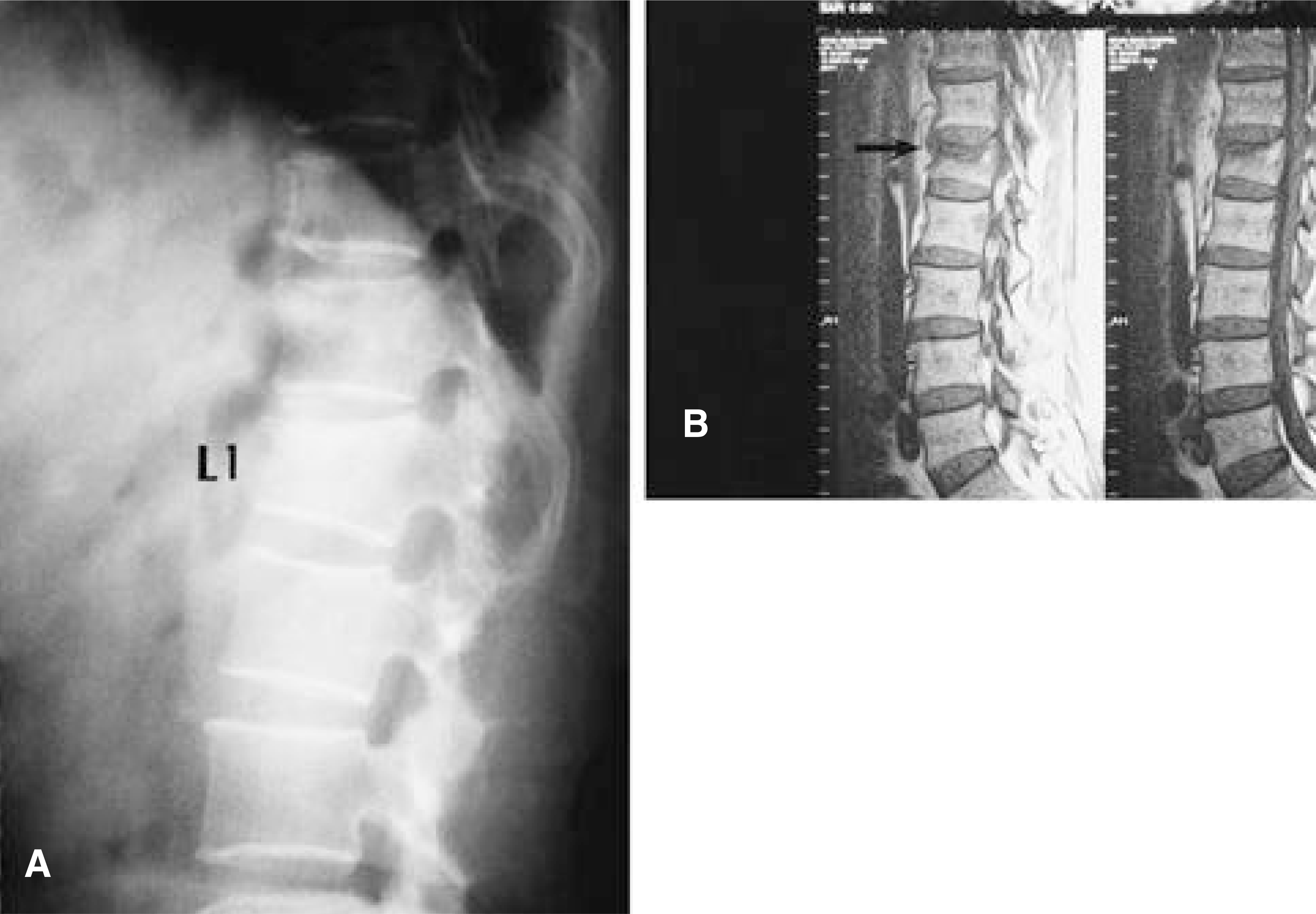
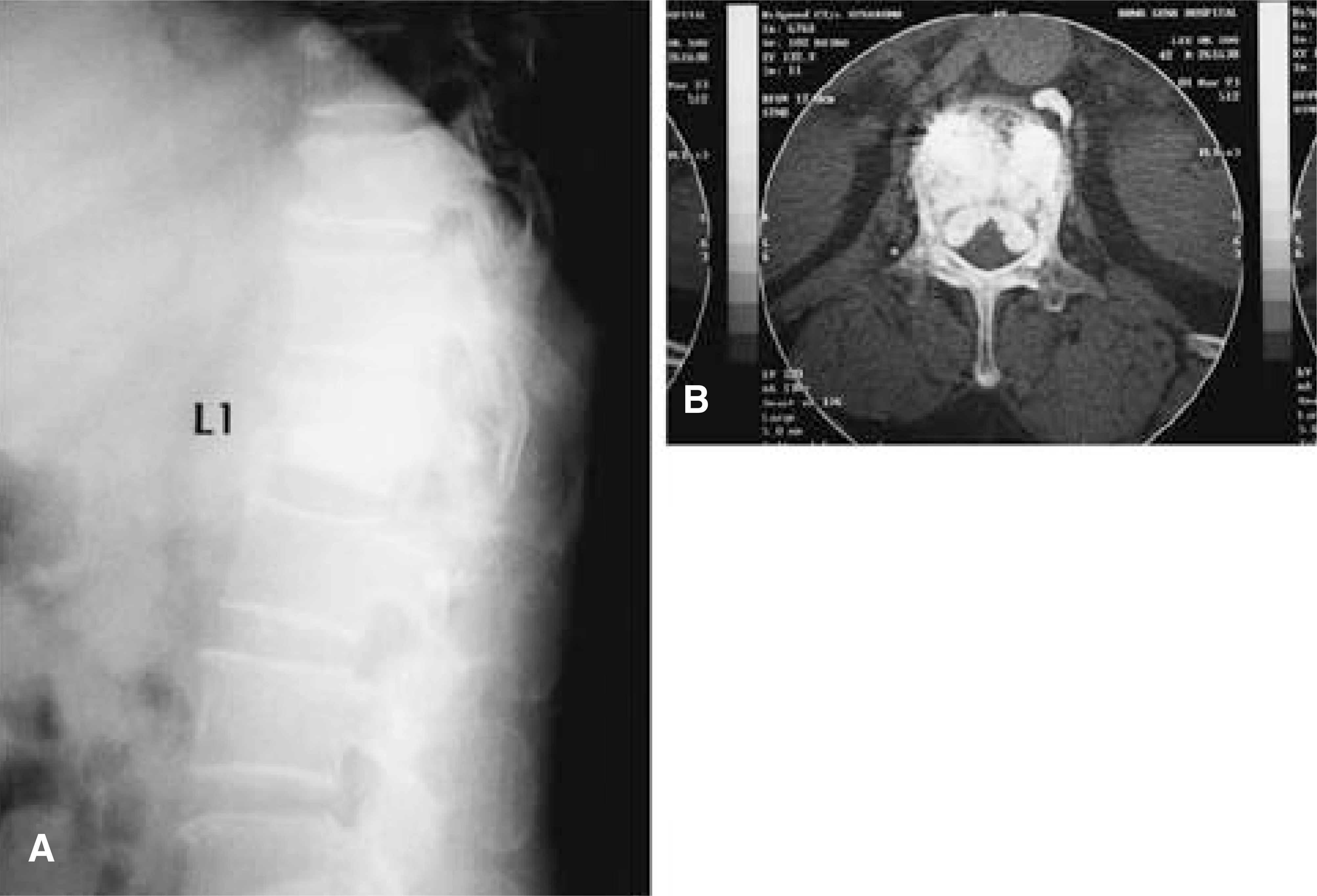
Fig. 1-A.
Lateral radiography of lumbar spine. It shows compression fracture at the L1 level. Fig. 1-B. Sagittal T1-weighted image shows low signal in the marrow space of L1.

 XML Download