Abstract
Study Design
A retrospective analysis was performed to identify the diagnostic and therapeutic factors related to postoperative compressive neuropathy by hematoma after posterior spinal decompressive surgery.
Objectives
To document by analysis the clinical course of postoperative compressive neuropathy by hematoma, the efficacy of early surgical decompression, and to recommend methods of prevention.
Summary of Literature Review
Various diagnostic and treatment modalities have been applied to postoperative compressive neuropathy after spinal surgery. However, the timing of surgical decompression remains controversial.
Materials and Methods
Five cases of postoperative compressive neuropathy after posterior spinal decompressive surgery, which occurred from May 1996 to May 2000, were investigated in terms of causes, clinical courses, and management profiles after early surgical decompression, and final outcome.
Results
Five cases (2.14%) among 234 patients were managed by re- decompression including the evacuation of hematoma. Four cases, which had been managed by earlier surgical decompression showed neurologic improvement after 2 postoperative weeks, and achieved favorable clinical results without grave neurologic sequelae. However, in one case, in which surgical decompression had been delayed, weakness of the peroneii remained.
Go to : 
REFERENCES
1). Bertalanffy H and Eggert HR. Complications of anterior cervical disectomy without fusion in 450 consecutive patients. Acta Neurochir. 99:41–50. 1989.
2). Carlson G, Abitbol JJ and Garfin SR. Prevention of complication in surgical management of back pain and sciatica. Orthop Clin N Am. 22:345–351. 1991.
3). Cho JL, Lee KH, Youn WK and Lee CW. Transpedicular screw fixation in lumbar spinal stenosis. J of Korean Spine Surg. 1:182–190. 1994.
4). Choi CH and Kim NH. A study of electrodiagnostic changes after decompression of chronic cauda equina compression in dogs. J of Korean Orthop Assoc. 32:163–176. 1997.
5). Dickman CA, Shedd SA, Spetzler RF and Shetter AG. Spinal epidural hematoma associated with epidural anesthesia: Complications of systemic heparinization in patients receiving peripheral vascular thrombolytic therapy. Anesth. 72:947–950. 1990.

6). Floman Y, Wiesel SW and Rothman RH. Cauda equina syndrome presenting as a herniated lumbar disc. Clin Orthop. 147:234–237. 1979.
7). Foo D and Rossier AB. Preoperative neurologic status in predicting surgical outcome of spinal epidural hematoma. Surg Neurol. 15:389–401. 1981.
8). Jonas AF. Spinal fractures: Opinions based on observations of sixteen operations. JAMA. 57:859–865. 1911.
9). Kim HS, Hong KD, Ha SS and Lee SW. Cauda equina syndrome following lumbar spine surgery. J of Korean Orthop Assoc. 32:1773–1781. 1997.
10). Kim YM, Won CH, Seo JB, Choi ES and Lee HS. Lumbar disc herniation with cauda equina syndrome after self traction therapy. J of Korean Spine Surg. 6:469–474. 1999.
11). Lee HM, Kim NH, Park BM and Lee DW. Neurologic complication after spine surgery. J of Korean Orthop Assoc. 29:954–964. 1994.
12). Markham JW, Lynge HN and Stahlman GEB. The syndrome of spontaneous spinal epidural hematoma. J Neurosurg. 26:334–342. 1967.

13). McLaren AC and Bailey SI. Cauda equina syndrome: A complication of lumbar discectomy. Clin Orthop. 204:143–149. 1986.
14). Park BM and Won YY. Clinical observation on 8 cases of cauda equina syndrome. J of Korean Orthop Assoc. 23:184–192. 1988.
15). Wagner S, Forsting M and Hacke W. Spontaneous res -olution of a large spinal epidural hematoma: Case report. Neurosurg. 38:816–818. 1996.
16). Wiesel SW. Neurologic complications after lumbar laminectomy: A standard approach to the multiply oper -ated lumbar spine. 1st ed.Garfin SR, editor. Baltimore: Williams and Wilkins;p. 64–74. 1989.
17). Yun HK, Jeon HS, Cho KN and Choi JH. Thoracolumbar epidural hematoma complicated by cauda equina syndrome: complication of systemic heparinization following epidural anesthesia. J of Korean Orthop Assoc. 33:1120–1125. 1998.
Go to : 
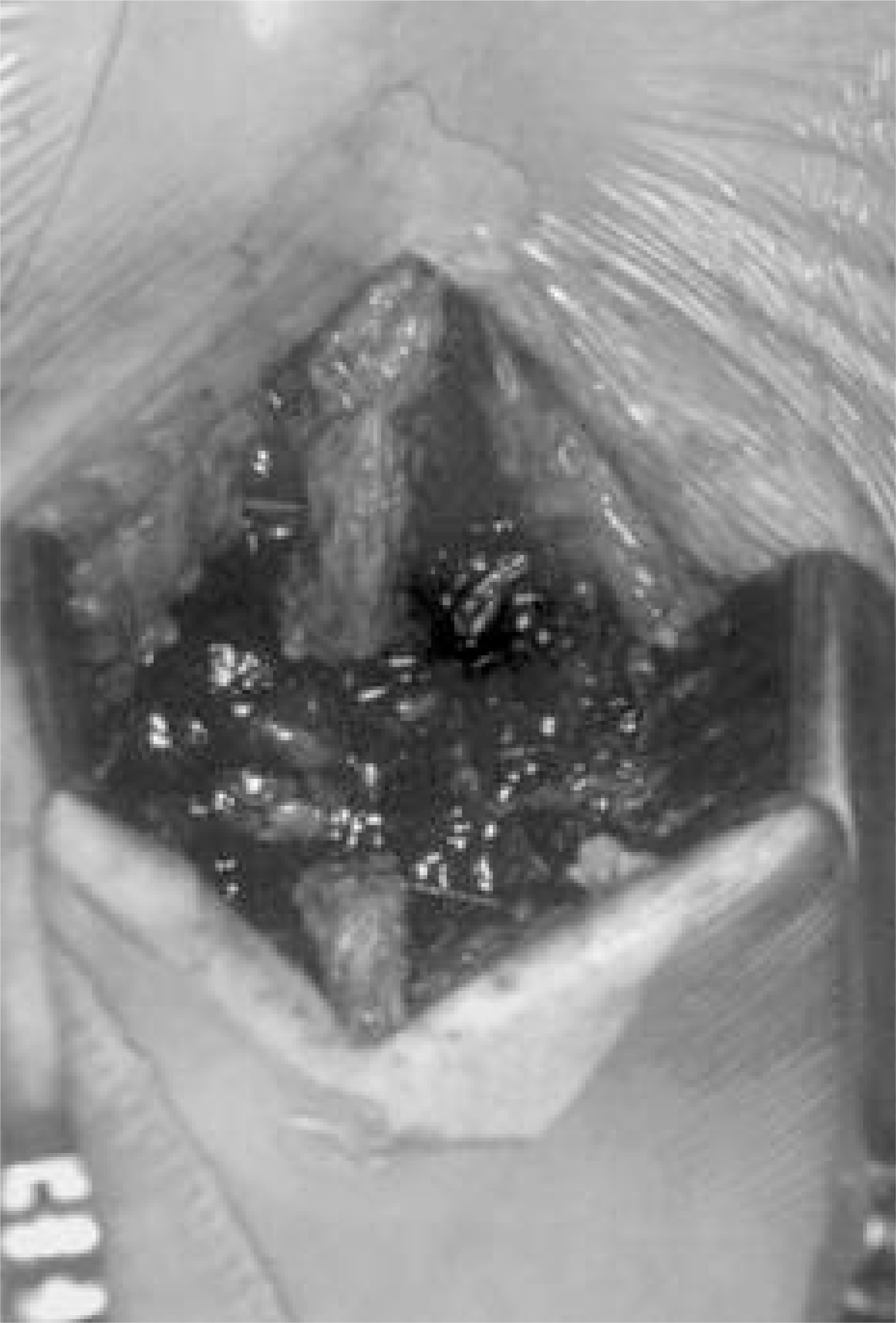 | Fig. 3.Abundant blood clots compressing dura were evacuated within 24 hours after initial decompressive surgery |
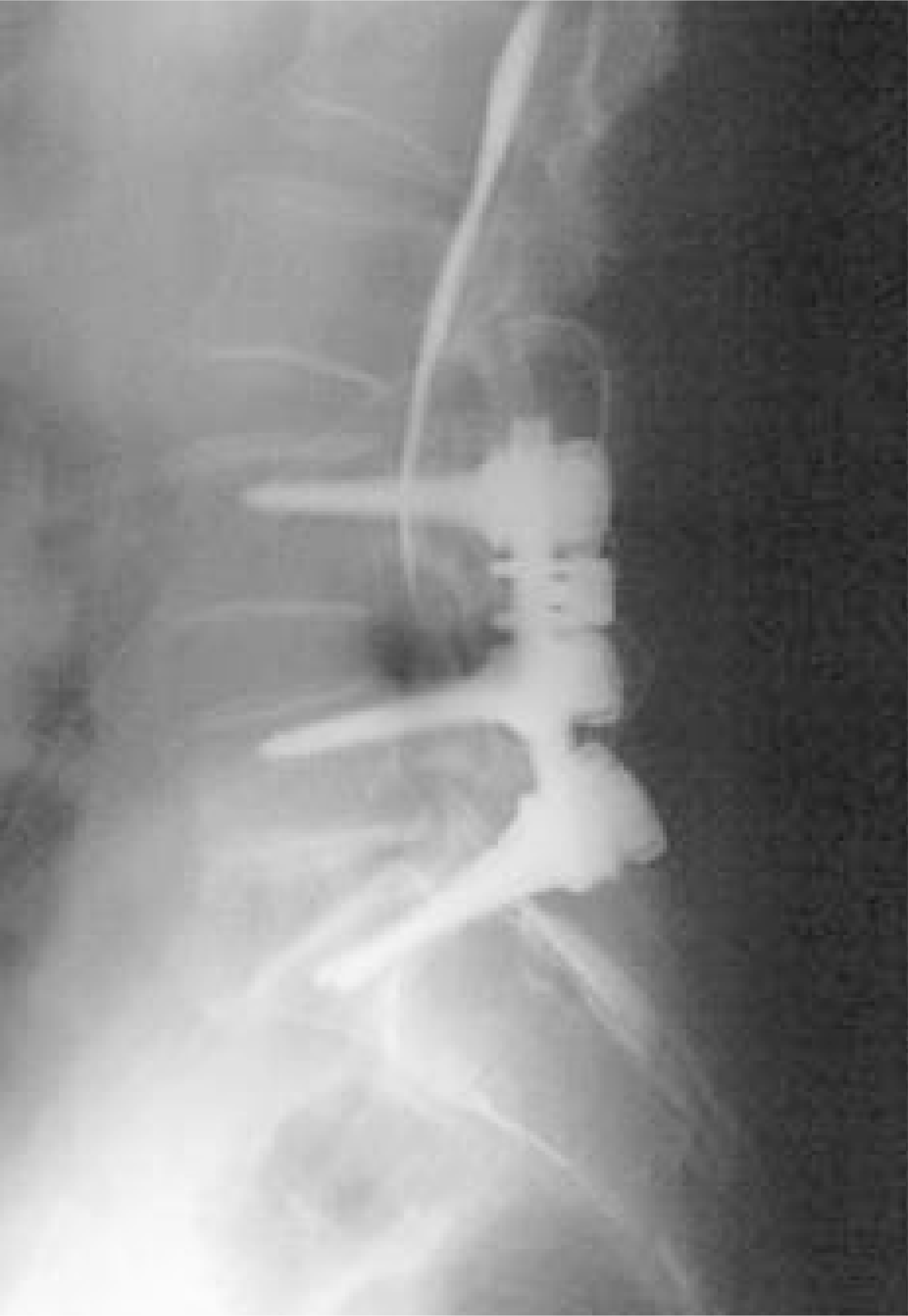
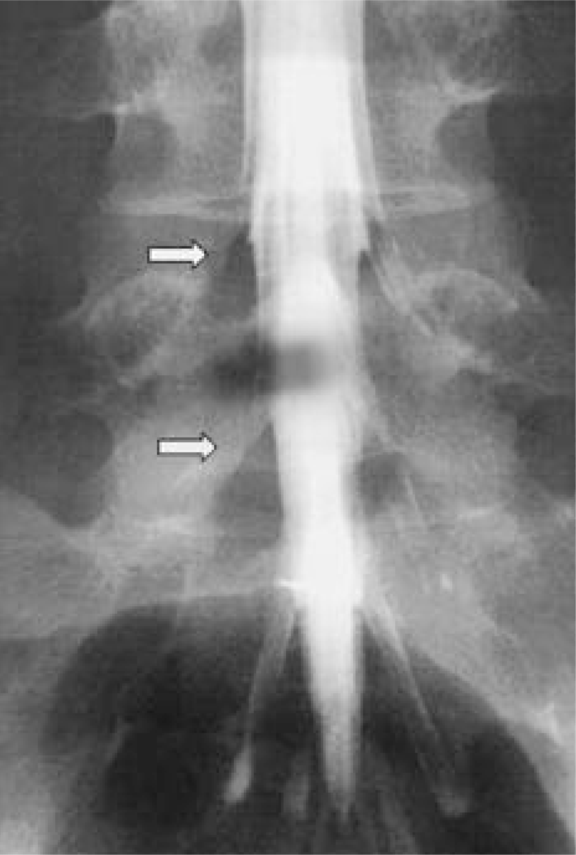 | Fig. 4.Right L5 & S1 roots were not visualized in myelogram performed on 11th postoperative day (white arrows) |
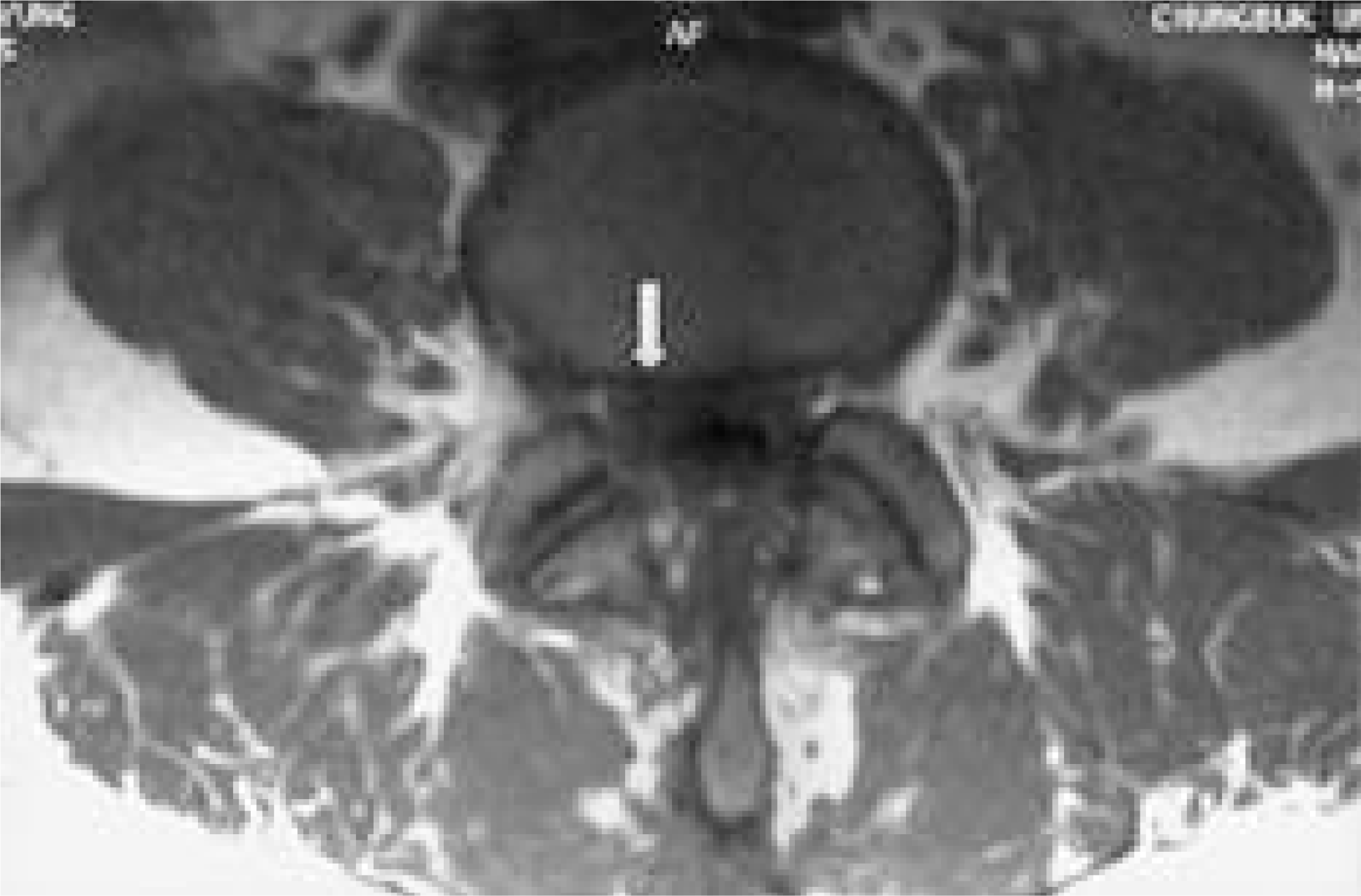 | Fig. 5.MRI performed on postoperative fourth month shows severe scar adhesion by granulation of hematoma at right L5 root (white arrow) |
Table 1.
Profile of Compressive Neuropathy by Hematoma after Spine Surgery
| Case | Sex/Age | Spine problem | Level of decompressive op.§ | Level of compressive neuropathy | Interval to reoperation |
|---|---|---|---|---|---|
| 1 | F/65 | Spinal stenosis | L4-S1 | Rt. L5, S1 root | 24hr |
| 2 | M/38 | L2 bursting Fx.∗ | L2 | Cauda equina | 20hr |
| 3 | F/35 | Traumatic disc rupture | L3-4 | Cauda equina | 16hr |
| 4 | F/24 | HIVD‡ | L4-5, L5-S1 | Rt. L5, S1 root | 8month |
| 5 | M/51 | CSM† | C3-7 | Lower cervical cord & root | 24hr |
Table 2.
Profile of Postoperative Neurologic Signs of Five Cases
| Case | Motor signs (Rt. / Lt.) | Sciatica | Sensory change | D.T.R∗ | Anal reflex | Ankle clonus | |
|---|---|---|---|---|---|---|---|
| Knee | Ankle | ||||||
| 1 | TA† 2+ / 4+ EPH‡ 3 / 4+ Peronei 3 / 4+ | Rt. | Rt. L5 & S1 area ↓ Perianal sense (++) | +/+ | +/+ | ++ | -/- |
| 2 | TA 0 / 0 EPH 0 / 0 Peronei 0 / 0 | Both | Both L3-S1 area ↓ Perianal sense (±) | -/- | -/- | ± | -/- |
| 3 | TA 2 / 0 EPH 0 / 0 Peronei 0 / 0 | Both | Both L4-S1 area ↓ Perianal sense (-) | -/- | -/- | - | -/- |
| 4 | TA 3+ / 5 EPH 2 / 5 Peronei 1+ / 5 | Rt. | Rt. L5 & S1 area ↓ Perianal sense (++) | +/+ | -/+ | ++ | -/- |
| 5 | Finger flexor 3 / 3 Finger abductor 2 / 3 | - | Both C5-C8 area ↓ Perianal sense (++) | ++/++ Biceps jerk Triceps jerk | ++/+++++++ | ++ | -/- |
Table 3.
Profile of Neurologic Signs at Final Follow-up of Five Cases
| Case | Follow-up period | Motor signs (Rt. / Lt.) | Sensory signs | D.T.R∗ | Final complaint | |
|---|---|---|---|---|---|---|
| Knee | Ankle | |||||
| 1 | PO§14 month | TA† 4+ / 5 EPH‡ 4+ / 5 Peronei 5 / 5 | symmetric intact Perianal sense (++) | +/++ | ++/++ | Low back pain |
| 2 | PO 22 month | TA 4+ / 5 EPH 4+ / 5 Peronei 4+ / 5 | Rt. S1 area ↓ Perianal sense (++) | +/++ | +/++ | Both L/E∫ weakness |
| 3 | PO 17 month | TA 5 / 4+ EPH 5 / 5 Peronei 5 / 5 | symmetric intact Perianal sense (++) | ++/++ | +/+ | Low back pain |
| 4 | PO 16 month | TA 3+ / 5 EPH 4- / 5 Peronei 3- / 5 | Rt. L5 area ↓ Perianal sense (++) | +/++ | +/++ | Rt. foot drop |
| 5 | PO 12 month | Finger flexor 5 / 5 Finger abductor 5 / 5 | Both C6-C7 area ↓ Perianal sense (++) | ++/++ Biceps jerk Triceps jerk | ++/++++++ | Both hand tingling sense |


 PDF
PDF ePub
ePub Citation
 Print
Print


 XML Download