Abstract
Purpose
To compare clinical results and radiologic changes after arthroscopic and microscopic discectomy of lumbar disc herniation in teenagers who have no degenerative change.
Materials and Methods
From Jan 1990 to A ug 2001. 70 lumbar disc herniations were performed in patients below 20 years old who were admitted to our department, among these 67 cases (49:male, 18:female) were evaluated for at least 1 year. Their average age was 18.1years (13 ~ 20 years). Forty- six received microscopic discectomy and 21 arthroscopic discectomy. Mean follow-up duration was 26.4 months (12~ 88 months).
Results
Clinical results and disc height change were compared between the arthroscopic and microscopic discectomy groups using the criteria of MacNab, and the relationship between disc height change and clinical results, excised disc volume, operative technique, body mass index and symptom duration were investigated. Clinically there was no significant difference between the two groups (p=0.425), and their results were the same as those of adults. A t the 1 year- follow up, disc height changes showed no correlation with the method of operation (p=0.996) or the volume of the excised disc. Postoperative disc height in teenagers of lumbar disc herniation who showed no degenerative change significantly decreased with time, but no significant relation was observed between disc height changes and clinical results, operative technique, excised disc volume, body mass index, involved disc site or symptom duration between the two groups.
Go to : 
REFERENCES
1). Adams S, Yoshinori S and Hansjoerg L. Does percutaneous nucleotomy with discectomy replace conventional discectomy? Clin Orthop. 238:24–34. 1989.
2). Chung JY and Joo DC. A prospective randomized study of arthroscopic and microscopic discectomy in protruded lumbar disc herniation. J of Korean Orthop Assoc. 35:119–125. 2000.
3). Clarke N.M.P and Cleak, D.K.Intervertebral lumbar disc prolapse in children and adolescents. J. Pediatr Orthop. 3:202–206. 1983.
4). Dabbs VM and Dabbs LG. Correlation between disc height narrowing and low-back pain. Spine. 15:1366–1369. 1990.

5). Epstein N. Surgically confirmed cauda equina and nerve root injury following percutaneous discectomy at an outside institution: A case report. J of Spine Disorders. 3:380–383. 1990.
6). Epstein JA and Lavine LS. Herniated lumbar intervertebral disc in teenage children. J Neurosurgery. 21:1070. 1964.
7). Fusek I. The clinical picture and the findings of operation in lumbar intervertebral disc herniation in adolescent. Cs Neurol. 33:199. 1970.
8). Grobler JJ, Simons EH and Barrington TW. Intervertebral disc herniation in the adolescent. Spine. 4:267–278. 1979.

9). Hahn JF and Mason L. Low back pain in children, in Hardy R(ed): Lumbar disc disease. New York: Raven Press;p. 217–228. 1982.
10). Hijikatat S. Percutaneous nucleotomy: A new concept technique and 12 years experience. Clin Orthop. 238:9–23. 1979.
11). Javid MJ, Nordby EJ, Ford LT, Hejna WJ, Whisler WW, Burton C, Millett DK, Wiltse LL, Widell EH Jr, Boyd RJ, Newton SE and Thisted R. Safety and effica - cy of chymopapain in herniated nucleus pulposus with sciatica. JAMA. 249:2489–2494. 1983.
12). Kambin P and Lawrence FC. Arthroscopic discectomy versus nucleotomy techniques. Clin in Sports Med. 337:49–57. 1997.
13). Kambin P and Lin Z. Arthroscopic discectomy of the lumbar spine. Clin Orthop. 337:49–57. 1997.
14). Kang CN, Kim JO, Kim DO and Koe OH. Analysis for prognostic factors of surgical treatment in lumbar disc herniation. J of Korean Orthop Assoc. 32:1090–1097. 1997.
15). Knustsson B. Comparative value of electromyographic, myelographic and clinical neurological examination in diagnosis of lumbar root compression syndrome. Acta Orthop Scand. (Suppl, 49):1961.
16). Kurihara A and Kataoka O. Lumbar disc herniation in children and adolescents. Spine. 5:443–451. 1980.

18). McCulloch JA. Focus issue on lumbar disc herniation: Macro-and microdiscectomy. Spine. 21(24s):45–56. 1996.
19). O'Connell JEA. Intervertebral disk protrusion in child -hood and adolescence. Br J Surg. 47:611. 1960.
20). Park BM, Kim NH, Guan SO and Yang GH. Clinical research for surgical treatment of lumbar disc herniation. J of Korean Orthop Assoc. 19:41–48. 1984.
21). Parisini P, Silvestre MD, Greggi T, Miglietta A and Paderni S. Lumbar disc excision in children and adolescents. Spine. 26:1997–2000. 2001.

22). Silver HR, Lewis PJ, Clabeaux DE, and Asch HL. Lumbar disc excisions in patients under the age of 21 years. Spine. 19:2387–2392. 1994.

23). Suk SI, Lee CK, Lee CS, Yoon KS, Kim WJ and Chang BS. Clinical experience of automated percutaneous lumbar discectomy. J of Korean Orthop Assoc. 25(2):500?509–1900.

24). Tibrewal SB and Pearcy MJ. Intervertebral disc heights in normal subjects and patients with disc herniation. Spine. 10:452–454. 1985.
25). Wahren H. Herniated nucleus pulposus in a child of twelve years. Acta Orthop Scand. 16:40–42. 1945.

26). Watts C, Knighton R and Roulhac G. Chymopa pain treatment of intervertebral disc disease. J Neurosurg. 42:374–383. 1975.
27). Williams RW. Mcirolumbar discectomy. A 12-year stati -cal review. Spine. 11(8):851–852. 1986.
Go to : 
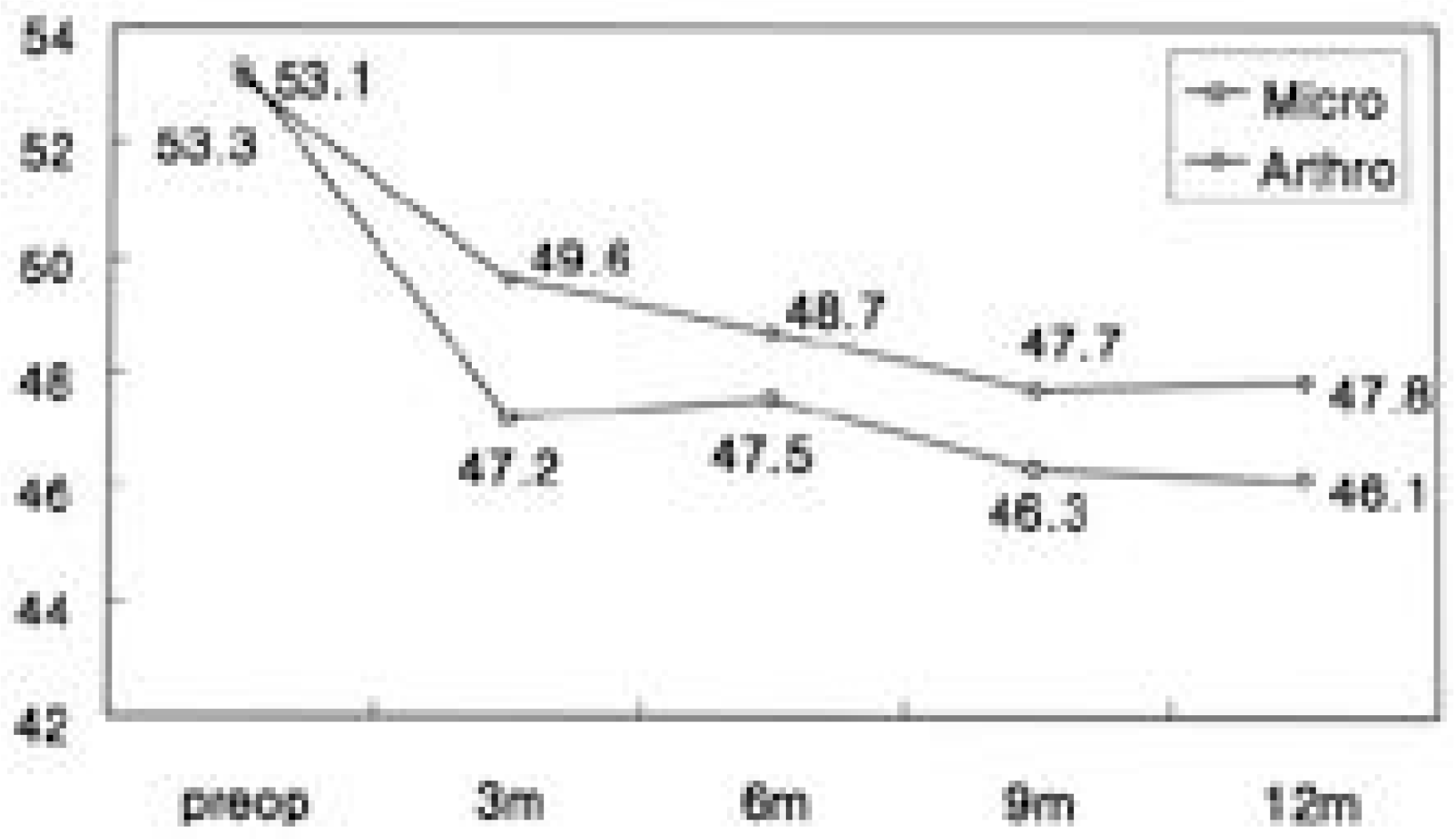 | Fig. 2.Comparision of mean disc height change between arthroscopic and microscopic discectomy. (by Farfan’s method) |
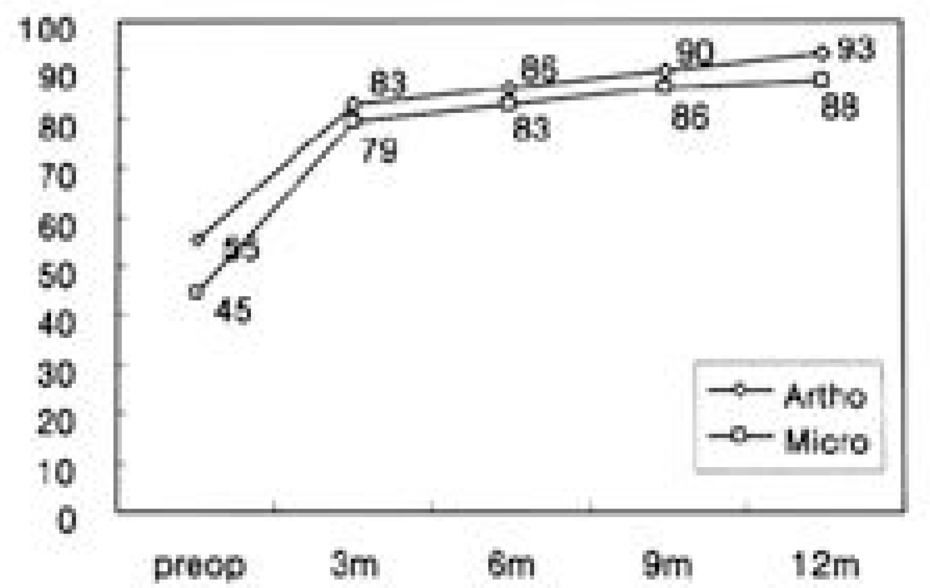
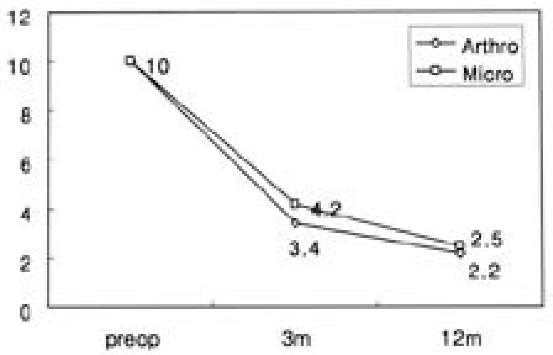 | Fig. 3.Comparison of changes of VAS(visual analogue scale) between arthroscopic and microscopic discectomy. |
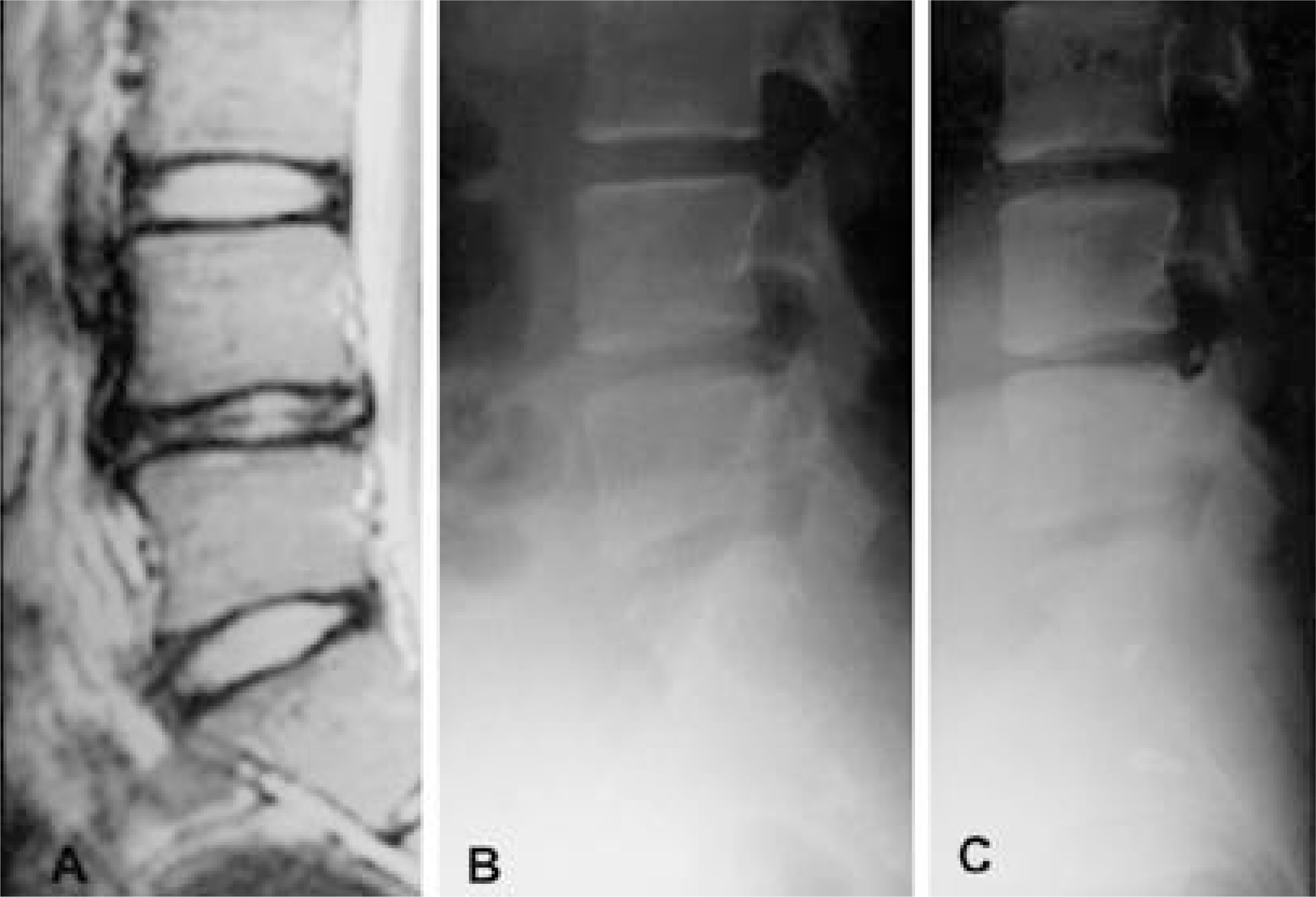 | Fig. 4.14-years-old female with Rt leg radiating pain. Preoperative MRI (A) showed the protruded disc herniation at L4-5. Lateral radiograph at 24 months follow-up after arthroscopic disc excision (C) showed decreased disc height in comparision with preoperative radiograph (B). |
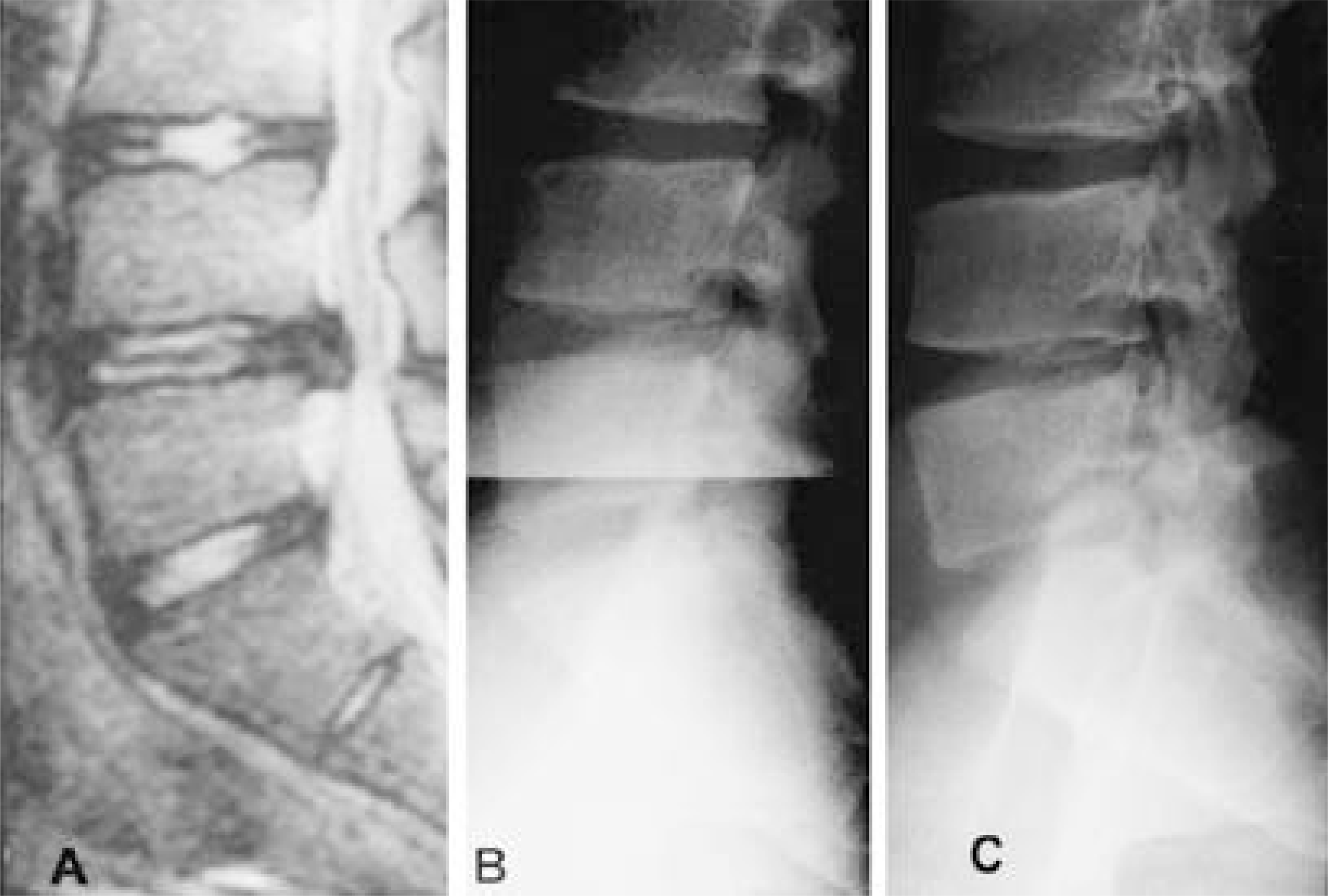 | Fig. 5.17-years-old male with Rt leg radiating pain and positive straight leg raising. Preoperative MRI (A) showed the extruded disc herniation at L4-5. Lateral radiograph at 26 months follow-up after microscopic disc excision (C) showed decreased disc height in comparision with preoperative radiograph (B). |
Table 1.
Objective clinical results of Arthroscopic & Mircoscopic discectomy
Table 2.
Comparison of the clinical results between Arthroscopic and Microscopic discectomy groups (by Macnab’s criteria)
| Results | Arthroscopic group | Microscopic group |
|---|---|---|
| Excellent | 14(66.7%) | 25(54.3%) |
| Good | 5(23.8%) | 16(34.8%) |
| Fair | 2(9.5%) | 4(8.7%) |
| Poor | 0(0%) | 1(2.2%) |
Table 3.
Comparison of mean disc height change between Arthroscopic and Microscopic discectomy (by Farfan’s method)
| Arthroscopic group | Microscopic group | |
|---|---|---|
| Preop | 53 | 53 |
| Follow-up | 48 | 46 |
| Decreased rate | 9.5% | 13.2% |
| (p<0.05) |
Table 4.
Subjective clinical results of Arthroscopic and Microscopic discectomy


 PDF
PDF ePub
ePub Citation
 Print
Print


 XML Download