

 PDF
PDF ePub
ePub Citation
 Print
Print


Abstract
Design
A retrospective study was performed in isthmic and degenerative spondylolisthesis patients who had undergone posterolateral fusion (PLF) only (group Ⅰ) or posterolateral fusion (PLF) with posterior lumbar interbody fusion (PLIF) (group Ⅱ).
Objectives
The objective of this study was to help in the selection of a surgical treatment option for spondylolisthesis.
Summary of Literature Review
Irrespective of whether groupⅠ or groupⅡ, satisfactory results have been reported in the surgical treatment of spondylolisthesis. However, isthmic and degenerative types have not been investigated in terms of outcome.
Material and Methods
We analyzed 112 patients (Isthmic: groupⅠ(32), groupⅡ(22), Degenerative : groupⅠ(37), groupⅡ(21)) who underwent surgical treatment for spondylolisthesis between A pril 1995 and December 2000. Kirkaldy-Willis criteria, radiologic union state, reduction ratio of slippage, change of disc space and change of segmental angle were analyzed as indicators of outcome.
Results
We found the following by radiologic analysis: In isthmic spondylolisthesis, groupⅡ was better than groupⅠ in terms of reduction ratio of slippage (reduction loss:3.38% vs. 2.3%, P=0.15), change of segmental angle (reduction loss : 2.11 °vs. 1.6°, P=0.15), bone union (83% vs. 92%, P=0.45) and change of disc space (reduction loss : 2.83 mm vs. 1.9 mm, P=0.02). In the degenerative spondylolisthesis, groupⅡ did not show significant difference from groupⅠ in terms of reduced slippage (reduction loss:3.8% vs. 3.85%, P=0.47), change of segmental angle (reduction loss: 2.73。 vs. 2.64。, P=0.43), bone union (80% vs. 87%, P=0.72) or disc height (reduction loss: 3.2 mm vs. 3.14 mm, P=0.45).
In terms of clinical outcome, groupⅡ was better than groupsⅠ in cases of isthmic spondylolisthesis (fair≤:85% vs. 93%, P=0.72), however, groupsⅡ was not better than groupsⅠ in cases of degenerative spondylolisthesis (fair≤:83% vs. 85%, P=0.23).
REFERENCES
1). Boos N, Marchesi D, Zuber K and Aebi M. Treatment of severe spondylolisthesis by reduction and pedicular fixation: A 4-6 Year Follow Up Study. Spine. 18:1655–1661. 1993.
2). Bridwell KG, Sedgewick TA, O’ Brien MF, Lenke LG and Baldus C. The role of fusion and instrumentation in the treatment of degenerative spondylolisthesis with spinal stenosis. J Spinal Disorder. 6:461–472. 1993.

3). Cho DY, Kim EH, Koh ES and Woo BC. The change of segmental sagittal angle in Low-grade spondylolisthesis after pedicular screw fixation with or without PLIF-PLIF+PLF versus PLF groups. J Korean Orthop Assoc. 30:842–851. 1995.
4). Cloward RB. Posterior lumbar interbody fusion updated. Clin Orthop. 193:20–37. 1985.
5). Enker P and Steffee AD. Interbody fusion and instrumentation. Clin Orthop. 300:90–101. 1994.
6). Esses S, Natout N and Kip P. Posterior interbody arthrodesis with a fibular strut graft in spondylolisthesis. J Bone Joint Surg. 77-A:172–176. 1995.
7). Esses SI, Sach BL and Dreyzin V. Complications associated with the technique of pedicle screw fixations. Spine. 18:2231–2239. 1993.
8). Fredrickson BE, Baker D. Mcholick WJ, Yuan HA and Lubicky JP. The natural history of spondylolysis and spondylolisthesis. J Bone Joint Surg. 66-A:699–707. 1984.
9). Garfin SR. Summation. Spine. 19(20S):2300S–2305S. 1994.
10). Heim SE. Transpedicle instrumentation in the degenerative spine. Clin Orthop. 337:97–110. 1997.
11). Herkowitz HN and Kurz ST. Degenerative lumbar spondylolisthesis with spinal stenosis. J Bone Joint Surg. 72-A:802–808. 1991.
12). Horowtch A, Peek RD, Thomas JC, Widell EH, Dimartino PP, Spencer CW, Weintein J and Wiltse LL. The pedicle screw fixation system, early clinical results. Spine. 14:461–467. 1989.
13). Kim SS, Denis F, Lonstein JE and Winter RB. Factors affecting fusion rate in adult spondylolisthesis. Spine. 15:979–983. 1990.
14). Kirkaldy-Willis WH, Paine KWE and Cauchoiz J. Lumbar spinal stenosis. Clin Orthop. 99:30–52. 1974.
15). Lenke LG, Birdwell KH, Bullis D, Betz RR, Baldus C and Schoenecker PL. Results of in situ fusion for isthmic spondylolisthesis. J Spinal Disorder. 5:433–441. 1992.
16). Lin PM. Posterior lumbar interbody fusion technique: Complication and pitfalls. Clin Orthop. 193:90–102. 1985.
17). Lombardi JS, Wiltse LL, Reynolds J, Widell EH and Spencer C Ⅲ. Treatment of degenerative spondylolisthesis. Spine. 10:821–827. 1985.
18). Lorenz M, Zindrick M, Schwaegler P, Vrbos L, Collatz MA, Behal R and Cram R. A comparison of single level fusions with and without hardware. Spine. 16:S455–S458. 1991.
19). Ma GWC. Posterior lumbar interbody fusion with posterior elements as chip bone graft. Clin Orthop. 193:57–63. 1985.
20). Matthiass HH and Heine J. The surgical reduction of spondylolisthesis. J Bone Joint Surg. 13:39–48. 1931.
21). Newman PH and Stone KH. The etiology of spondylolisthesis. J Bone Joint Surg. 45-B:39–59. 1963.
22). Steffee AD and Sitkowski DJ. Posterior lumbar interbody fustion and plates. Clin Orthop. 227:99–102. 1988.
23). Suk SI, Lee CK, Kim WJ and Kim HG. Adding poste - rior lumbar interbody fusion to pedicle screw fixatin and posterolateral fusion after decompression in spondylolytic spondylolisthesis. J Korean Orthop Assoc. 30:1638–1646. 1995.
24). Taillard W. Le Spondvloisthesis chez I'ecfant et I'adolescent. Acta Orthop Scand. 24:115. 1954.
25). Valkenburg HA, Haanen HCM. The epidemiology of low back pain. In: White AA, Gordon SL, des, Proces Am Assoc Orthop Surg Symposium on Low Back Pain. 9–22. 1982.
26). Verlooy J, De Smedt K and Selosse P. Failure of a modified posterior lumbar interbody fusion technique to produce adequate pain relief in isthmic spondylolytic grade l spondylolisthesis patients. Spine. 18:1491–1495. 1993.
27). Wang JM, Kim DJ and Yun YH. Posterior pedicular screw instrumentation and anterior interbody fusion in adult lumbar spondylolysis or grade I spondyloilisthesis with segmental instability. J Spinal Disord. 9(2):83–8. 1996.
28). Wiltse LL, Newman PH and Macnab I. Classification of spondylolysis and spondylolisthesis. Clin Orthop. 117:23–29. 1976.
Fig. 1.
Isthmic Spondylolisthesis with PLF Fig. 1. A. Lateral radiograph of symptomatic isthmic spondylolisthesis in a 48-year-old female. Fig. 1.B. Postoperative radiograph demonstrated correction of deformity. Fig. 1. C. Last follow-up radiograph demonstrated loss of the correction.
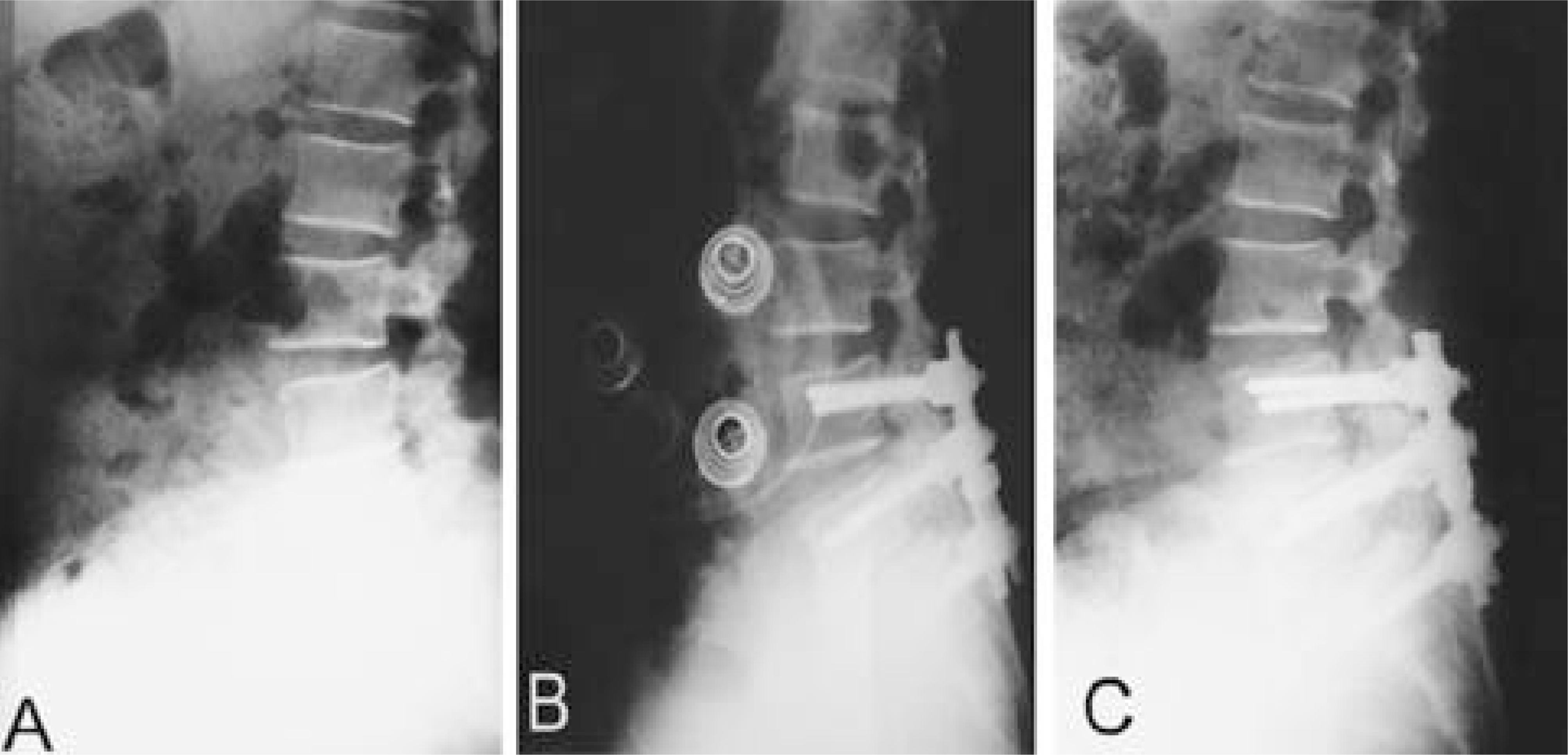
Fig. 2.
Isthmic Spondylolisthesis with PLF and PLIF Fig. 2. A. Lateral radiograph of symptomatic isthmic spondylolisthesis in a 53-year-old female. Fig. 2. B. Postoperative radiograph demonstrated full correction of deformity. Fig. 2. C. Last follow-up radiograph demonstrated well-consolidated interbody fusion without loss of the slippage.
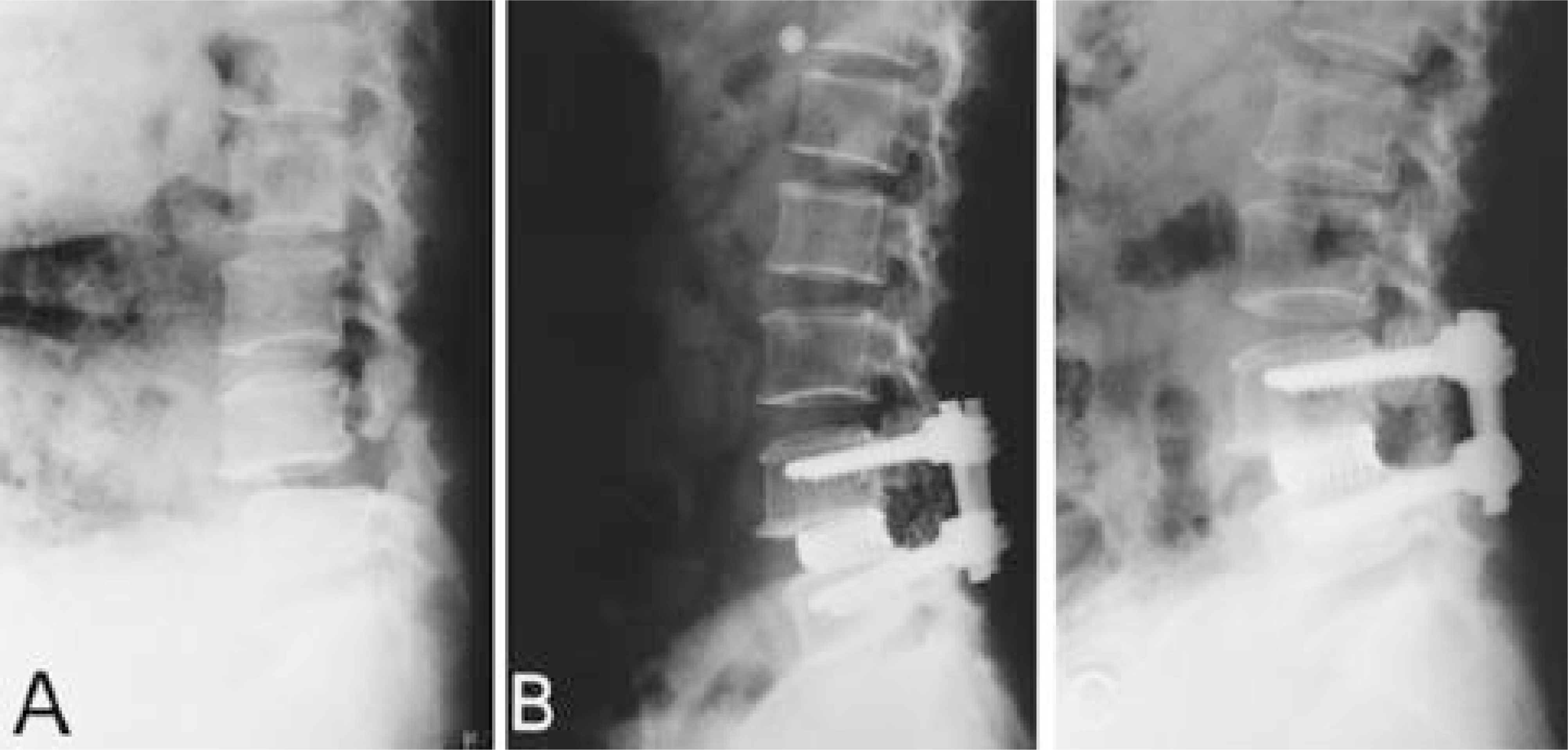
Fig. 3.
Degenerative Spondylolisthesis with PLF Fig. 3. A. Lateral radiograph of symptomatic degenerative spondylolisthesis L4 on L5 in a 52-year-old female. Fig. 3. B. Postoperative radiograph demonstrated correction of deformity. Fig. 3. C. Last follow-up radiograph demonstrated loss of disc height.
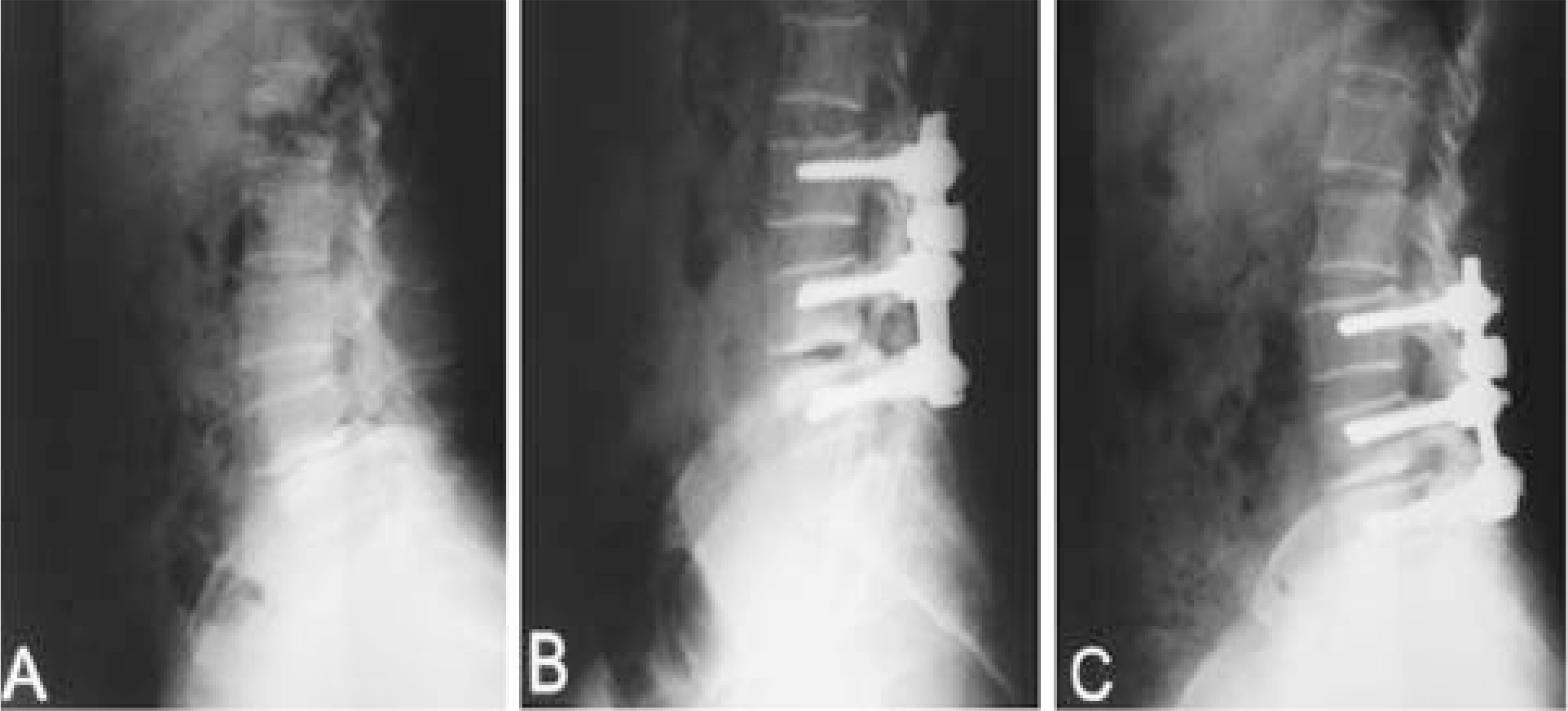
Fig. 4.
Degenerative Spondylolisthesis with PLF and PLIF Fig. 4. A. Lateral radiograph of symptomatic degenerative spondylolisthesis in a 66-year-old female. Fig. 4. B. Postoperative radiograph demonstrated correction of deformity. Fig. 4. C. Last follow-up radiograph demonstrated loss of the correction and disc height.
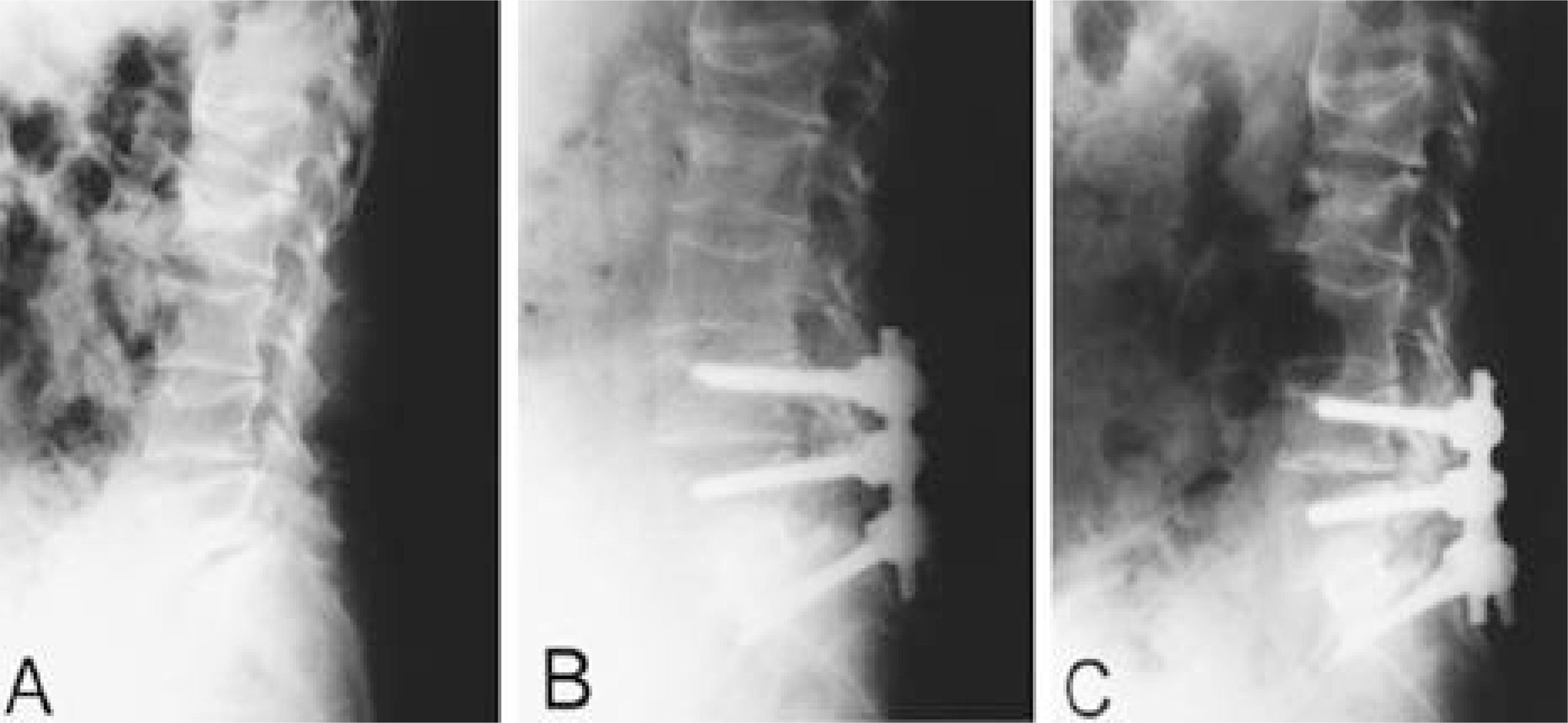
Table 1.
Patient data
Table 2.
Kirkaldy-Willis 평가기준
Table 3.
Radiologic Measurements
 XML Download