

 PDF
PDF ePub
ePub Citation
 Print
Print


Abstract
Objectives
The purpose of this study is to compare the outcomes of short segment fusion and long segment fusion in posterior facet fracture dislocation in the lumbar spine.
Summary of Literature Review
There are many controversies exist about the treatment of fracture dislocation in lumbar spine.
Material and Methods
Sixteen patients with lumbar fracture dislocation were studied retrospectively. The patients divided two groups; group one treated with one level above and below the fracture segment fixation, group two treated with two level above and below the fracture segment fixation. Two groups were compared with neurologic recovery, bladder function recovery and radiologic changes of deformities.
Go to : 
REFERENCES
1). Bohlman HH. Treatment of fracture and dislocations of the thoracic and lumbar spine. J Bone Joint Surg. 67-A:165–169. 1985.
2). Chapman JR, Anderson PA. Thoracolunbar spine fractures with neurologic deficit. Orthop Clin North Am. 25:595–597. 1994.
3). Choi WS, Baik CH, Cho SS, Park HJ, Koh DH. Clinical analysis of unstable thoracolumbar fractures and fracture-dislocation using transpedicular screws. J Korean Ortho Surgery. 26:719–727. 1991.
4). Denis. The three column spine and it's significance in the classification of acute thoracolumbar injuries. Spine. 8:817–831. 1983.
5). Denis F, Barkus JK. Shear fracture dislocation of the thoracolumbar spine associated with forceful hyperex -tension. Spine. 17:156–161. 1992.
6). Denis F, Ruiz H, Searls K. Comparison between square-ended distraction rods and standard round-ended distraction rods in the treatment of thoracolumbar spinal injuries. Clin Ortho. 189:162–167. 1984.
7). Dick W. Internal fixation of thoracic and lumbar spine fractures. 1st ed. Toronto: Hans Huber Co;p. 75–86. 1989.
8). Dick W, Kluger P, Margel F. A new device for internal fixation of thoracolumbar and lumbar spine fracture. Paraplegia. 23:225–232. 1985.
9). Dickson JH, Harrington RP, Erwin WD. Harrington instrumentation in the fractured unstable thoracic and lumbar spine. J Bone Joint Surg. 55-A:422–426. 1973.
10). Edward CC, Levine AM. Early rod sleeve stabilization of the injured thoracic and lumbar spine. Orthop Clin North Am. 17:121–145. 1986.
11). Farcy J, Weidenbaum M, Michelson CB, Hoeltzel DA, Athansiou KA. A comparative biomechanical study of spinal fixation using Cotrel-Dubousset instrumentation. Spine. 12:877–881. 1987.

12). Frankel HL, Hancock DO, Hyslop G. The value of postural reduction in the initial management of closed injuries of the spine with paraplegia and tetraplegia. Paraplegia. 7:179–192. 1969.
13). Gertzbein SD. Multicenter spine fractured study. Spine. 17:528–539. 1992.
14). Krag MH. Biomechanics of thoracolumbar spinal fixation. Spine. 16:S84–S99. 1991.
15). McLain RF, Sparling E, Benson DR. Early failure of short-segment pedicle instrumentation for thoracolumbar fractures. J Bone Joint Surg. 75-A:162–167. 1993.
16). Shaffrey CJ, Saffrey ME, Whitehill R, Nockels RP. Surgical treatment of thoracolumbar fractures. Neurosurgery Clin North Am. 8:519–540. 1997.
17). Stauffer ES. Internal fixation of fracture of the thoracolumbar spine. J. Bone Joint Surg. 66-A:1136–1138. 1984.
18). Stauffer ES. Thoracolumbar spine fractures without neurologic deficit. AAOS monography series, 49-80. 1993.
19). Tasdemirogle E, Tibbs PA. Longterm followup results of thoracolumbar fractures after posterior instrumentation. Spine. 20:1704–1708. 1995.
Go to :
Figures and Tables%
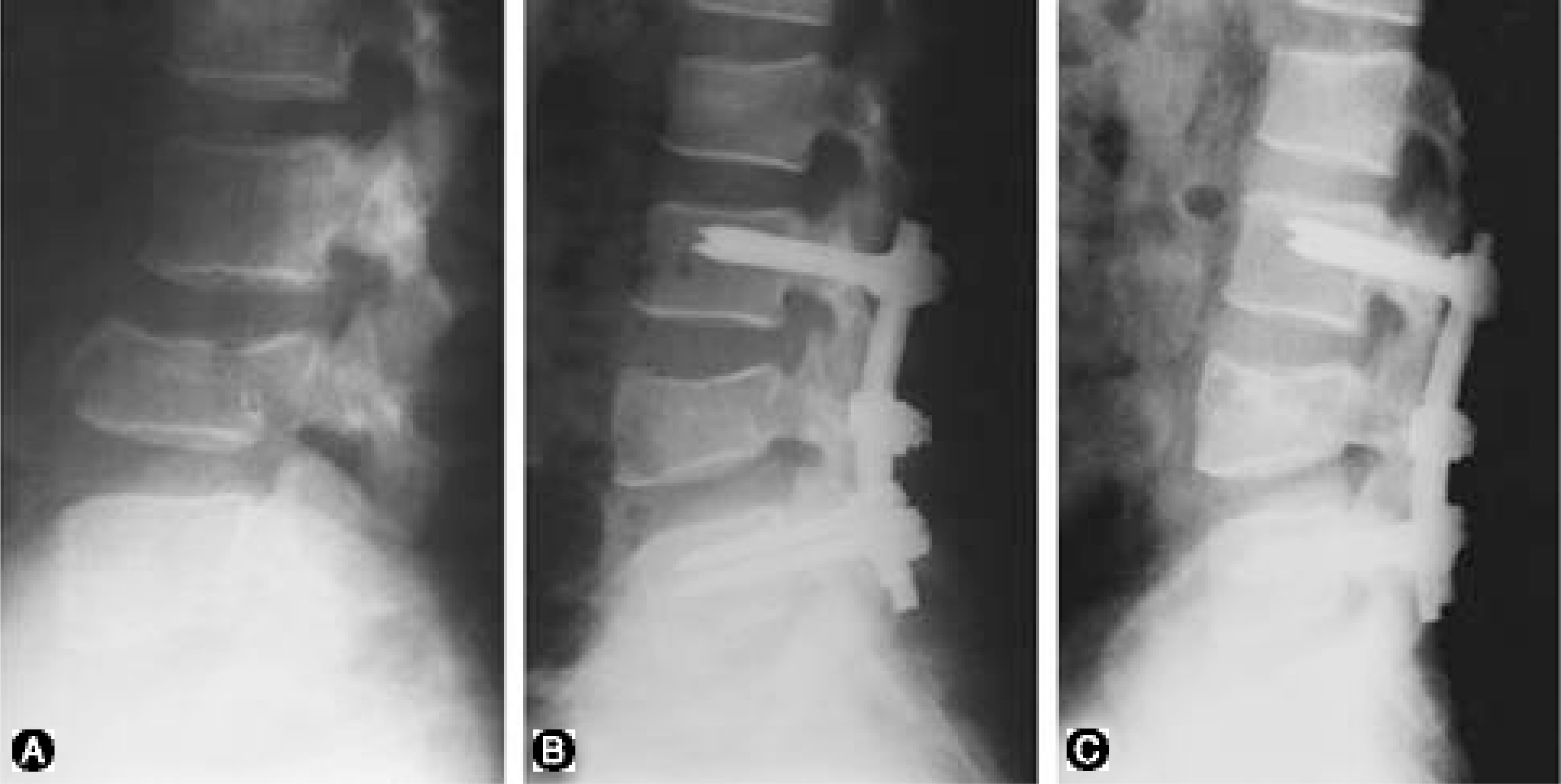 | Fig. 1-AInitial lateral radiograph shows L4-5 fracture dislocation. Fig. 1-B : Immediate postoperative radiograph, treated posterolateral fusion with above and below 1 level fixation, shows satisfactory reduction. Fig. 1-C : Twelve months after operation, lateral radiograph shows 3° loss of sagittal angle |
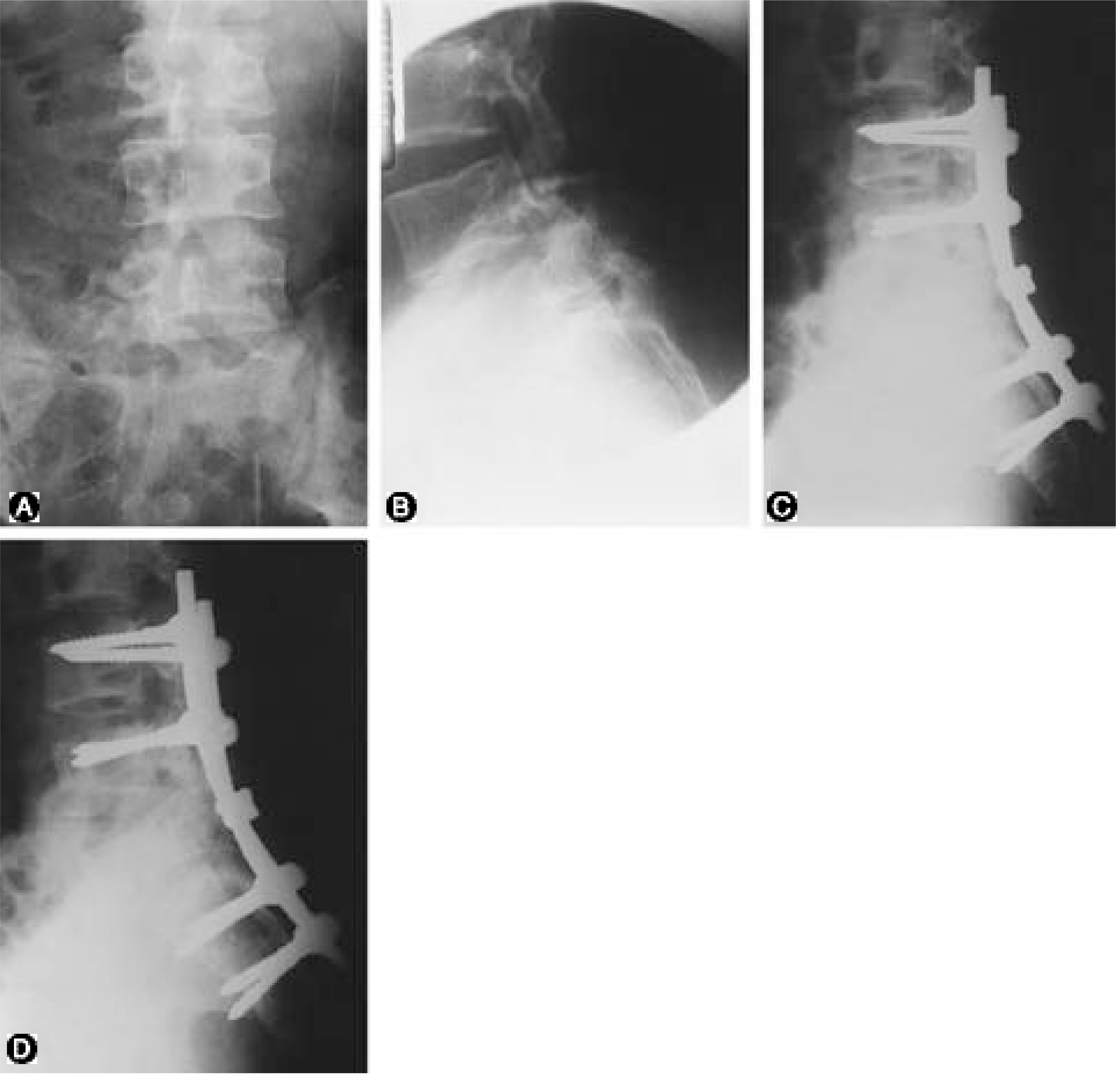 | Fig. 2-AB. Initial anteroposterior(A) and lateral(B) radiographs show L5-S1 fracture dislocation. Fig. 2-C. Immediate postoperative radiograph, treated posterolateral fusion with above and below 2 level fixation, shows satisfactory reduction. Fig. 2-D. 5 years after operation, lateral radiograph shows 5° loss of sagittal angle. |
Table 1.
Level of Fracture-Dislocation
| Level | No (%) |
|---|---|
| L2/L3 | 3(18.75) |
| L3/L4 | 4(25.00) |
| L4/L5 | 6(37.50) |
| L5/S1 | 3(18.75) |
 XML Download